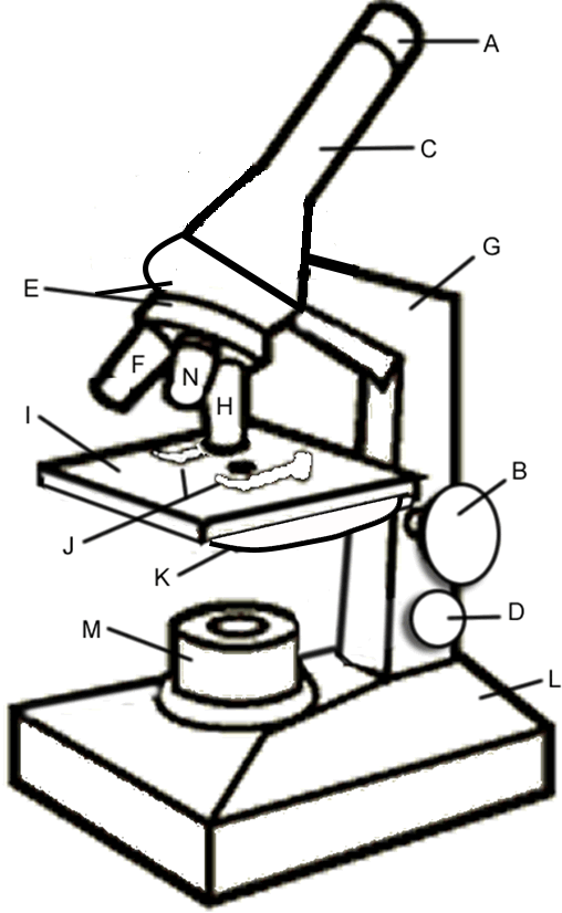
Biology lesson plans worksheets tutorials and resources for teachers and students. The arm is the correct place to grip the microscope when carrying it while supporting the base with the palm of your other hand.
Microscope Coloring
Free microscope worksheet parts labeling diagram 1 page printable features high quality microscopic worksheets life science.
Microscope labeling worksheet biology corner. Leave a Reply Cancel reply. Carry by the base and arm with both hands. Resource comes as both a PDF for printing and a.
Most biology classes will cover microscope use at the beginning of the year. Required fields are marked. Label a microscope worksheet.
Answer key to the worksheet on mitosis in an onion root labels the graphic and describes the phases of the cell cycle. A lens inside the eyepiece A usually has a magnification of 10x. Home Worksheet Microscope Labeling Worksheet Answers Coloring Sheet Plant Cell Answer Key Animal Worksheet Biology Corner Drawing Cells Parts.
Each row of desks uses the same cord. Plant Cell Diagram Biology Corner. The word microscope is derived from the greek words micro small and.
Store with cord wrapped around microscope and the scanning objective clicked into place. Saved by Help Teaching. This simple worksheet pairs with a lesson on the light microscope where beginning.
FREE Microscope Parts Worksheet - This free microscope diagram printable is perfect for reviewing parts of microscope before your life science students or biology classes take out the microscopes for their next lab. Mitosis internet lesson cells alive. 1 86 Answer Key Science 1-7 71 57 43 29 14 0 1.
Plug your microscope in to the extension cords. Microscope Lab Worksheet Name. Slides contain images with drag and drop labels for students to practice labeling.
View Lab Report - iLab4_WK4_BIOS135_MD_NOMAN from BIOS 135 at DeVry University New York. This resource contains 1 worksheet for students to label the parts of a microscope and 1 worksheet for students to complete a chart detailing the functions of each microscope part. This activity has been designed for use in homes and schools.
G Labeling Scientific Tools Microscope G F E D C B A 1 Illuminator 2 Stage 3 Eyepiece 4 Focus Fine 5 Lense 6 Focus Course 7 Base Determine which letter best matches each microscope piece. Microscope Labeling The first part of this investigation students examine slides of mitosis in an. Your email address will not be published.
Microscope The Biology Corner Name microscope labeling. This is a slide activity for students to learn the parts of the muscle such as the epimysium perimysium endomysium and microscopic structures like the A and I bands actin myosin and sarcolemma. The Microscope This mitosis investigation was created during the 2020 pandemic for remote learning.
Microscope tips guides ideas science activity. Make sure all backpacks and junk are out of the aisles. Plant Cell Lab microscope observation of onion and elodea Plant Cell Lab Makeup can be done at home or at the library Plant Cell Virtual.
In this internet lesson you will review the steps of mitosis and view video simulations of cell division. Images of cells showing the major structures and organelles including a diagram that maps the process by which proteins are made and exported. 500x see also microscope ppt for students to learn the parts and function of the microscope microscope labeling student worksheet.
Color the arm green and the base red. Follow Biologycorner on Facebook. Examine data that compares states that.
Please label the microscope diagram below. In biology corner microscope labeling worksheet answers biology microscope worksheet answers the compound microscope worksheet answers biology if8765. The body tube C allows the light to pass upward to where the users eye is.
Meiosis internet lesson answer key the biology corner.
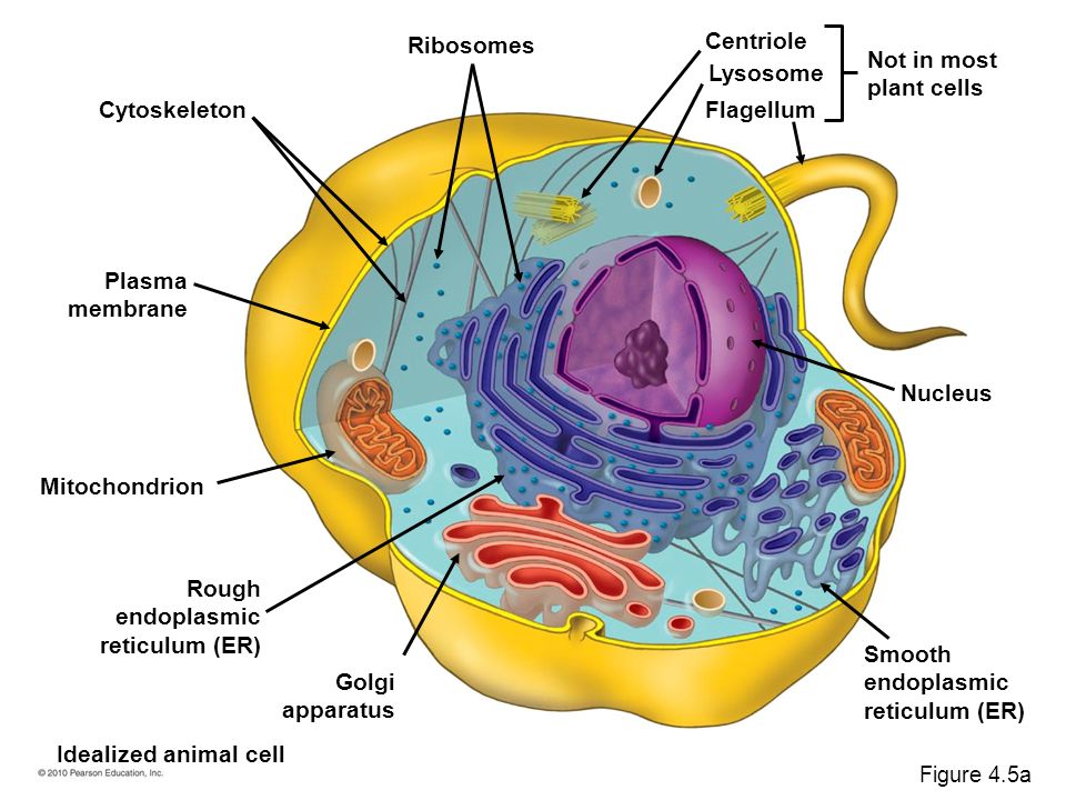
As observed in the labeled animal cell diagram the cell membrane forms the confining factor of the cell that is it envelopes the cell constituents together and gives the cell its shape form and existence. Ib Biology Topic 2 3 5 Animal Cell Vs Plant Cell.
2010 Pearson Education Inc Chapter 4 A Tour Of The Cell 6 Sections 1 Microscopic World Of Cells 2 Membrane Structure 3 Nucleus Ribosomes Genetic Ppt Download
Solved Rag Each Cell Structure To The Appropriate Bin If Chegg Com - The functional and structural units of all living things.

Animal cell structure labeled mastering biology. They also comprise other membrane-bound organelles and cellular structures. A thread-like gene-carrying structure found in the nucleus of a eukaryotic cell and most visible during mitosis and meiosis. Ribosomes flagella plasma membrane.
As stated before animal cells are eukaryotic cells with a membrane-bound nucleus. _____ serve as intracellular highways for transporting vesicles and organelles. They are also required for cellular locomotion via flagella and cilia.
Most of the cells are microscopic in size and can only be seen under the microscope. Strands containing DNA genes and associative protein. Also the main.
Cell membrane is made up of lipids and proteins and forms a barrier between the extracellular liquid bathing all cells on the exterior and the cell organelles floating in the cells cytoplasm. Eukaryotic Cell Structure Sciencetopia. Animal cell structure labeled diagram science structures labeled diagrams animal cell biology pictures animal cell diagram homeschool Label Gallery Get some ideas to make labels for bottles jars packages products boxes or classroom activities for free.
_____ are responsible for cell locomotion and the cells structural characteristics. An animal usually has a centrosome with a pair of centrioles involved in cell division. Large organelle the contains most of the genes that control the cell.
Furthermore these cells exhibit the presence of DNA inside the nucleus. Animal Cell Structures And Functions Mastering Biology. 700x502 px Eukaryotic Cell Structure Animal Cell Plant And Animal Cells Eukaryotic Cell - Ideas about cell structure have changed considerably over the years.
This is an animal cell. Animal cells have a basic structure. August 9th 2013 055415 AM.
Plankton are organisms drifting in oceans and seas. A Labeled Diagram Of The Animal Cell And Its Organelles Biology Wise. Cell Structure and Function - Illinois Community College Board 109 Cell Structure and Function By Lori Armstrong Course BIO 111 Introduction to Biology Description This module will introduce the student to the structures of.
Simple Animal Cell Diagram Labeled images similar and related articles aggregated throughout the Internet. Biology animal cell labeled parts information recently was sought by people around us maybe one of you. So it is called as the structural and functional unit of life.
Mod6pdf - Read File Online - Report Abuse. Animal Cell Structures And Functions Mastering Biology Describe and interpret drawings and photographs of typical animal and plant cells. Plankton are organisms drifting in oceans and seas.
44 Labeled Biology Animal Cell Structure Pics. Animal cell structure mastering biology free Pictures Images and Stock Photos. The shape of animal cells also varies with some being flat others oval or rod-shaped.
Appendage that propels the cell. Weve gathered our favorite ideas for Animal Cell Labeled Explore our list of popular images of Animal Cell Labeled Photos Collection with high resolution. Free Animal Cell.
Structure Of Animal And Plant Cell Download Scientific Diagram. Animal Cell - All the living organisms are made up of cells and it is the smallest unit of life. Double membrane that encloses the nucleus.
There are also more intriguing shapes such as curved spherical concave and rectangular. A structure in an animal cell composed of cylinders of microtubule triplets arranged in a 9 0 pattern. Animal Cell Structures And Functions Mastering Bio.
An easy and convenient way to make label is to generate some ideas first. Identify the part of the cell that is highlighted. You already know that animal cells consist of a cell membrane nucleus and a fluid cytoplasm.
You should make a label that represents your brand and. High Resolution Cross Section of Ascaris Giant Roundworm Parasitic Worm in Humans Captured by a scientific microscope and Canon 5D Mark IV. People nowadays are already accustomed to using the internet on mobile phones to search for image and video information to be used as inspiration for one of them in category information and according to the title of this post we will will share biology animal cell labeled parts Images.
It helps in carrying out the functions such as respiration nutrition digestion excretion etc. Choose the best description of the cell cytoskeleton-The cell cytoskeleton is a dynamic network of fibres that can be quickly dismantled and reassembled to change cell shape and the position of cell components-Cytoskeleton is made of actin and tubulin and they are organized similarly to muscle cells. Below the basic structure is shown in the same animal cell on the left viewed with the light microscope and on the right with the transmission electron.
Animal cell definition with cell size and shape. Cancer Stem Cells Cancer Stem Cells. Plant Vs Animal Cells Review Article Khan Academy.
Mitochondrion Definition Structure And Function Biology Dictionary. The cell from latin cella meaning s. Animal Cells And The Membrane Bound Nucleus.
2 3 Eukaryote Questions Answered.
Parts Of A Leaf Labelled Diagram. Recently Viewed and Downloaded.

Leaf Structure Labelled With Annotations Leaf Structure Leaves Science Images
Show with another labelled arrow the direction the energy comes from.
Leaf diagram biology labelled. Leaves stems and roots are organs consisting of different types of tissues Plant leaves are the main organ. On the stem they are little apart while on the branches they are overlapping. Leaf Cell Definition And Types Biology Dictionary.
The foliage leaves are characterised by green colour thinness and flatness. Labelled Leaf Diagram Spanish Science Biology Flora Plant Mps Ks2 Structure Of A Leaf Youtube Structure Of A Typical Leaf With Diagram Gcse Plant Biology Diagrams To Label Teaching Resources Labeled Parts Of A Leaf Diagram Draw A Leaf And Label It S Parts Brainly In Enjoy. Leaf Anatomy Vector Illustration Diagram Biological Macro Scheme.
In addition to supporting the flower the stem enables water and nutrients to flow from the soil into the leaf for the process of photosynthesis to take place. Draw A Labelled Diagram Of Cross Section Leaf Lamina To Show Chloroplasts From Science Life Processes Class 10 Himachal Pradesh Board Class 7 Science Diagram Of A Section Leaf You Ib Biology Notes 9 1 Plant Structure And Growth. Leaves are borne on the main stem as well as on the branches.
Answers to this Paper must be written on the paper provided separately. Photosynthesis Leaf Diagram Labeled. Cell Structure of a Leaf Labelled diagram.
8CA Label the Internal Structure of a Leaf Labelled diagram. Parts of a leaf their structure and functions with diagram describe the structure of leaf with help a neat well labelled diagram brainly in draw the labelled diagram of parts a simple leaf brainly in draw a labelled diagram of internal structure dicot leaf qs study. KS3 English Writing and literary techniques.
By means of labelled arrows drawn on the diagram above show the pathway taken by each of the two raw materials to a cell in the centre of the leaf. The leaf is a flattened lateral outgrowth of the stem in the branch developing from a node and having a bud in its axil. Parts of a plant.
Cross Section of a Leaf Labelled diagram. I It is green compressed with a wide lamina. Leaf Definition Parts And Types With Diagram Botany.
Iii Mesophyll tissue is present and is composed of palisade parenchyma and spongy parenchyma. Features of a Leaflet Labelling Labelled diagram. Leaf Diagram Labeled.
You will not be allowed to write during the first 15 minutes. Labeled Leaf Diagram Biology Written By JupiterZ Wednesday November 21 2018 Add Comment Edit. Leaf Epidermis Images Stock Photos Vectors Shutterstock.
The leaves take up water and carbon dioxide and convert them into carbohydrates in. Structure of a Leaf - Photsynthesis Made Simple Lilly Figueroa A stem and leaf diagram shows numbers in a table format. Give one word names for the processes by which these raw materials move across the leaf as seen in the diagram.
ICSE Biology Previous year Question Paper 2018 Solved for Class 10 General Instructions. Labelled diagram of nerium leaf transverse section. Cross Section Of A Leaf Worksheet.
Labelled diagram of a leaf Basic. Labelled Diagram Of Pumpkin Leaf Building And Troubleshooting. Diagram of a flowering plant with label.
Recently Viewed. Required fields are marked. Draw A Well Labelled Diagram Of A Typical Leaf Biology.
Cross Section of a Leaf Labelled diagram. Your email address will not be published. The structure of a leaf has adaptations so that it can carry out photosynthesis effectively.
It is normally green in colour and manufactures food for the whole plant. A leaf is a plant organ that is flat thin and usually green in color. Identifying characteristics of the internal structure of dorsiventral or dicot leaf.
Whats people lookup in this blog. Copy of 10xBi1 Label the Internal Structure of a Leaf Labelled diagram. Leaves are arranged in a spiral manner with a phyllotaxy of 25 ie sixth leaf will come above the first leaf.
Ii Leaf-blade is enriched with reticulate venation. Flower anatomy flowers consist of reproductive parts and modified leaves. Functions of leaves The function of a leaf is photosynthesis.
Plants primary teaching resources and printables sparklebox. A way to transport water to the leaf and glucose to other parts of the. It is noticeably differentiated into palisade and spongy parenchyma.