Modified from Evans HE 1993. Cornea - the clear dome-shaped tissue covering the front of the eye.

Eye Anatomy Labeled
Parts of the eye labeled vector illustration diagram Parts of the eye labeled vector illustration diagram.
Labelled diagram of eyelid. The third eyelid or nictitating membrane is a mobile protective and glandular structure lying between the cornea and the lower eyelid in the medial portion of the inferior conjunctival sac Figure 8-1. Connective tissue plate containing sebaceous glands that provide oily base for tear film. Educational beauty and nursing information.
Facts About The Eye To understand more in detail about our eye and how our eye functions we need to. Please refer to the back of this handout for descriptions. Iris - the colored part of the eye - it controls the amount of light that enters the eye by changing the size of the pupil.
The eyelids upper and lower are two movable structures composed of skin and two types of muscle. This diagram demonstrates the conceptual split between the anterior lamella skin and orbicularis right side and posterior lamella tarsus and conjunctiva left side of the eyelid. The highest peak on the upper eyelid lies slightly nasal and the lowest contour of the lower eyelid rests slightly lateral.
Instead it is made up of two separate segments fused together. There are seven extraocular muscles the levator palpebrae superioris superior rectus inferior rectus medial rectus lateral rectus inferior oblique and superior oblique. Supplied by seventh cranial facial nerve.
Their purpose is to protect the eye from foreign bod-ies and limit the amount of light entering the eye. Gives firmness to the distal portion of upper lid. The diagrams great for when youre making up new eye looks but it wouldve better if you labelled the inner crease.
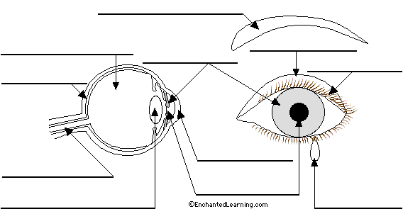
95 10 6 votes Eye Anatomy labeled In this image you will find Eyelid Lacrimal caruncle Tear duct Lateral rectus muscle Sclera Choroid Retina Macula lutea Fovea centralis Optic nerve and retinal blood Medial rectus muscle Ora Serrata Ciliary body and muscle Suspensory ligaments Posterior chamber Anterior chamber Cornea. Figure 8-1 Diagram of the eye showing normal position of the third eyelid. The upper eyelid is made up of 3 compartments lamella each of which contain a mixture of skin muscle fat tarsal plate and conjunctiva.
Lens - a crystalline structure located just behind. More detail on upper eyelid anatomy available here. This limbus is the border between the cornea and the sclera as shown in this labelled diagram.
The extraocular muscles are located within the orbit but are extrinsic and separate from the eyeball itself. When seventh nerve is damaged this muscle malfunctions and lower eyelid droops exposing cornea to drying. Eyelid eyelashes pupil lacrimal gland and other anatomical parts.
Healthy visual sensory organ. The upper eyelid normally rests as 1-2mm below the corneal limbus and is highest just nasal to the pupil 1. Label the Eye Diagram.
In addition they serve to distribute tears that lubricate the surface of the eye Fig. An eyelid is a thin fold of skin that covers and protects the eye. 165636852 stock photos online.
Contrary to popular belief the eyes are not perfectly spherical. The size of the pupil determines the amount of light that enters. Hover the diagram to zoom.
The upper eyelid is a tri-lamellae structure anterior middle and posterior. The pupil is the opening in the centre of the iris that regulates the amount of light entering the eye. The eyelids are thin mobile folds that cover the eyeball anteriorlyThey offer protection from excessive light or injury and maintain lubrication by distributing tears over the surface of the eyeball.
The average aperture of the eyelids measures about 30 mm in horizontal width and approximately 10 mm in vertical height. Diabetes and Healthy Eyes Toolkit Parts of the Eye To understand eye problems it is helpful to know the different parts of the eye. The eyelids are split into upper and lower portions which meet at the medial and lateral canthi of the eyeThe opening between the two eyelids is called the palpebral aperture or opening.
The eyelids comprise of an upper and lower eyelid joined at the medial and lateral canthi. Millers Anatomy of the Dog. December 31 2008 at 407 pm 123108 Christine.
New users enjoy 60 OFF. The eyelid margin. They act to control the movements of the eyeball and the superior eyelid.
Human Eye Diagram. The human eye eyeball diagram parts and the eyelids are folds of skin that can close to cover the eyeball in this manner it can protect the eyeball from very bright light and injury the eyelids are also able to wipe away any particles that make contact with the outer layer of the eyeball and keeps the cornea moist diagram of the eye view more eye diagrams mission. The gray line which is the muscle of Riolan the most superficial aspect of.
This article explores this anatomy in more detail and is key to understanding ptosis. Download 4228 Eye Diagram Stock Illustrations Vectors Clipart for FREE or amazingly low rates. Read the definitions then label the eye anatomy diagram below.
Controls forceful eyelid closure.