Click the bone below for the answers. Ahcdw7sol5 Pdf 5 Award 1 00 Point Problems Adjust Credit For All Students Correctly Label The Following.

Module 2 Skeletal System Flashcards Quizlet
Correctly label the following bones and anatomical features of the lateral view of the skull.

Correctly label the following parts of bone cells quizlet. Lysosomes osteocyte ruffled border stem cells secretary vesicles periosteum osteoclast nuclei osteoblast osseous ue prev 37 of 50 next. Most organelles of the cell such as mitochondria are packed into the spaces between the myofibrils. 36 Correctly Label The Following Parts Of Bone Cells.
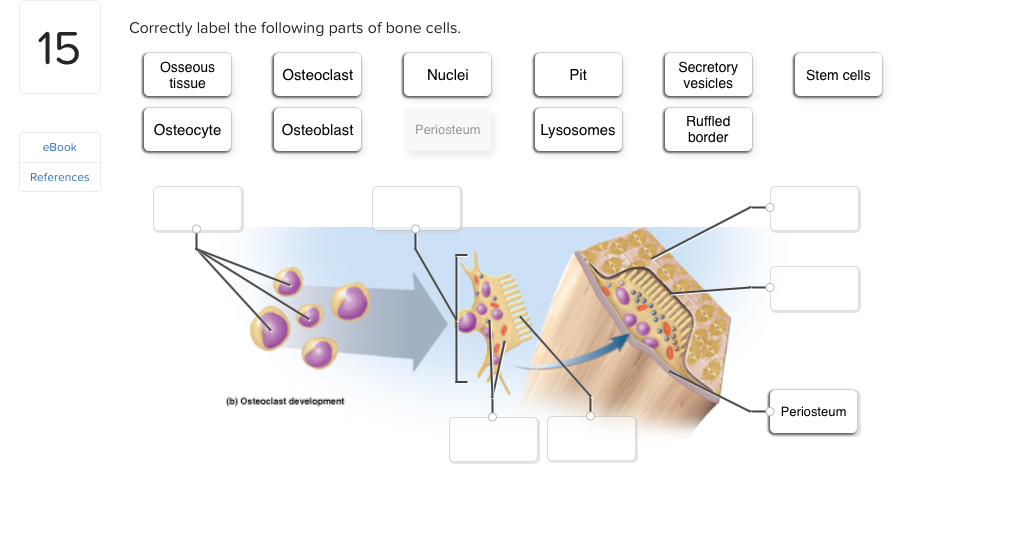
Osseous Tissue Show transcribed image text Correctly label the following parts of bone cells Osseous tissue Secretory vesicles Osteoclast Nuclei Pit Stem cells Ruffled border Osteocyte Osteoblast Periosteum Lysosomes eBook References b Osteoclast development Periosteum Correctly label the following parts of bone cells. Shannan muskopf october 16 2020. Correctly label the following parts of a skeletal muscle fiber.
Correctly label the following parts of bone cellsOsseous Tissue Show transcribed image text Correctly label the following parts of bone cells Osseous tissue Secretory vesicles Osteoclast Nuclei Pit Stem cells Ruffled border Osteocyte Osteoblast Periosteum Lysosomes eBook References b Osteoclast development Periosteum Correctly label the following parts of bone cells. Aconnos flexible IoT development platform - Personalized solutions for powering the Internet of Things. Lacrimal bone nasal bone temporal bone occipital bone parietal bone sphenoid bone ethmoid bone frontal bone.
Long Bone Labeled - Solved. A P Module 6 Flashcards Quizlet. The outer surface of.
The long bones are those that are longer than they are wide. Chap11 Muscle Tissue. Correctly label the following areas of the thoracic cavity in the newborn and the adult.
Correctly label the following parts of bone cells quizlet. Long bones contain yellow bone marrow and red bone marrow which produce blood cells. When axial loading of long bones occurs an electropositive convex surface and an electronegative concave surface are created.
A very large cell formed in bone marrow its function is to absorb and remove unwanted tissue They resorb dissolve the bone. Muscle fibers have multiple flattened or sausage-shaped nuclei pressed against the inside of the sarcolemma. They are primarily spongy bone that is covered with a thin layer of.
Correctly label the following parts of bone cells Osseous tissue Secretory vesicles Osteoclast Nuclei Pit Stem cells Ruffled border Osteocyte Osteoblast Periosteum Lysosomes eBook References b Osteoclast development Periosteum. Frontiers Sarcoplasmic Hypertrophy In Skeletal Muscle A Scientific Unicorn Or Resistance Training Adaptation Physiology. Correctly label the following parts of bone cells.
Correctly label the following parts of bone cells. Written By Kimberly T Kirkpatrick Sunday June 20 2021 Add Comment Edit. Long bone labeled find out more about long bone labeled.
Correctly label the following parts of a skeletal muscle fiber quizlet. Long Bone Labeling Long Bone Diagram Labeled Quizlet - 32 Correctly Label The Following Anatomical Parts Of A Long. Unit 3 part 1 x section bone.
The labels include proximal epiphysis. Long Bone Labeling Quizlet 32 Correctly Label The Following Anatomical Parts Of A Flat Bone Labels For Your Ideas. Once a tunnel of bone is formed around a blood vessel the bone building cells of the periosteum lay down bone in concentric circles that fill in the tunnel.
Periosteum Compact Bone Spongy Bone. Drag each label into the proper position to identify the type of bone. Correctly label the following parts of bone.
There also are bands of fibrous connective tissuethe ligaments and the tendonsin intimate relationship with the parts of the skeleton. Bone building cells beneath the endosteum lay down bone to form ridges around a blood vessel. Correctly label the following parts of a skeletal muscle fiber.
Correctly Label The Following Anatomical Parts Of. Place the labels in the proper position to designate the location of the structures or events Those needed in small amounts are classified as micro minor or trace minerals Recruitment is the process by which a muscle produces more force to pick It might seem impossible to you that all custom-written essays research papers. Correctly label the following parts of bone cells Osseous tissue Secretory vesicles Osteoclast Nuclei Pit Stem cells Ruffled border Osteocyte Osteoblast Periosteum Lysosomes eBook References b Osteoclast development Periosteum.
A P Module 6 Flashcards Quizlet. I like the game correctly label the following parts of bone cells - You should try it out and see what you think.
The bones of the human skeleton can be grouped into five types. Contains small blood lie at right angles to the long axis of the bone and connect structural unit of compact bone.
Cartilage Bone Ossification The Histology Guide
From the quiz author.

Label parts of compact bone. The bone is arranged in concentric layers around those canals. Circumferential lamellae are located between osteons. Label parts of compact bone Learn with flashcards games and more for free.
The hollow region in the diaphysis is called the medullary cavity which is filled with yellow marrow. No reproduction but without the prior written consent of Mewton Trabeculae Long bone Spongy bone Spicules Compact bone 100 Chwe tonnonomen. The walls of the diaphysis are composed of dense and hard compact bone.
Compact bone is formed from a number of osteons which. Compact bone is the denser stronger of the two types of osseous tissue Figure 636. A flat bone has a sandwichlike construction with two layers of compact bone enclosing a middle layer of spongy bone.
The basic units of compact bone are called osteons or Haversian systems. Prowany the photo Reset Zoom. The two layers of compact bone and the interior spongy bone work together to protect the internal organs.
Weve gathered our favorite ideas for Compact Bone Labeled Explore our list of popular images of Compact Bone Labeled Photos Collection with high resolution. Label the parts of a long bone. A moderate blow to the skull can fracture the outer layer of compact bone but the diploe may absorb the impact and leave the inner layer of compact bone unharmed.
These are cylinder-shaped structures that have a mineral matrix and are home to osteocytes mature bone cells that are trapped in the matrix. Label c lengthwise long bone growth during infancy and youth is exclusively through. Lamellae are formed by osteons that align themselves in a parallel orientation to form layers along the long axis of the bone.
Compact bone dense bone in which the bony. The diaphysis takes the brunt of the force a long bone must support and is made up primarily of compact bone a dense strong bone composed of minerals including calcium phosphorus and magnesium as hard as many types of rock. Compact bone also known as cortical bone is a denser material used to create much of the hard structure of the skeletonAs seen in the image below compact bone forms the cortex or hard outer shell of most bones in the bodyThe remainder of the bone is formed by cancellous or spongy bone.
Label the parts of compact bone tissue. In long bones as you move from the outer cortical compact bone to the inner medullary cavity the bone transitions to spongy bone. Label parts of compact bone.
Trace the route taken. Ap i compact bone labeling. There is a printable worksheet available for download here so you can take the quiz with pen and paper.
Mature compact bone is lamellar or layered in structure. Both surfaces of a flat bone are covered with periosteum and the marrow spaces amid the spongy. 3 C h a p t e r 6 L a b H o m e w o r k.
This is an online quiz called Structure of Compact Bone. Expert answer 100 5 ratings previous question next question transcribed image text from this question. Endosteum Periosteum Central canal Perforating canal Sharpeys fibers Osteocyte Osteon Circumferential lamellae Concentric lamellae Interstitial lamellae Lacuna Collagen fibers.
It makes up the outer cortex of all bones and is in immediate contact with the periosteum. Label parts of compact bone learn with flashcards games and more for free. A long bone has two parts.
The diaphysis is the tubular shaft that runs between the proximal and distal ends of the bone. Compact bone is composed of spicules called trabeculae Spongy bone houses red and yellow bone marrow. If the outer layer of a cranial bone fractures the brain is still protected by the intact inner layer.
Correctly label the following anatomical parts of a flat bone. Compact Bone Diagram Labeled 13 photos of the Compact Bone Diagram Labeled bone marrow diagram compact bone diagram quiz compact bone slide labeled diagram long bone labeled compact bone model Human Anatomy bone marrow diagram compact bone diagram quiz compact bone slide labeled diagram long bone labeled compact bone model. The diaphysis and the epiphysis.
The extracellular matrix of bone consists of a. The spongy layer in the cranium is called diploe. Compact bone compact bone is the denser stronger of the two types of bone tissue figure 612.
Copyright Meducation All rights reserved. There is a printable worksheet available for download here so you can take the quiz with pen and paper. What differences between compact and spongy bone can be seen with the naked eye.
Compact bone is the hard outer bone. The remainder is spongelike cancellous bone. Flat bones like those of the cranium consist of a layer of diploë spongy bone lined on either side by a layer of compact bone Figure 3.
Long bones like the tibia and fibula are those bones whose length exceeds their width. Compact Bone Definition. It is permeated by an elaborate system of interconnecting vascular canals the haversian systems which contain the blood supply for the osteocytes.
This is an online quiz called Label the parts of a long bone.
3 - the cell. Learn bone names skeletal with free interactive flashcards.

Skeletal System Bone Names Flashcards Quizlet
Palatine Bone 2.

Skeletal system bone names quizlet. Learn vocabulary terms and more with flashcards games and other study tools. 1 - the skeleton. What are the major components of the musculoskeletal system.
QUIZ - SKELETAL SYSTEM. The skeletal system in an adult body is made up of 206 individual bones. Clavicle Femur Fibula Hip bone Humerus Mandible Patella Pubis Radius Ribs Sacrum Skull Sternum Tibia Ulna 15 Create custom quiz.
The soft spots on an infants skull are called. Axial skeleton and appendicular skeleton. Lacrimal Bone 2.
Axial skeleton anatomy The adult axial skeleton. Bones of the Skeleton. These bones can be grouped in two divisions.
Megatarsal calcaneous carpal sesamoid. Name the parts of the human heart. The adult human skeleton usually consists of 206 named bones.
Maxilla Bone 1. The axial skeleton runs along the bodys midline axis and is made up of 80 bones in the following regions. Skeletal System Anatomy.
Start studying Skeletal System. Vomer Bone 1. 2 - the brain.
These bones can be grouped in two divisions. A bone like the femur or humerus is classified as an. The two key components of the musculoskeletal system are the skeleton and skeletal muscle.
It does not matter if you do not know the answer just see how far you can get and the email your result to me at athomassonmyerscoughacuk Good Luck. 6 - the heart. Do you know the bones of the skull.
Learn vocabulary terms and more with flashcards games and other study tools. What are the two major parts of the skeletal system. Can you name the main anatomical areas of the brain.
The adult human skeleton usually consists of 206 named bones. Nasal Bone 2. Learn skeletal system bones names with free interactive flashcards.
The axial skeleton and the appendicular skeleton. The skeletal system supports our body weight and helps us to stand. Regardless of age or sex the skeletal system can be broken down into two parts known as the axial skeleton and the appendicular skeleton.
Axial skeleton and appendicular skeleton. 5 - the axial skeleton. 4 - the skull.
Learn the anatomy of a typical human cell. Bone cartilage ligaments tendons adipose epithelia and nerves Bone cartilage ligaments haversian systems osteons and endomysium. Learn vocabulary terms and more with flashcards games and other study tools.
The skeleton of the face is formed by 13 bones. Start studying Skeletal System - bone names. Test your knowledge of the bones of the full skeleton.
Irregular bones making up your spine. Long bone connecting your arms to your rib cage. Fontanels osteoclasts foramens haversian canals.
The skeletal system consists of. Choose from 500 different sets of skeletal system bones names flashcards on Quizlet. 7 - the muscles.
Some Other Bones. How about the bones of the axial skeleton. Start studying Skeletal System - bone names.
Aka shoulder blades a part of our pectoralshoulder girdle. Have a go at this quiz to see how much you already know about bones types of bones and joints. The largest of the tarsal bones the heel is called the.
Ossified bone intramembranous bone compact bone long bone. The bones of the face are Zygomatic Bone cheekbone 2. What is the most important part in skeletal system.
Mandible Bone 1. Inferior conchae 2. Can you identify the muscles of the.
Quizzes on human skeletal system anatomy bone anatomy and bone markings. Visit Kenhub for more skeletal system quizzes. A part of your pectoral or shoulder.
Choose from 500 different sets of bone names skeletal flashcards on Quizlet. These bones are arranged into two major divisions.
The Inner Architecture of the Upper FemurThe spongy bone of the upper femur to the lower limit of the lesser trochanter is composed of two distinct systems of trabeculæ arranged in curved paths. Your leg bones are very large and strong to help support the weight of your body.
Femur Bone Anatomy Labeled Diagram Quiz Color Coded Parts Skeletal System Lower Extremity Ezmed

A long bone is one that is cylindrical in shape wrist bones anatomy function and injuries.
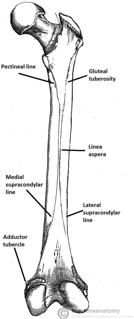
Labelled diagram of the femur bone. In a fan-like radiation to the opposite side of the bone. Unlabelled diagram of the human skeleton. Diagram Of Femur - Getting Started of Wiring Diagram - Radius along with ulna connects elbow to forearm.
Cranium Mandible jaw Clavicle collar bone Scapula Ribs Humerus Vertebrae Ulna Pelvis Femur Patella Tibia Fibula. Bone Diagram Forehead Frontal bone Nose bones Nasals Cheek bone Zygoma Upper jaw Maxilla Lower jaw Mandible Breast bone Sternum Upper arm bone Humerus Lower arm bone Ulna Thigh bone Femur Collar bone Clavicle Toe bones Phalanges Ankle bones Tarsals Kneecap Patella Shin bone Tibia Calf bone Fibula Foot bones. Start learning with our skeleton diagrams bone labeling exercises and skeletal system quizzes.
By most measures the two femurs are the strongest bones of the body and in humans the largest and thickest. Femur Bone Anatomy. It subsists of a head and neck and two bony processes the greater and lesser trochanters.
Label Major Bones of the Skeleton Share Share. Scapula Clavicle Sternum Thoracic Cage Humerus Vertebral Column Coccyx Radius Ulna Carpals Metacarpals Phalanges of the Hands Femur Patella. An immense amount of strength is required to break through a tree trunk attributed to their.
Femur upper bone of the leg or hind leg. In humans the neck of the femur connects the shaft and head at. This is called the diaphysis.
The femur is a type of long bone located in the thigh and is the largest bone of the skeletal system. - Long short flat irregular and sesamoid. Long Bone Diagram Labled - labelled image of femur long bone of the thigh showing.
The human leg consists of 8 bones 4 per leg. Types of bones learn skeleton anatomy. Labelled image of femur long bone of the thigh showing.
Health diagram bone skeleton leg knee science anchor chart human human body. Femur Diagram Femur Bone Parts. Long bones grow more than the other classes of bone throughout childhood and so are responsible for the bulk of our height as adults.
Femur Bone Anatomy. 9780199135653pdf - Read File Online - Report Abuse. Osteons of a long bone can be compared to a tree trunk.
Bones in the Body - Labelled diagram. Labelled Radius Bone. - There is a printable worksheet available for download here so you can take the quiz with pen and paper.
The femur or thigh bone is the proximal bone of the hindlimb in tetrapod vertebrates. We will walk through the anatomy of the femur using the 2. Long Bone Diagram Labelled.
Femur human bone marrow hip 3d human skeleton diagram vector hip. The femur is a type of long bone located in the thigh and is the largest bone of the skeletal system. The femur is the only bone located within the human thigh.
These pictures of this page are abouttoe bones diagram. Femur Definition Function Diagram Facts Britannica. The head forms a ball-and-socket joint with the hip at the acetabulum being held in place by a ligament within the socket and by strong surrounding ligaments.
Label the parts of a long bone. There was a previous EZmed post see below on the anatomy of the femur where we labeled all of the main parts of the bone on a color-coded diagram. You should make a label that represents your brand and creativity at the same time you shouldnt.
Find femur bone Stock Images in HD and millions of other royalty-free stock Related. U2022 femur u2022 ribs u2022 skull. It is both the longest and the strongest bone in the human body extending from the hip to the knee.
The other having origin in the lateral outer portion of the shaft and arching. One which has its origin in the medial inner side of the shaft and curving upward. On Femur Diagram Unlabeled.
Long Bone Diagram Labled. Right femur in relation to the hip bone patella tibia and fibula. This post discusses social security disability benefits and bone fractures.
Long bone diagram timothyakeller flickr. The proximal prospect of the femur links with the acetabulum of the pelvis to form the hip joint. You can use learn mode and.
Its best to save yourpicture as ajpg file. The head of the femur articulates with the acetabulum in the pelvic bone forming the hip joint while the distal part of the femur articulates with the tibia and patella forming the knee joint. The bone that goes from your pelvis to your knee is called the femur say.
Long Bone Diagram - labelled image of femur long bone of the thigh showing. Labelled diagrams of a transverse section of a bone Whale skeleton. The radius or radial bone is one of the two large bones of the forearm the other being the ulna.
Labeled Diagram and Quiz Femur Bone Anatomy.
The diaphysis and the epiphysis. B Bone lining cells BLC exhibiting scarce cytoplasm are situated on the osteoid surface Otd.
Correctly Label The Following Parts Of Bone Cells Chegg Com
Fat cells are also found within the bone marrow.

Label parts of bone cells. Most blood cells are produced in the red marrow of bones. Assignment Lab Ex 7 8 Graded Correctly label the following parts of bone cells. Correctly label the following parts of a chemical synapse.
Aconnos flexible IoT development platform - Personalized solutions for powering the Internet of Things. Labeled parts of bone cells including osteogenic cellstem cellsosteocytesresorption bayosteoclast. Labeled parts of bone cells including osteogenic cellstem cellsosteocytesresorption bayosteoclast osteoblasts and fusion.
The diaphysis and the epiphysis The diaphysis is. Labeled parts of bone cells including osteogenic cellstem cellsosteocytesresorption. Correctly label the following parts of bone cells osseous tissue secretory vesicles osteoclast nuclei pit stem cells ruffled border osteocyte osteoblast periosteum lysosomes ebook references b osteoclast development periosteum.
Correctly label the following parts of bone cells Osseous tissue Secretory vesicles Osteoclast Nuclei Pit Stem cells Ruffled border Osteocyte Osteoblast Periosteum Lysosomes eBook References b Osteoclast development Periosteum. Correctly label the following anatomical parts of osseous tissue-Spicules-Trabeculae-Spongy Bone-Central Canal-Lacuna-Collagen Fibers-Endosteum-Perforating Canal-Nerve and Blood vessels-Periosteum -Collagen Fibers-Osteon. Bones Of The Skull Skull Osteology Anatomy Geeky Medics.
Correctly label the following anatomical features of the clavicle. The diaphysis is the tubular shaft that runs between the proximal and distal ends of the bone. The hollow region in the diaphysis is called the medullary cavity which is filled with yellow marrow containing fat storage cells.
I like the game correctly label the following parts of bone cells - You should try it out and see what you think. Found in bone marrow its function is to produce red blood cells white blood cells and platelets. Types of Bone Cells.
Jump to Expert Tutor Answer. The Compact bone tissue covers the outer part of the bone structure and provides toughness and strength to the structure of bone. Osteogenic cell Stem cells Osteocyte Mitochondrion Secretory vesicles Osteoblast Lysosomes Osteoclast Nucleus Osseous tissue Rough ER A y development Reset Zoom Prev 2 of 15 Next.
A vertebra is considered a long bone. There is a printable worksheet available for download here so you can take the quiz with pen and paper. Temporal Bone Anatomy Parts Of Temporal Bone And Temporal.
Because of the complexities of a bones function from providing strength and support for the body to serving as a site for development and storage of blood cells there are many disorders and diseases that can affect bone. Mastoid Part Of The Temporal Bone. Correctly label the following parts of bone cellsOsseous Tissue Show transcribed image text Correctly label the following parts of bone cells Osseous tissue Secretory vesicles Osteoclast Nuclei Pit Stem cells Ruffled border Osteocyte Osteoblast Periosteum Lysosomes eBook References b Osteoclast development Periosteum Correctly label the following parts of bone cells Osseous tissue.
Solved Correctly Label The Following Parts Of Bone Cells. The epiphyseal plate is the only mechanism by which the diaphysis can increase in length cartilage cells produced by mitosis on the epiphyseal side of the. Correctly label the following parts of bone cells Osseous tissue consists of cells and matrix like any other connective tissue and the matrix consists of fibers and ground substance.
Determine which of the following functions apply to the skeletal system. Your Skills Rank. A layer of bundles of collagen fibrils situated between osteoblasts Ob and calcified bone surface B constitutes the osteoid Otd.
Correctly label the following anatomical features of the vertebrae. A radiograph X-ray of a childs hand will show epiphyseal. Osteogenic cells osteoblasts osteocytes and osteoclasts.
Correctly label the following parts of bone cells. Answer to correctly label the following parts of bone cells osseous tissue secretory vesicles osteoclast nuclei pit stem cells ruf. Temporal Bone Anatomy Parts Of Temporal Bone And Temporal.
Transparent Skeletal System Png Parts Of Skeleton System. There are four kinds of bone cells. Correctly label the following parts of bone cells.
Ambesonne Human Anatomy Diagram Of Human Skeleton System. Then they control calcium and mineral deposition A long bone has two parts. Structure of an adult human long bone Correctly label the following anatomical features of the vertebrae Assignment Lab Ex 7 8 Graded Correctly label the following parts of bone cells The second part of the word.
This is an online quiz called Parts of Bone Label Game. Comments 1 Hope that helpsThanks. The bones are a core founding component of a living body that holds the structure of muscles and organsThe bones of the skeletal system are composed of two types of tissues ie compact and spongy bone tissue.
A long bone has two parts. 342928 students got.