A scientist isolated the plasma membrane from some animal cells and put them in a solution of chemicals that stabilised the membranes. The proportions of proteins lipids and carbohydrates in the.

Draw A Well Labelled Diagram Of Fluid Mosaic Model Of Plasma Membrane Plz Help Brainly In
When she added a small amount of a salt solution she discovered that although the membranes.
Draw a labelled diagram of fluid mosaic model of plasma membrane. Draw A Labelled Diagram Of The Fluid Mosaic Model Of The Plasma Who Gave The Fluid Mosaic Model Of Plasma Membrane Quora Drawing The Fluid Mosaic Model Of Cell Membrane Structure Youtube Fluid Mosaic Model Bioninja Fluid Mosaic Model Of Cell Membranes Video Khan Academy The. Correct answer to the question. The cell membrane consist of a phospholipid bilayer as well as integral.
The model has evolved somewhat over time but it still best accounts for the structure and functions of the plasma membrane as we now understand them. Glycoproteins receptor proteins on the outside. This biology video tutorial discusses the fluid mosaic model of the plasma membrane.
I II 10 nm a Identify structures labelled I and II. E has centriole P has no centriole. Labeled Diagram Of The Fluid Mosaic Model Of The Plasma Membrane images similar and related articles aggregated throughout the Internet.
The drawing below shows the structure of a virus. Cholesterol embedded in the membrane. Peripheral extrinsic proteins associated with the membrane.
Draw a labelled diagram showing. Draw a well labelled diagram of fluid mosaic model of plasma membrane. How To Draw Plasma membrane Fluid Mosaic Model Of Plasma Membrane By Chetan Pachauri - YouTube.
Plasma membranes range from 5 -10 nm in thickness. Draw a well labelled diagram of fluid mosaic model of plasma membrane. - 36223521 krishankewal577222 krishankewal577222 02032021 Chemistry Secondary School answered Draw a well labelled diagram of fluid mosaic model of plasma membrane.
Draw a labelled diagram showing the fluid-mosaic model of a biological membrane. Nicolson in 1972 to explain the structure of the plasma membrane. Draw a labelled diagram to show the fluid mosaic structure of a plasma membrane indicating the hydrophilic and hydrophobic regions.
How to draw the structure of fluid mosaic model how to draw fluid mosaic model of plasma membraneHello Friends in this video I tell you about how ca. Draw a labelled diagram of the fluid mosaic model of the plasma membrane. Total 5 marks 3.
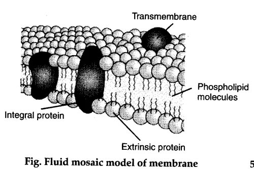
The mosaic model of membrane structure describes the structure of the plasma membrane as a mosaic of components including phospholipids proteins carbohydrates cholesterol and proteins that gives the membrane a fluid character. Protein channels integral intrinsic membrane proteins. Find an answer to your question iDraw a well labelled diagram of Fluid mosaic model of cell membrane.
The fluid mosaic model was first proposed by SJ. Draw a labelled diagram of the fluid mosaic model of the plasma membrane. Singer and Garth L.
Plants have a rigid cell wall that surrounds the plasma. Its function is to protect the integrity of the interior of the cell by allowing certain substances into the cell while keeping other substances out.

Draw It Neat How To Draw Plasma Membrane Cell Membrane
Membrane Structure and Function All cells have a plasma or cell membrane which contains the cell.
Labelled diagram of cell surface membrane. It also serves as a base of attachment for the cytoskeleton in some organisms and the. The Formation of Cell Membranes is Crucial to Life. We are pleased to provide you with the picture named Cell Membrane DiagramWe hope this picture Cell Membrane Diagram can help you study and research.
In a plant cell the cell wall is made up of cellulose hemicellulose and proteins while in a fungal cell it is composed of chitin. While bacterial membranes mostly act as protective barriers they can also form protrusions to make membrane. The cell membrane plasma membrane is a thin semi-permeable membrane that surrounds the cytoplasm of a cell.
Thus both surfaces of the plasma membrane are. In cell biology a vesicle is a structure within or outside a cell consisting of liquid or cytoplasm enclosed by a lipid bilayer. The cell membrane consists of all 4 categories of macromolecules.
Although fixed cells lose membrane potential which is observed in living cells owing to ion dynamics we hypothesized that fixed cells still have a cell membrane surface charge due to cell membrane components and structure. Our LATEST youtube film is ready to run. When drawing and labeling a diagram of the plasma membrane you should be sure to includeThe phospholipid bilayer with hydrophobic tails and hydrophilic h.
Extracellular vesicles EVs can mediate local and longrange intercellular communication via cell surface signaling. In plant cells the membrane encapsulates the protoplasm. C describe with the aid of diagrams the fluid mosaic model of membrane.
If you were to zoom in on the cell membrane you would see a pattern of different types of molecules put together also known as. Just need a glimpse leave your valuable advice let us know and subscribe us. An additional non-living layer present outside the cell membrane in some cells that provides structure protection and filtering mechanism to the cell is the cell wall.
For more anatomy content please follow us and visit our website. As we just learned the main fabric of the membrane is composed of two layers of phospholipid molecules. Animals have been evolutionarily successful because of their flexible cell membranes which.
The plasma membranes of mammalian red blood cells erythrocytes have been particularly useful as a model for studies of membrane structure. This organelle is also referred to as plasma membrane. Images obtained through electron micrography reveal the bilayer structure of cell membranes.
Eukaryotic cells have a nucleus organelles and are surrounded by a cell membrane. The hydrophilic or water-loving areas of these molecules which looks like a collection of balls in an artists rendition of the model Figure 1 are in contact with the aqueous fluid both inside and outside the cell. A bacteria diagram in actual fact facilitates us to profit extra about this unmarried cell organisms which have neither.
Scanning electron micrograph SEM of adipocytes Ad Membrane Structure and Function Prokaryotic Cells. Can Cell Surface Membrane Form Vesicles. The cell membrane contains external structures which help it identify other cells and be recognized as well.
Can Cell Surface Membrane Form Vesicles. The plasma membrane is the most thoroughly studied of all cell membranes and it is largely through investigations of the plasma membrane that our current concepts of membrane structure have evolved. Start studying Some substances can cross the cell-surface membrane of a cell by simple diffusion through the phospholipid bilayer.
The graphic below shows an unlabeled animal cell. Cell Membrane Unlabeled Labeled. Labelled Diagram Definitions and Structure Structure of Plant Cells Cell Wall Plant cells are eukaryotic cells but unlike animal cells which have a cell membrane plant cells have cell walls.
PEG-NPs exhibited good serum stability in vitro and draining. Cell Membranes a outline the roles of membranes within cells and at the surface of cells b state that plasma cell surface membranes are partially permeable barriers Plasma membranes are partially permeable meaning they let some molecules through but not others. Cell membrane is a protective covering that acts as a barrier between the inner and outer environment of a cell in animals.
The cell membrane is a structure that is fluid-like in movement. We used 5 cell lines in this study ARO C32TG RT4 TK UM-UC-14. Finally an unlabeled version of the diagram is included at the bottom of the page in color and black and white.
Lets explore the fluid mosaic model of cell membranes now why is it called the fluid mosaic model well if we were to look at a cell membrane and just to be clear what were looking at if this is a cell right over here this is a cell and this is its membrane this is the membrane its kind of what keeps the cell the inside of the cells separated from whatever is outside the cell were looking. Labelled Diagram Definitions and Structure Animal Cells Animals are made of eukaryotic cells Animal Cells. The cell membrane has hydrophilic surfaces but is hydrophobic internally.
The fluid mosaic model of the cell membrane is how scientists describe what the cell membrane looks and functions like because it is made up of a bunch of different molecules that are distributed across the membrane.
Search Help in Finding Can you Label the Plasma Membrane - Online Quiz Version. The principal components of the plasma membrane are lipids phospholipids and cholesterol proteins and carbohydrates.

Membranes
D eztomhed tpx 181 points Label the types of plasma membrane proteins Integral membrane protein Glycoprotern Membrane channel protein Membrane channel pore Peripheral membrane Type here to.

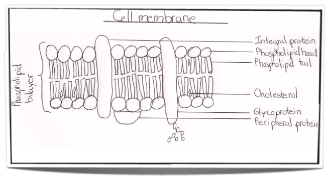
Label parts of plasma membrane. Label The Parts Of A Cell Membrane With The Term That Describes Each Part. A phospholipid is a lipid made of glycerol two fatty acid tails and a. The cell membrane of the cell is a phospholipid bilayer containing many different molecular components including proteins and cholesterol some with carbohydrate groups attached.
C A phospholipid phospholipid bilayer hydrophilic head hydrophobic tail D. Each method labels a distinct class of membrane molecules. This article describes four methods for labeling the plasma membrane with fluorescent probes.
The plasma membrane protects intracellular components from the extracellular environment. Labeling of Plasma MembraneExocytosisEndosomes. Another important group of integral proteins are cell recognition proteins which serve to mark a cells identity so that it can be recognized by other cells.
It is also simply called the cell membrane. Fluorescent dyes that label the plasma membrane intercalate into the membrane bilayer and therefore have significant hydrophobicity. The plasma membrane compartmentalizes the cell separating it from extracellular fluid and other cells.
Image modified from OpenStax Biology. All cells have a plasma membrane. Because of their amphipathic nature being composed of both hydrophilic and hydrophobic portion they.
Find the parts of a cell membrane. Diagram the plasma membrane and label each component. Start your trial now.
The fluid mosaic model of the plasma membrane. Plasma Membrane Teaching Resources Ib Cell Membrane Transport Review 1 3 1 4 Answered 2 Label The Following Diagram Outside Bartleby The D4h Sterol Bio Sensor Labels The Plasma Membrane In Fission Yeast Download Scientific Diagram Ib Cell Membrane Transport Review 1 3 1 4. Identify the labeled parts of the plasma membrane below from the diagram.
First week only 499. Structure and Composition of the Plasma Membrane The plasma membrane is an extremely pliable structure composed of 2 layers of back-to-back phospholipids a bilayer. Glycoprotein and glycolipids are found as part of the outer leaflet of cellular plasma membranes.
The plasma membrane provides the essential dynamic boundary and a vital selective interface between the cell compartment and its environment. Image by Author Lady of Hats Mariana Ruiz Villarreal in the Wikimedia Commons. It is semi-permeable and regulates the materials that enter and.
The principal components of the plasma membrane are lipids phospholipids and cholesterol proteins and carbohydrate groups that are attached to some of the lipids and proteins. Plasma membranes range from 5 to 10 nm in thickness. The plasma membrane of a cell is a network of lipids and proteins that forms the boundary between a cells contents and the outside of the cell.
Protein lipid and carbohydrate components of the membrane. The main function of the plasma membrane is to protect the cell from its surrounding environment. The cell membrane plasma membrane is a thin semi permeable membrane that surrounds the cytoplasm of a cell.
A diagram of the plasma membrane of a cell. The first important component of plasma membrane is the phospholipidis a type of lipid that is made up of a polar head group and a hydrophobic tail. The membrane is made up of lipids proteins and some.
The plasma membrane mediates cellular processes by regulating the materials that enter and exit the cell. Identify the labeled parts of the plasma membrane below from the diagram. This image is created by Wikimedia Commons user LadyofHats Mariana Ruiz who released it into the public domain.
This is an online quiz called Can you Label the Plasma Membrane There is a printable worksheet available for download here so you can take the quiz with pen and paper. Label the plasma membrane. The phospholipid bilayer separates the cytoplasm below from the extracellular fluid above.
The plasma membrane should be the easiest cell membrane to label because it is the most accessible but the process is not always completely straightforward. Cholesterol is also present between the phospholipids which contributes to the fluidity of the membrane. Cell Membrane Detailed Diagram Labeled.
Those glycoproteins or glycolipids that contain sialic acid and N-acetylglucosamine residues can be labeled with wheat germ agglutinin WGA a plant lectin that exists as a dimer mwt 36000 and is normally cationic.
The cell membrane is an extremely pliable structure composed primarily of back-to-back phospholipids a bilayer. These are situated at the upper and lower parts of cell membrane.
Basic Cell Components Cell Structure Organelles

Proteins and lipids are the major components of the cell membrane.
What are the three main parts of the cell membrane. The main parts of human cell structure can be divided into the outer plasma membrane the usually central nucleus the cytoplasm and the different organelles that are found in the cytoplasm. Cholesterol is also present which contributes to the fluidity of the membrane and there are various proteins embedded within the membrane that have a variety of functions. It includes features from all cell types.
Cell membrane is made of 4 kinds of molecules namely phospholipids carbohydrates proteins and cholesterol. The plasma membrane contains channel carrier receptor enzymatic and cell recognition proteins that all contribute to the membranes functions. The cell membrane has Bilayered structure ie.
Cell membrane is semisolid in structure facilitating movement of cell organs to other places. M olecules of the cell membranes are mostly synthesized in two organelles. Within the cytoplasm lie intricate arrangements of fine fibers and hundreds or even thousands of miniscule but distinct structures called organelles.
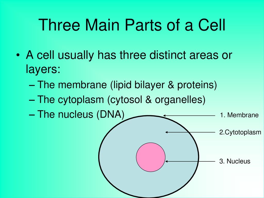
In the pages dealing with these organelles we will describe the synthesis processes in more detail. Thus lipid synthesis will be studied in the pages dedicated to the smooth endoplasmic. The three main parts of a cell are the plasma membrane the region containing the DNA and the cytoplasm.
It is selectively-permeable. The vesicle membrane then becomes part of the cell membrane. These allow the membrane to be rather fluid.
The exact mix or ratio of proteins and lipids can vary depending on the function of a specific cell. It is the outer end of two layers also called Polar End and considered as water-soluble. Specific examples of exocytosis include cells of the stomach and pancreas producing and secreting digestive enzymes through exocytosis Figure 3110 and endocrine cells producing and secreting hormones that are sent throughout the body.
The plasma membrane mediates cellular processes by regulating the. The channel proteins form small openings where certain molecules and solutes diffuse through and get into the cell. The three main functions of the cell membrane are.
The plasma membrane is the outer boundary of the cell and controls what enters and leaves it. Each layer consists of two parts. The cell membrane the nucleus and between the two the cytoplasm.
This membrane is called the fluid mosaic model as it is a mixture of phospholipids cholesterol proteins and carbohydrates. However not all cells have exactly the same basic parts. The plasma membrane protects intracellular components from the extracellular environment.
Endoplasmic reticulum and Golgi apparatus. A cell consists of three parts. 1 It regulates materials that come in and out of a cell.
Phospholipids are important components of cell membranes. Molecules of the cell membrane are arranged in a sheet. It consists of two layers of Phospholipids upper layer and lower layer.
The phospholipids form the major parts. The principal components of the plasma membrane are lipids phospholipids and cholesterol proteins and carbohydrates. Cell membrane is made of proteins and lipids making it easy for the cell for exchange of matter with other cells.
Most of the membrane is composed of phospholipid molecules. There is a difference between the structures of eukaryotic and prokaryotic cells.
Transport-MOVEMENT OF CHEMICALS INTO OR AROUND CELL. One of the most important functions of carbohydrates is to form a structure called the glycocalyx.

Ch 5 Study Guide Questions Diagram Quizlet
Some carbohydrate molecules function as the receptors for some hormones.

What is the function of carbohydrates in the cell membrane quizlet. These carbohydrate chains may consist of 260 monosaccharide units and may be either straight or branched. Transport proteins transport specific substances across the cell membrane and other cellular membranes. This means not only are they great at releasing energy into the human body but are also good at helping your body recognize cellsCarbohydrates can also be utilised to anchor proteins into the membrane.
They also act as a great cell marker helping guide. Carbohydrates are present only in the cell membrane. To protect the cell from its surroundings and control the transfer of substances in and out of the cell.
They are always found on the exterior surface of cells and are bound either to proteins forming glycoproteins or to lipids forming glycolipids. Phospholipids make up the basic structure of a cell membrane. This quiz will ask you questions about cell membranes their structure and their functions.
Membrane carbohydrates wherefunction Project from the exterior surface of the plasma membrane when attached to proteins or phospholipids within the membrane. They also help to take part in the formation of the membrane in the form of sugar molecules or more scientifically known as oligosaccharides. The membrane is the outside covering of a cell.
The Fluid Mosaic Model describes membranes as a fluid lipid bilayer with floating proteins and carbohydrates. Each of these molecules gives the cell membrane unique. A part of the cell membraneIt is a protein with a carbohydrate attached to itThey help to recognize substances trying to enter the cell.
They are phospholipids cholesterol proteins and carbohydrates. Carbohydrates are the third major component of plasma membranes. This communication helps the body recognize and remove harmful bacteria and pathogens and cancerous cells and bring about immune responses against allergy-causing substances.
Carbohydrates or sugars are sometimes found attached to proteins or lipids on the outside of a cell membrane. Enzymes of cell membrane are either ectoenzymes or endoenzymes and are of about 30 types. Do take up this quiz and prove how much you know about this barrier in a cell.
Glycocalyx from the neighbouring cells assists in the tight fixation of cells with one another. The membranes primary function is to keep unwanted substances out of a cell and at the same time ensure the wanted substances can move in and out of the cell. Function of membrane lipids.
Serve as recognition sites on the cell surface. What are the roles of carbohydrates in the plasma membrane. Carbohydrates are covalently linked to proteins glycoproteins or lipids glycolipids and also an important part of cell membranes and function as adhesion and address loci for cells.
Functions of Carbohydrates in Cell Membrane Carbohydrate molecules are adversely charged and do not allow the adversely charged substances to move in and out of the cell. Aside from providing energy the cell also uses carbohydrates for its various activities and processes. Carbohydrates located on the cell surfaces regulate communication between cells and other molecules.
If the cell membrane is like a city wall then the glycocalyx is. They are present as short unbranched or branched chains of sugars oligosaccharides attached either to exterior ectoproteins forming glycoproteins or to the polar ends of phospholipids at the external surface of the cell membrane. What are 3 functions of proteins in the cell membrane.
Four main molecules make up the mosaic structure of the cell membrane. Membrane proteins can function as enzymes to speed up chemical reactions act as receptors for specific molecules or transport materials across the cell membrane. A part of the cell membrane is a carbohydrate attached to a lipid itit is the cell markeridentifies what the cell is.
Membrane carbohydrates perform two main functions. Participate in cell recognition and adhesion either cell-cell signalling or cell-pathogen interactions and they have a structural role as physical barrier. Authors describe the synthesis and assembly of a glycan on a protein the formation of N-glycans the maturation of an N-glycan in different cellular compartments the structure of the glycocalyx and how it.
Start studying Carbohydrates in the Cell Membrane. What is the role of carbohydrates in the cell membrane quizlet. Role of lipids in the body Chemical messengers Storage and provision of energy Maintenance of temperature Membrane lipid layer formation Cholesterol formation Prostaglandin formation and role in inflammation The fat-soluble vitamins Function of membrane proteins.
Learn vocabulary terms and more with flashcards games and other study tools. Enzymatic activity-A protein built into the. This is a coat around the cell.
In the following paper authors describe glycans present on cell membranes as they affect the folding the spatial arrangement the behavior and the interaction with the substrate of some membrane proteins.