This tube connects are nasal cavity to our lungs. Read Label The Diagram Of The Respiratory System Below With The Following Parts PDF on our digital library.
Respiratory System Organs Read Biology Ck 12 Foundation
Label The Diagram Of The Respiratory System Below The Respiratory.

Label the diagram of the respiratory system below with the following parts then colour your diagram. Left bronchus alveoli right lung pharynx throat diaph m lett lung oral cavity nasal cavity epiglottis. Complete the labeling of the diagram of the upper respiratory structures sagittal section. Students insert several text boxes and add and format text inside.
Trachea that branches off to the left and right lungs. Click on a label below the map to select it and. The human respiratory system consists of a group of organs and tissues that help us to breathe.
You can read Label The Diagram Of The Respiratory System Below With The Following Parts PDF direct on your mobile phones or PC. Click an item in the list or group of pictures at the bottom of the problem and holding the button down drag it into the correct position in the answer box. Organs in the digestive system.
Drag The Labels Onto The Diagram To Identify Respiratory System Human Respirato. Label the diagram of the respiratory system below with the following parts then colour your diagram. Art labeling activities art labeling activity.
Read LABEL THE DIAGRAM OF THE RESPIRATORY SYSTEM BELOW WITH THE FOLLOWING PARTS PDF direct on your iPhone iPad android or PC. Start studying respiratory system label. Drag each label to the appropriate location on the flowchart.
Show transcribed image text Label the diagram of the respiratory system below with the following parts then colour your diagram. Click the trashcan to clear all your answers. Label the following topographic map.
Nose nasal cavity larynx trachea. Image 37789 is a 1125 by 1408 pixel PNG Uploaded. Air enters this part of the respiratory system.
View Original Image at Full Size. HLT 100- Respiratory System Labeling Diagram 2. The other main parts of this system include a series of airways for air passages blood vessels and the muscles that facilitate breathing.
If you change your mind drag the item to the trashcan. Release your mouse button when the item is place. Label the diagram of the respiratory system below with the following parts then colour your diagram.
Label the diagram of the respiratory system below with the following parts then colour your diagram. Chapter 3 - systems S m Label the diagram of the system below with the following parts then colour your diagram. With a labeled diagram you can see all of the main structures of an organ system together on one page - great for helping you to memorise the appearance of several structures and their relations.
Label parts 1 5 in order. Start studying respiratory system label. Unlabeled diagrams can then help you to put your memory to the test.
Label the diagram of the respiratory system below with the following parts then colour your diagram. Learn vocabulary terms and more with flashcards games and other study tools. Draw The Human Respiratory System And Label The Following Parts Respiratory System Quiz Study Guide Name Date Period.
Below youll find the respiratory system labeled and unlabeled on two. Green Label the diagram of the respiratory system below with the following parts then colour your diagram. The main organ of the respiratory system.
Drag the labels onto the diagram to identify the major components of the respiratory. Label the parts of the respiratory system. Lungs are the primary organs of the respiratory system which help in the exchange of gases.
View img_0005jpg from BIO MISC at Flint Hills Technical College. Label the diagram with the parts of the respiratory system. As per our directory this eBook is listed as LTDOTRSBWTFPPDF-252 actually introduced.
Label the three accessory organs in the diagram to the right. Label the diagram of the respiratory system below with the following parts then colour your diagram. Transcribed Image Textfrom this Question.
Labeled diagram of the lungsrespiratory system. Label The Diagram Of The Respiratory System Below With The Following Parts - PDF-LTDOTRSBWTFP25-2 Download full version PDF for Label The Diagram Of The Respiratory System Below With The Following Parts. Solved Drag The Labels Onto The Diagram To Identify Respi Respiratory System Drawing At Paintingvalley Com Explore Art Labeling Quiz 28 Respiratory System Gas Exchange Worksheet Respiratory.
Discussed in the video of the respiratory system.
2 Label the following parts of the nephron on Figure 2518. By dilatory Plays Quiz not verified by Sporcle.

Ch 32 Test Prep For Ap Courses Biology For Ap Courses Openstax
The first and primary sort of water and ion reabsorption in the kidney where all glucose in the blood is reabsorbed.
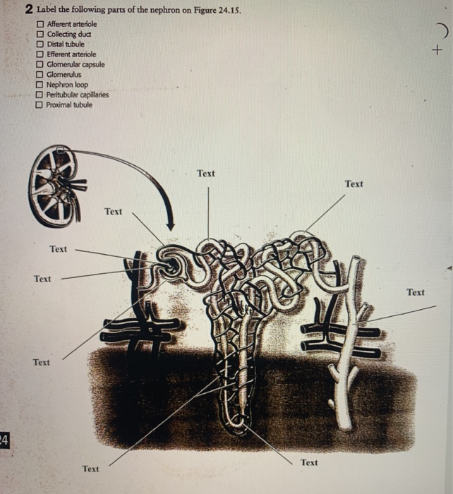
Label the following parts of the nephron. Labels can be used more than once. Correctly label the following parts of the pancreas and its passages. 30 Label The Blood Vessels And Parts Of The Nephron By Selecting The Letter For The Correct Structure Written By Kim M Grant Wednesday July 28 2021 Add Comment Edit.
Label the blood vessels and parts of the nephron by selecting the letter for the correct structure. Correctly label the following parts of a renal corpuscle. Asked Oct 12 2019 in Biology by Suchita.
Correctly label the following parts of the gallbladder and bile passages. Dehydration Page 9 of 11. Science Quiz Labeling Parts of the Nephron Random Science Quiz Can you name the Label Parts of the Nephron.
The hormone aldosterone acts in this part of the nephron and has a big impact on Na and K levels in the filtrate. Gallbladder Hepatic ducts Common hepatic duct Cystic duct. Label the following diagram of a nephron and provide a brief description of the function of the structure beside each label.
Renal corpuscle and renal tubule TF. In which of the following regions of the nephron is water actively transported. Afferent article testarteriole Cartikleting duct Glomdar chule Dstal tubule Glomerus Nephron loop Pomaltube ROURE 2518 Structure of the nephron.
Glomerulus Bowman capsule renal artery collecting duct b What happen to glucose that enters the nephron along with the filtrate. Tubular parts of a Nephron converts the filtrate into urine The Bowmans capsule Glomerular capsule. Correctly label the following components of the urinary system.
Correctly label the following anatomical structures of the female urethra and urinary bladder. Glomerulus Bowmans capsule Renal artery Collecting duct. Rate 5 stars Rate 4 stars Rate 3 stars Rate 2 stars Rate 1 star.
Each nephron is composed of two parts. Which of the following indicates the parts of a renal tubule in the correct sequence from beginning to end. A Draw the structure of a nephron and label the following on it.
A cup-like sac at the beginning of the tubular component of a nephron in the mammalian kidney. A renal corpuscle and a renal tubule. ロEfferent arteriole Nephron loop Proximal tubule Afferent arteriole Cortical collecting duct Dstal tubule Glomerular capsule Glomerulus Nephron Afferent arteriole Arcuate artery Collecting duct Distal convoluted tubule purple Efferent arteriole Interlobular artery Loop.
There is a printable worksheet available for download here so you can take the quiz with pen and paper. Describe how the following events would change urine composition and outputvolume. Transcribed Image Textfrom this Question.
It surrounds the glomerulus. Click hereto get an answer to your question Draw the structure of a nephron and label the following. Get the ad-free and most optimal full-featured Sporcle experience.
The mammalian nephron is a long tube-like structure its length varying from 3555 mm long. ロEfferent arteriole Nephron loop Proximal tubule Afferent arteriole Cortical collecting duct Dstal tubule Glomerular capsule Glomerulus. At one end the tube is closed folded and expanded into a double-walled a cuplike structure called the Bowmans capsule or renal corpuscular capsule which encloses a cluster of microscopic blood vessels called the glomerulus.
Of the nephron from which it is reabsorbed by dropping each label onto the appropriate areas of the nephron. This is an online quiz called Label a Nephron. The Bowmans capsule also called the glomerular capsule is the beginning of a nephron.
Label the structures of a nephron in the figure. The juxtaglomerular apparatus is a structure of the nephron where the DCT contacts the afferent arteriole. 2 Label the following parts of the nephron on Figure 2518.
2 Label the following parts of the nephron on Figure 2518. A small intertwined group of capillaries within the nephrons of the kidney that filter the blood to make urine. Draw the diagram of nephron and label the following parts.
Ii The part that terminates in balloon-like slructures. You should be able to label a diagram showing.

Draw A Diagram Showing Human Respiratory System Label The Following Parts A Alveolus B Trachea Brainly In
It involves in inhalation of oxygen and release of carbon-dioxide.

Label the following parts of the human respiratory system in the diagram. Iv part which separates chest cavity from abdominal cavity. Label parts of the respiratory system displaying top 8 worksheets found for label parts of the respiratory system. Any particle whose size is more than 6 µ gets filtered in the nose.
Ii Why is the rate of breathing in aquatic organisms much faster than in terrestrial. Start studying The Respiratory System Label. External nostrils For the intake of air.
Students will label the parts of the respiratory system on the diagram list the structures on the data table and then describe the function of each structure. Human respiratory system is one of the vital systems in our body. Learn vocabulary terms and more with flashcards games and other study tools.
Balloon-like structures where exchange of gases takes place. In this article we will discuss about the parts of human respiratory systems. Asked Jun 22 2019 in Class VII Science by navnit40 -4936 points excretion in humans.
Diagram to Label with Data Table. Bronchus nasal cavity epiglottis pleural membrane lung larynx pharynx trachea diaphragm Esophagus Using arrows and the following parts trace the flow of air in the human respiratory system. C baloon-like structures where exchange of gases takes place.
Draw a diagram of human excretory system and label the following parts. Jan 28 2021 - In this assignment students color the various parts of the respiratory system and then answer some follow up questions to describe the functions of the respiratory system. Play this quiz called respiratory system labeling interactive and show off your skills.
Pharynx It is a passage behind the nasal chamber and. Kidney ureter urinary bladder and urethra. THE HUMAN RESPIRATORY SYSTEM espiratory System Label the following parts of the human respiratory system on the diagram.
Iii Balloon-like structures where exchange of gases takes place. B Draw a diagram of human excretory system and label the following. I Draw a diagram of human respiratory system and label the following.
Inhalation of oxygen takes place from nasal cavity passes through larynx and trough trachea and primary bronchi reaches lungs. I part where air is filtered by fine hair and mucus. B Nostrils are lined with mucus.
A part where air is filtered by fine hair and mucus. Nasal chamber which is lined with hair and mucus to filter the air and remove dust and dirt. Use the mouse or tap the screen to label this diagram of the complete human respiratory system combining the head and thorax chest regions.
B part which terminates in balloon-like structures. The Respiratory System Human Anatomy 85 terms. These hairs serve to filter the air that is entering the respiratory system.
Larynx bronchioles glottis alveoli nasal cavity. Trachea Bronchi and Diaphragm. Part which terminates in balloon-like structures.
Students save the finished document with a descriptive name and or print. I The part where air is filtered by fine hair and mucus. The students enjoy coloring the structures as well.
Draw a diagram of human respiratory system and label the following. A Draw a diagram of human respiratory system and label the following. Iv part which separates chest cavity from abdominal cavity.
Start studying respiratory system label the organs. Iii balloon-like structures where exchange of gases takes place. Hairs are present in the nasal cavity.
Click hereto get an answer to your question Draw a neat diagram of human respiratory system and label its following partsRings of cartilage lung bronchi alveolar sac. A Draw a diagram of human respiratory system and label the following. I Draw a diagram of human respiratory system and label.
Part which separates chest cavity from abdominal cavity. Part where air is filtered by fine hair and mucus. D part which separates chest cavity from abdominal cavity.
A Draw a diagram of human respiratory system and label. The respiratory tract in humans is made up of the following parts. Ii Give reasons for the following.
Identify and label the following diagram of the human respiratory system. A Lungs always contain residual volume of air. This assignment is geared toward 9th10th grade Biology students.
From lungs oxygen is distributed to different parts of body. I part where air is filtered by fine hair and mucus ii part which terminates in balloon-like structures iii balloon-like structures where exchange of gases takes place. Mouth nose trachea windpipe bronchi bronchioles alveoli air sacs pleural membranes diaphragm ribs and intercostal muscles.
Ii part which terminates in balloon-like structures.
To draw the inside of a tooth start about a half an inch inside the tooth and draw a line around following the shape of the tooth. ToothStructureToothAnatomyAndPhysiologyToothDiagramThe teeth are the hardest substances in the human body.
Draw A Labelled Diagram To Show The Internal Structure Class 12 Biology Cbse
Tissues that Surround and Support Teeth.
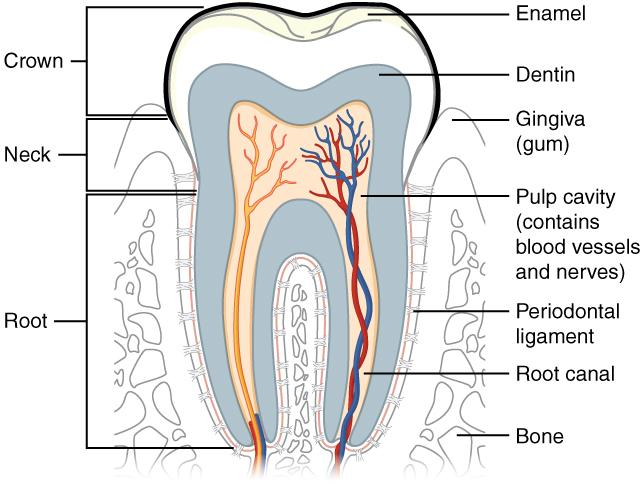
Label the following diagram of a tooth. The middlemost four teeth on the upper and lower jaws. The tooth as discussed in class is a made up of different parts that help the teeth be strong and perform its faction with ease. A tooth diagram worksheet with labels.
These 3 differentiated worksheets will help children be able to label the parts of a tooth and are great for different ability levels. When the auto-complete results are available use the up and down arrows to review and Enter to select. Answer to Label the following diagram of a toothABCDEFGHIJKLM.
The crown and roots. In man the design of the teeth is a reflection of eating habits as humans tend to be meat eaters so teeth are formed for cutting tearing and grinding food. These worksheets will really give your students something to get their teeth into.
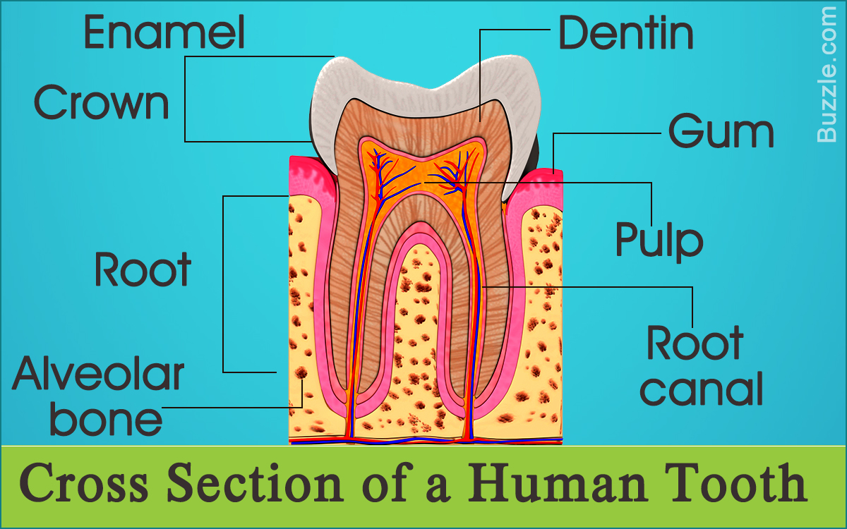
The enamel dentin cementum and pulp. The human teeth dental chart illustrates the types and working surfaces of the four classes of teeth. A great educational tool for professionals patients and students this print-quality tooth diagram is available in various sizes.
The periodontium consists of. 22Teach children about their teeth with this free printable activity. Tooth Parts Tooth Label Parts Worksheet Label the Parts of the Tooth Worksheet Unlabeled Tooth Diagram Tooth Label Parts Worksheet.
It holds the teeth in place. Crypt is a cavity in the alveolar bone that contains developing tooth germ of a permanent tooth. Besides being essential for chewing the teeth.
A super easy drawing of a tooth. Healthy dental hygiene is something that everyone should strive to have. Diagram of the Tooth Numbering System viewed as if looking into the mouth Buccal Facial Surface Occlusal Surface Incisal Surface Right Left Maxillary Arch Upper Jaw Mandibular Arch Lower Jaw Adult Dentition Permanent teeth 1-32 Child Dentition Primary teeth A-T Wisdom Teeth 1 16 17 and 32 Central Incisor Lateral Incisor Cuspid 1st Bicuspid Bi-Rooted.
See 10 Best Images of Parts Of The Tooth Worksheet. Every tooth consists of three parts. The enamel dentin and odontoblast layers.
Kids can color and label the different parts of a tooth diagram as they learn about enamel crowns roots and more. The quiz below is designed to help you practice labeling the parts of a tooth. And lines showing divisions of the crown neck and root.
It also includes a completed diagram with all the labels filled in and is great for kids to. A tooth is a hard bony appendage that develops on the jaw to pulverize food. The enamel dentin cementum pulp root periodontal ligaments etc are important parts of the tooth structure.
Inspiring Parts of the Tooth Worksheet worksheet images. May 7 2015 - An illustrated worksheet to teach your class the different types of teeth. View Standard Image License.
Learn vocabulary terms and more with flashcards games and other study tools. Also every tooth made of several layers. Human Tooth Anatomy With Labeled Diagrams About Press Copyright Contact us Creators Advertise Developers Terms Privacy Policy Safety How YouTube works Test new features 2021 Google LLC.
For young students you might just stop there or have them label the outside of the tooth. Information About the Human Tooth Anatomy With Labeled Diagrams. Give it a try.
A normal adult mouth has 32 teeth which except for wisdom teeth have erupted by about age 13. The jaw bone also called the alveolar bone is the bone that contains the tooth sockets and surrounds the teeths roots. Start studying Tooth Structure- Label.
Anatomical crown is that portion of tooth which is covered by enamel. The structures labeled in this diagram include. The crown neck and root.
Pulp cavity including arteries veins and nerves. Repeat that step again to add one more part. Bodytomy provides labeled human tooth diagrams to help you understand the human tooth anatomy.
A clinical crown is that portion of tooth which is visible in mouth. Neck The neck also called the dental. Its perfect for dental hygiene month or anytime the kids need to brush up on knowledge about their teeth.
Our teeth help us to tear and crush food. Draw a labelled diagram to show the internal structure of a mammalian tooth with two roots. Clinical parts structures of a human tooth.
The crown refers to the part of a human tooth that is visible to us. Cusp is an elevated point on the crown portion of the tooth. Canines 4 total.
The periodontium anchors teeth to surrounding tissues and supports teeth during its function. A cusp has four ridges and an apex. Ideal for science lessons or to teach the importance of tooth brushing.
Touch device users can explore by touch or with swipe. Incisors 8 total.
You can refer to your textbook in order to label the. The inner layer of the heart wall is called endocardium.
Draw A Diagram Of Cross Section Of The Human Heart And Label The Following Parts Studyrankersonline
29Draw the diagram of human heart and label the following parts a which receives the blood from the lungs b the chamber which is having the thickest walls c the blood vessel which carries the blood to the lungs d the blood vessel which brings the deoxygenated blood to the heart.

Draw the diagram of human heart and label the following parts. 6 2 1 Draw And Label A Diagram Of The Heart Showing The Four. A heart diagram labeled will provide plenty of information about the structure of your heart including the wall of your heart. I Receives deoxygenated blood from vena cava.
Draw the diagram of human heart and label the following parts which i receives deoxygenated blood from vena cava ii send deoxygenated blood to lung through pulmonary artery iii receives oxygenated blood from lungs and iv sends oxygenated blood to all parts of the body through aorta. Atria and Ventricles The human heart comprises four chambers. 29Draw the diagram of human heart and label the following parts a which receives the blood from the lungs b the chamber which is having the thickest walls c the blood vessel which carries the blood to the lungs d the blood vessel which brings the deoxygenated blood to the heart.
B What does the blood consist of. We will review the anatomy function and order of blood through the human heart using pictures of the atria ventricles tricuspid valve mitral valve pulmonary valve aortic valve superior and inferior vena cava pulmonary arteries and veins and aorta. Start studying Label the Heart.
A Draw a diagram of cross-section of the human heart and label the following parts. The lower two chambers of the heart are called ventricles. Draw the diagram of sectional view of human heart and label on it 1chamber wch receives deoxygenated blood from vena cava 2 chamber wch pumps oxygenated blood into aorta 3 blood vesel wch brings oxygenetd blood to the heart 4 chamber wch receives oxygeneted blood from lungs.
A Draw The Diagram Of Human Heart And Label The Following Parts. The blood vessel that receives deoxygenated blood from the other parts of the body c. Part which prevents the back flow of blood.
Draw the top vein slightly smaller than the bottom veinStep 3. Draw the diagram of sectional view of human heart and on it name and label the following parts. Label the parts of the heart if youd to reference it for anatomy.
Well-Labelled Diagram of Heart. The wall of the heart has three different layers such as the Myocardium the Epicardium and the Endocardium. Find an image that displays the entire heart and click on it to enlarge itStep 2 Find a piece of paper and something to draw with.
B Give reasons for the following i The muscular walls of ventricles are thicker than the walls of atria. Start with the pulmonary veins. Please log in or register to add a comment.
The heart wall is made up of three layers. Ii Send deoxygenated blood to lung through pulmonary artery. The chambers of the heart that pumps out deoxygenated blood.
There are two of them. Basic Anatomy Of The Heart Download Scientific Diagram. I Right ventricle ii Aorta iii Left atrium iv Pulmonary arteries.
Exterior of the Human Heart. Draw the diagram of sectional view of human heart and label the following parts. Iii Receives oxygenated blood from lungs.
Asked Mar 19 in Science by Sandhya01 591k points Draw the diagram showing the sectional view of the human heart. They will be to the lower left of the Aorta. The middle layer of the heart wall is called myocardium.
If youre trying to identify parts of the heart for a class youre taking its good practice to draw the heart yourself and label each segment. Right and left atria. The anatomy of the heart is made easy in this post using labeled diagrams of the main cardiac structures and vascular system.
Easy Diagram Of Heart. Right atrium left atrium right ventricle and left ventricle. Heres more about these three layers.
As you can see in the above diagram heart consists of various parts. Observing a diagram of the heart as the one here will help comprehend the different parts of the human heart. Iv Sends oxygenated blood to all parts of the body through aorta.
The upper two chambers of the heart are called auricles. This will help you to understand the functioning of its different parts. A The chamber of the heart that pumps out deoxygenated blood.
B The blood vessel that carries away oxygenated blood from the heart. Learn vocabulary terms and more with flashcards games and other study tools. Ii Arteries have thick elastic walls.
The Heart 7th Grade Circulatory System. Ii Chamber of the heart that receives deoxygenated blood. So as we discuss the various parts you keep checking out the parts simultaneously in the above given labeled diagram of the human heart.
Label the following parts. The blood vessel that carries away oxygenated blood from the heart. C The blood vessel that receives deoxygenated blood from the lower part of our body.
Step 1 To find a good diagram go to Google Images and type in The Internal Structure of the Human Heart. The heart is made up of two chambers. A Draw The Diagram Of Human Heart And Label The Following Parts.
A Draw the diagram of human heart and label the following parts which. The outer layer of the heart wall is called epicardium.