The ventricles are located in the lower part of the heart while the atria are located in the upper part of the heart. Heres more about these three layers.
Understanding Human Heart With Heart Diagram Edrawmax Online
The walls of the heart are composed of an outer epicardium a thick myocardium and an inner lining layer of endocardium.

Heart diagram labeled in order. Fattoria Estense Silver Label Farmacia Style Bottle Previously labeled as Aceto Balsamico di Modena Aged 10 years In July 2009 Aceto Balsamico di Modena Balsamic Vinegar of Modena obtained the Protected Geographical Indication status from the European Union. Cardiac conduction is the rate at which the heart conducts electrical impulses. The right atrium receives systemic blood relatively low in oxygen and pumps it into the right ventricle which pumps it into the pulmonary circuit.
The function of heart is quite complex but you can understand things better through the heart diagram labeled below. Well-Labelled Diagram of Heart. Frogs heart removed from the body and in human beings who have transplanted heart the heart continues to function.
Weve gathered our favorite ideas for Label Heart Diagram Explore our list of popular images of Label Heart Diagram Photos Collection with high resolution. Enchanted Learning Free Heart Anatomy Diagram. The outer layer of the wall of the heart.
The upper two chambers of the heart are called auricles. Enchanted Learning Free Heart Anatomy Diagram. The heart wall is made up of three layers.
Lastly we can revisit the original diagram shown at the beginning of this post and you should be able to understand and label the entire image. Conducting System of Human Heart With Diagram Cardiac function does not require intact innervations. Selecting or hovering over a box will highlight each area in the diagram.
Anatomy of the Heart. For every use a template has been designed with a motive of making it easy for the user to get the print of it without making a new one of his own. This diagram depicts Labeled Heart with parts and labels.
Enchanted Learning Free Heart Anatomy Diagram. The shape of the human heart is. The perfusion of the isolated heart in the laboratory will also continue to beat at regular intervals.
The human heart consists of a pair of atria which receive blood and pump it into a pair of ventricles which pump blood into the vessels. The heart is made up of two chambers. It can be used by a teacher or student for academic purpose by a friend or relative for mutually sending and exchanging cards or for baby toys or printing on dresses etc.
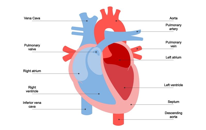
Red arrows demonstrate flow of oxygenated blood through the left side of the heart. It provides information about different chambers of the heart and valves that help transfer blood from one part of your heart to another. In this interactive you can label parts of the human heart.
Blue arrows demonstrate flow of deoxygenated blood through the right side of the heart. The wall of the heart has three different layers such as the Myocardium the Epicardium and the Endocardium. Labeled Heart Diagram - Labeled Heart Chart - Human anatomy diagrams and charts explained.
The base of the heart is located along the bodys midline with the apex pointing toward the left side. Keep reading to learn more about how your heart works. Epson LabelWorks LW-300 Label Maker C51CB69010 The Epson LabelWorks LW-300 label printer is where organization meets imagination.
Because the heart points to the left about 23 of the hearts mass is found on the left side of the body and the other 13 is on the right. No longer restricted by simple options you can choose from a huge range of symbols frames and fonts. Heart diagram labeled Fattoria Estense.
Four Chambers of the Heart and Blood Circulation. The lower two chambers of the heart are called ventricles. A heart diagram is a popular design used by different people for various uses.
When autocomplete results are available use up and down arrows to review and enter to select. 30 The Heart Labeled And Functions Images. Two ventricles and two atria.
A heart diagram labeled will provide plenty of information about the structure of your heart including the wall of your heart. Label the heart. The inner layer of the heart.
Function and anatomy of the heart made easy using labeled diagrams of cardiac structures and blood flow through the atria ventricles valves aorta pulmonary arteries veins superior inferior vena cava and chambers. With dozens of tapes in a variety of styles borders sizes and colors your creativity can be boundless. Drag and drop the text labels onto the boxes next to the diagram.
Includes an exercise review worksheet quiz and model drawing of an anterior view frontal section of the heart in order to match the anatomy to the picture and test yourself. As you read this article try scrolling back up and seeing if you can spot the chambers. Pin on HOLIDAY Hearts.
The muscular middle layer of the wall of the heart. Heart nodes and nerve fibers play an important role in causing the heart to contract. There are the left and right ventricle and the left and right atrium.
Labeled heart diagram Epson LabelWorks. The human heart consists of four chambers. A detailed explanation of the heart along with a well-labelled diagram is given for reference.
The middle layer of the. The outer layer of the heart wall is called epicardium. The main artery carrying oxygenated blood to all parts of the body.
This status is established to protect the producers of certain products as well as to.