Arguably the most important tendon is the Achilles tendon which allows the calf muscles to move the ankle joint. It is often caused by overuse fatigue or improper use of muscles.

Foot Bones Anatomy Conditions And More
The calf muscle may be tight placing stress on the tendons at the top of the foot that pull the foot upward.
What is the muscle on the top of your foot called. The ankle joint or tibiotalar joint is formed where the top of the talus the uppermost bone in the foot and the tibia shin bone and fibula meet. Only two of these muscles are located on the dorsal aspect top of the foot. Extensor tendonitis is a common problem that causes pain across the top of the foot.
If they become inflamed due to overuse or. This is rare and characterized as an inflamed sinus tarsi. The tibialis anterior muscle is the muscle located in the front part of the shin bone of your lower leg.
It flexes and extends the foot ankle and knee. Function of the Tibialis Anterior Muscle. When the muscles tighten contract they pull on the tendons which in turn move the bones.
The muscles at the top of the foot fan out to supply the individual toes. The muscles in the sole of the foot are located in four layers. One of the large muscles of the leg it connects to the heel.
Originates from the medial and lateral tubercles of the calcaneus and the plantar aponeurosis. Bones and Joints of the Foot and Ankle The Ankle Lateral side of the ankle Joint capsule. You can pull any muscle in your body but this is the most common in feet lower back and neck.
Another very common cause of the condition is the foot rubbing against a shoe. This is the muscle that allows the trunk of your body to twist this is controlled by the external oblique muscle on the opposite side of the direction that youre twisting. Collectively they are referred to as the intrinsic muscles of the foot because they are entirely contained within the foot.
A pulled muscle also known as a muscle strain is a result of a torn or overstretched muscle. It is homologous with the abductor digiti minimi of the hand. One of the large muscles of the leg it connects to the heel.
Gastrocnemius calf muscle. You can divide the foot muscles into groups based on their locations either in the sole of the foot the bottom or plantar area or the dorsum on the top of the foot. Extensor tendinitis involves inflammation of the tendons near the middle top of the foot.
Gastrocnemius calf muscle. Most of the muscles are in the sole. This form of foot tendonitis is caused by inflammation or irritation of the tendons that pull the toes up.
The abductor digiti minimi muscle is located on the lateral side of the foot. Adapted screenshot from Kenhub anatomy video CFCF via Wikimedia Commons cc Gluteus medius is responsible for abduction and rotation of the hip. Your external obliques sit on either side of your rectus abdominis and are actually the largest of your abdominal muscles.
This muscle extends from the back of the knee. The extensor tendons located in the top of the foot are needed for flexing or pulling the foot upward. If muscles are overworked or overstressed they can become torn or strained.
Typically tendonitis of the extensor tendons develops due to repeated friction across the top of the foot or excessive pressure from a poorly-fitting shoe. The muscle courses from an area just below your knee down the front of your shin and finally attaches to the top of your foot. Strains usually manifest as pain especially with movement or pressure.
The Muscles of the Foot. The extensor hallucis brevis and the extensor digitorum brevis. What does this mean.
The tendons that run along the top of the foot and pull the foot upwards become inflamed and painful. It flexes and extends the foot ankle and knee. The gluteus medius muscle sits as a deeper layer of muscle beneath the gluteus maximus and is also sometimes referred to as your upper glutes.
The ankle joint is both a synovial joint and a hinge joint. Hinge joints typically allow for only one direction of motion much like a door-hinge. The muscles of the dorsum top of the foot You can learn more about each individual group of muscles in the foot using this table.
This small thin muscle is absent in about. The tendons are thick bands that connect muscles to bones.
Draw a neat labeled diagram of non-striated muscle. May 31 2021 Reading time.
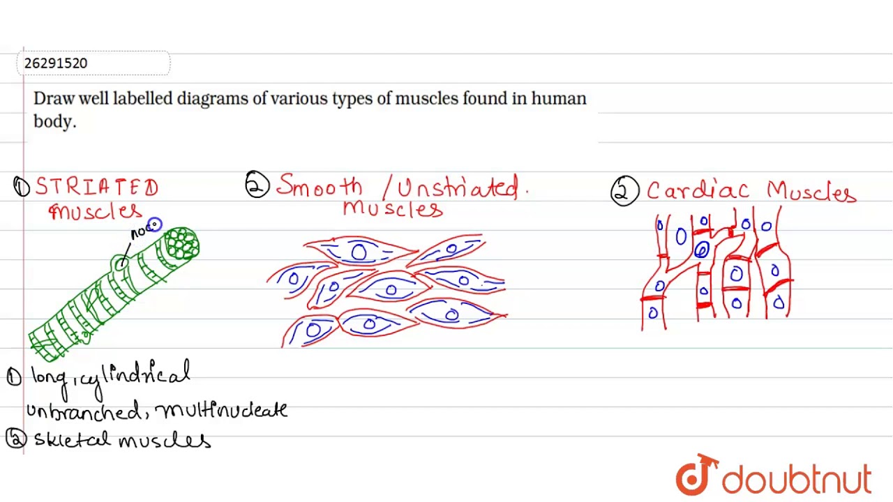
Draw Well Labelled Diagrams Of Various Types Of Muscles Found In Human Body Youtube
Draw a labelled diagram of your preparation.
Draw a labelled diagram of striated muscle. This type of involuntary non-striated muscle is also found in the tracts of the urinary respiratory and reproductive systems. Jul 10 2018 - This website is for sale. When smooth muscle contracts it helps the organs carry out their functions.
Draw A Labelled Diagram Of Smooth Muscle Explain Brainlyin. Each muscle fibre has a single nucleus in the central thick portion. Structural Organization in Animals.
Striations hence these muscles are called striated muscles Fig53a. The term smooth muscle refers to a muscle of the human body that is part of an involuntary muscle group. How to draw diagram of striated muscle tissue step by step for beginners Hello Friends in this video I tell you about how to draw diagram of striated.
Write two differences between striated and smooth muscles. Smooth muscle It has spindle-shaped non-striated uninucleated fibres and occurs in walls of internal organs. Simple Labeled Simple Striated Muscle Diagram Ditulis JupiterZ Sabtu.
Structural Organization in Animals. Model-question-paper23-2-20091pdf - Read File Online - Report Abuse. Muscles are made up of highly specialized thin and elongated cells called muscle fibres.
Dimitrios Mytilinaios MD PhD Last reviewed. Skeletal muscle and cardiac muscleSkeletal muscle is the tissue that most muscles attached to bones are made of. Click hereto get an answer to your question Draw a neat labelled diagram of non - striated muscle.
Hence the word skeletal. In this video I am gonna to show you how to draw the diagrams of cardiac straited smooth muscle for class 1st to 10th. These muscles are also called non-striated or non straight muscles.
5 minutes Striated musculature is comprised of two types of tissues. Smooth muscles never connect with skeleton. Draw a neat labelled diagram of non - straited muscle.
Draw A Diagram Of Cardiac Muscle And Label Any Two Parts Write. Memorizing Complex Images And Charts Studyprof. Draw a labelled diagram of unstriated muscle tissue and mention its occurrence features and functions.
How to draw labelled diagram of striated muscle tissueHello Friends in this video I tell you about how to draw diagram of striated muscle tissue step. A Labeled Diagram Of A Striated Muscle Striated Beef Muscle A Smooth Muscle Images Stock Photos Vectors Shutterstock Muscle Tissue Drawing At Paintingvalleycom Explore Collection Of. Striated musculature Author.
CWhere do you find Kuffer cells. Smooth Muscle Diagram Labeled Class 9. It is the pen diagram of Skeletal Smooth and Cardiac Muscle for class 10 11 and 12.
Welcome to knowledge palace. The structure of the smoothstriatedcardiac muscle tissue with a diagram. Human Muscle Anatomy Quiz.
Draw a well labelled diagram of cardiac muscle found in the human body. Medwritebiz is your first and best source for all of the information youre looking for. It is involutary in action.
How to draw striated muscle in easy way how to draw well labelled diagram of striated muscleHello Friends in this video I tell you about how to draw. Their contractions are voluntary in nature and thus are not. Cardiac muscle It has striated branched uninucleated fibres and occurs only in the walls of the heart.
Discussion Striated muscles constitute the main component of musculature of our body primarily attached to bones via tendons hence are also called skeletal muscles. Achudhan Karunaharamoorthy Reviewer. Muscular System Drawing At Paintingvalley Com Explore Collection.
When you are taking anatomy and physiology you will be required major muscles anatomy quiz for anatomy and physiology class. In this video I have shown the simplest way of drawing Muscle drawing. The muscle fibres are made up of long narrow spindle shaped cells that are generally shorter than striated muscle cells.
I The cells are long and spindle-shaped. We hope you find what you are searching for. Draw a neat labelled diagram of non - striated muscle.
They average a length of about 02 mm. It is divided into two subgroups. From general topics to more of what you would expect to find here medwritebiz has it all.
Striated Muscle Diagram Labeled. Often called striated muscles due to presence of alternate dark and light bands straitions. Ii They do not have striations.
These muscles are also called non-striated or non straight muscles. Hence the word skeletal.

Draw Well Labelled Diagrams Of Various Types Of Muscles Found In Human Body Youtube
Cardiac muscles are involuntary muscles that contract these muscles control the contraction and relaxation of the heart.
Draw a labelled diagram of striated muscle tissue. The three types of muscle fibres arestriated muscles smooth muscles unstriated muscle draw a labelled diagram of a neuron. It controls the movement of an organism. Cells are stimulated and contract as multiple individual units.
Smooth Muscle Diagram - Draw A Labelled Diagram Of Smooth Muscle Explain Brainly In. Write two differences between striated and smooth muscles. Lower back bones and muscles.
Skeletal muscle is the tissue that most muscles attached to bones are made of. The three types of muscle fibres arestriated muscles smooth muscles unstriated muscle draw a labelled diagram of a neuron. The cytoplasm in the muscle fibers is called sarcoplasm.
Draw a neat labelled diagram of non - striated muscle. A smooth muscle is quite important to the human body. Muscular tissue is a specialized tissue in animals which applies forces to different parts of the body by contraction.
Mlck myosin light chain kinase pi phosphate. Smooth muscle has a fusiform shape which resembles a football or spindle. How to draw labelled diagram of striated muscle tissueHello Friends in this video I tell you about how to draw diagram of striated muscle tissue step.
ANAT2241 Muscle Tissue - Embryology from embryologymedunsweduau Identification of parenchyma collenchyma and sclerenchyma tissues in plants striped smooth and cardiac muscle fibers and draw their labeled diagrams. Draw a labelled diagram of a neuron. Draw a labelled diagram of unstriated muscle tissue and mention its occurance features and functions.
The Muscle Tissue Patrick Steele In skeletal muscle a single type of somatic nervous system traverses to muscle where it stimulates organelle in the muscle cells in order to release calcium. Cbse class 9 science chapter 5 tissues exercise questions with solutions to help you to revise complete answer. A Labeled Diagram Of A Striated Muscle Striated Beef Muscle A.
I need to make a labeled model of one and i cant seem to fine one anywhere. MUSCULAR TISSUE ANIMAL TISSUE CHAPTER-6 TISSUE CLASS. Muscle tissues are of three types striated smooth and cardiac.
Each muscle fibre has a single nucleus in the central thick portion. When smooth muscle contracts it helps the organs carry out their functions. Smooth muscles never connect with skeleton.
Skeletal muscle and cardiac muscle. The muscle fibres are made up of long narrow spindle shaped cells that are generally shorter than striated muscle cells. They average a length of about 02 mm.
It contains a network of membrane called the sarcoplasmic reticulum. Smooth Muscle Diagram Labeled Class 9 - Draw and label a diagram of a reflex arc - IB Biology Syllabus Make a labelled diagram to highlight two differences between striated and cardiac muscles. Ii They do not have striations.
The structure of the smoothstriatedcardiac muscle tissue with a diagram. Smooth muscle is found throughout the body around various organs and tracts. Draw a well labelled diagram of cardiac muscle found in the human body.
Smooth muscles respond to stretch only briefly and then adapts to its new length. Striated skeletal muscle brings about voluntary movements of body parts eg movement of arms. Get the answers you need now.
I The cells are long and spindle-shaped. Give one important functional difference amongst the muscle tissues and draw a labelled diagram of the muscle tissue which never shows fatigue. Structural Organization in Animals.
Smooth muscle diagram diagram of smooth muscle cell brainlyin smooth muscle tissue anatomy and physiology diagrams Learn vocabulary terms and more with flashcards games and other study tools. Muscle Tissue Drawing At Paintingvalleycom Explore Collection Of 108 Smooth Muscle Anatomy And Physiology My Friends Told Me About You Guide Skeletal Muscle Tissue Labeled. Cardiac muscle on the other hand is the muscle found on the walls of the heart.
How to draw labelled structure of striated muscle tissueHello Friends in this video I tell you about how to draw structure of striated muscle tissue s. Striated musculature is comprised of two types of tissues. Draw a labelled diagram of unstriated muscle tissue and mention its occurrence features and functions.
Microscopically both the skeletal and cardiac musculature have a striated appearance due to their. It is made up of thin and elongated cells called muscle fibers. 30 unlabeled muscle diagram worksheet.
Match column i with column ii. They average a length of about 02 mm.

Draw Well Labelled Diagram Of Different Types Of Muscles Found In Human Body Science Tissues 13320635 Meritnation Com
Smooth muscle tissue diagram labeled tissue photos and wallpaper upaaragonco.

Draw a neat labelled diagram of striated muscle. Smooth Muscle Diagram. Some muscles skeletal muscles will not contract unless stimulated by neurons. In these organs muscles serve to move substances.
In simple words muscular force is the force produced by contracting muscle cells. Discussion Striated muscles constitute the main component of musculature of our body primarily attached to bones via tendons hence are also called skeletal muscles. The muscular system is composed of specialized cells called muscle fibers.
Smooth Muscle Diagram Class 11. Beta 2 receptors are also on small coronary arterioles thus increasing hormonally induced blood flow within the musculature of the heart. Each muscle fibre has a single nucleus in the central thick portion.
Draw a neat labelled diagram of non - striated muscle. Structural Organization in Animals. Cardiac muscle It has striated branched uninucleated fibres and occurs only in the walls of the heart.
These muscles are also called non-striated or non straight muscles. Structural Organization in Animals. Click hereto get an answer to your question Draw a neat labelled diagram of non - striated muscle.
Their contractions are voluntary in nature and thus are not controlled by autonomic nervous system. The muscle fibres are made up of long narrow spindle shaped cells that are generally shorter than striated muscle cells. Most Downloads Size Popular.
Smooth Muscle Diagram Smooth Muscle Diagram Class 11 Draw A Neat Labelled Diagram Of Non Striated Muscle Learn How To Draw Smooth Muscle Pictures Using These Outlines Or Print Just For Coloring Smooth muscles are involved in many. How to draw labelled diagram of striated muscle tissueHello Friends in this video I tell you about how to draw diagram of striated muscle tissue step. In this video I have shown the simplest way of drawing Muscle drawing.
Smooth Muscle Diagram Class 11 Draw A Neat Labelled Diagram Of Non Striated Muscle Learn How To Draw Smooth Muscle Pictures Using These Outlines Or Print Just For Coloring Novellav Toknot. Muscles are attached to bones or internal organs and blood vessels. It is involuntary in action.
Smooth muscle It has spindle-shaped non-striated uninucleated fibres and occurs in walls of internal organs. Documents similar to class 11 biology chapter 20 notes. Vascular smooth muscle refers to.
Get 10 free Shutterstock images - PICK10FREE. Muscles allow a person to move speak and chew. They are made up of muscle cellthey are elongated and large sizeso they are called as muscle fibrethey contain special protein called contractile protein.
Draw A Neat Labelled Diagram Of Non Straited MuscleHeres a quick rundown of the key. Cardiac skeletal and smooth muscles are the three types of muscles found in the human body. How to draw striated muscle in easy way how to draw well labelled diagram of striated muscleHello Friends in this video I tell you about how to draw.
Striations hence these muscles are called striated muscles Fig53a. Draw a labelled diagram of your preparation. Smooth muscle is under involuntary control and is innervated by.
How to draw structure of striated muscle tissue striated muscle tissue diagramHello Friends in this video I tell you about how to draw structure of. Draw a neat labelled diagram of non - straited muscle. There are more than 600 muscles present in a human.
Almost all the animals have muscles. At the simplest level muscles allow us to move. Transcribed image text from this question.
Their predominant function is contractibility. It is the pen diagram of Skeletal Smooth and Cardiac Muscle for class 10 11 and 12. It is involutary in action.
The structure of the smoothstriatedcardiac muscle tissue with a diagram. Smooth Muscle Diagram Labeled Class 9 Draw A Neat Labelled Diagram Of Non Striated Muscle. I need to make a labeled model of one and i cant seem to fine one anywhere.
Often called striated muscles due to presence of alternate dark and light bands straitions. Muscular system is an organ system Body performs all the kind of movements with the help of muscular system. Smooth Muscle Diagram Simple - Draw A Neat Labelled Diagram Of Non Striated Muscle.
Save my name email and website in this browser for the next time I comment.
Hamstring group Three muscles run down the back of your leg from your thigh to your knee the biceps femoris semitendinosus and semimembranosus and help you bend your knee and extend your hip. Muscle Diagram Back Posterior View.

Posterior View Of Body Muscles Diagram Quizlet
Back muscle diagrams labeled find out more about back muscle diagrams labeled.
Labeled muscle diagram posterior. The anterior and the posterior compartments of the arm. Smartdraw includes 1000s of professional healthcare and anatomy chart templates that you can. Tendonitis is the swelling of a tendon which is a thick cord attaching a muscle to a bone.
Jun 19 2017 - This Pin was discovered by David Clark. The muscles of the leg are important for. When autocomplete results are available use up and down arrows to review and enter to select.
Muscles of the leg include muscles of the thigh and foot. Five pairs of lumbar spinal nerves labeled l1 to l5 branch off your spinal cord and exit through small holes between the vertebrae. Posterior View Of Human Body Muscles Diagram.
Discover and save your own Pins on Pinterest. Anterior lateral and posterior compartment. Muscle contraction 1125 describe the structure of striated muscle fibres including the myofibrils i bands only actin h band only myosin you need to be able to draw and label these diagrams.
In human anatomy the muscles of the hip joint are those muscles that cause movement in the hip. Muscles in the body labelled diagram. Human anatomy and physiology diagrams.
Discover and save your own Pins on Pinterest. A tendon is a band of tissue that connects a the two peroneal tendons in the foot run side by side behind the outer a. Label and color these muscles of the posterior body.
Male muscular system full anatomical b. The gastrocnemius muscle has two large bellies called the medial head and the lateral. Muscles of the leg anterior lateral posterior teachmeanatomy.
Back Muscles Diagram Labeled. Leg Muscle Diagram Unlabeled - posterior muscles unlabeled Study Resources Pinterest. 820 x 1267 jpeg 122 кб.
As part of the iliopsoas psoas major contributes to flexion in the hip joint. Leg muscle anatomy posterior leg muscles diagram photo album. Gastrocnemius muscle large posterior muscle of the calf of the leg.
Related posts of muscles of the leg labeled. Get a handful labeled leg muscle diagrams to assist your study about humans leg muscle anatomy. To see a muscular system picture from the anterior front view click here.
Leg Muscle Diagram Labeled Muscles of the Leg - Anterior - Lateral - Posterior. Anatomy muscle chart diagram poster muscle diagram muscle. The extrinsic group originate from the torso and attach to the bones of the shoulder whereas the intrinsic ones.
Muscle diagram blank each of these muscles is a discrete organ constructed of skeletal muscle tissue blood vessels tendons and nerves. Jun 19 2017 - This Pin was discovered by David Clark. 12 photos of the back muscle diagrams labeledMuscles 6 muscular system labeled pinterest diagram of the muscles system the muscular system micro 28 images muscles of the unlabeled anatomy we gather some.
By Sport Fitness Advisor Staff. This diagram depicts hip muscles diagram. Arm Muscle Diagram Labeled - Posterior - Can a posterior placenta migrate to the back.
System diagram labeled 209 human muscular system diagram labeled. There are anterior muscles diagrams and posterior muscles diagrams. Muscular System Diagram Posterior Back View.
There are anterior muscles diagrams and posterior muscles diagrams. - Muscles of the head and neckThe supraspinatus plural supraspinati is a relatively small muscle of the upper back that runs from the supraspinous fossa superior portion of the scapula shoulder blade to the greater tubercle of the humerus. Labeled Anterior And Posterior Muscles Of The BodyThere are 20 forearm muscles which are arranged an anterior compartment that contains flexor forearm muscles in the posterior compartment of the forearm are popularly called the extensor the anterior growth causes the vertebral bodies and discs to bulge laterally toward the convexity and to.
Also referred to as ventral anterior cavity larger body cavity located anterior to the posterior dorsal body cavity. As a result the physician will be able to provide you with exact medication based on the report and thus you will be to. Tendon Diagram Labeled posterior muscles of the body diagram With images.
Detailed anterior lateral and posterior viewsmen sports fitness training. Resource site for teachers and students of anato. Touch device users.
This labeled human muscular system chart illustrates the major muscle groups in the back posterior view and the front anterior view. Human muscle diagram muscular system sketch at paintingvalley explore collection of. The leg muscles are organized in 3 groups.
Broadly considered human musclelike the muscles of all vertebratesis often divided into striated muscle smooth muscle and cardiac muscle. In this image you will find splenius capitis rhomboid minor rhomboid major levator scapulae semispinalis capitis trapezius latissimus dorsi deltoid rotator cuff muscle teres major erector spinae group iliocostalis spinalis teres minor supraspinatus infraspinatus subscapularis in it. This muscular system diagram shows the major muscle groups from the back or posterior view.
It originates at the back of the femur thighbone and patella kneecap. Posterior - Can a posterior placenta migrate to the back. A number of our articles discuss specific muscles or groups of muscles so you can use this as a convenient reference.