Understanding the Human Figure. Directory of leading providers of high resolution medical images and interactive human anatomy models.
Human Body Anatomy And Physiology Of Human Body
Find high-quality stock photos that you wont find anywhere else.
Anatomy and physiology picture of human body. Anatomy Books Made For Artists. Human Anatomy is the scientific study of form and shapes of human beings. Urine formation filtration reabsorption secretion excretion.
Advertentie 3D And Live Models From Various Angles And Body Postures And Color-Coded Muscle Diagrams. Advertentie Anatomy And Physiology Resources. Anatomy and Physiology-1-The Human Body.
The 1 Human Anatomy and Physiology Course - Learn About The Human Body With Illustrations and Pictures Award Winning Human Anatomy and Physiology Home Study Course - For Practitioners Students Medical ProfessionalsParamedics Academia. An introduction to the human body in 50 visual interactive chapters. Provides educational apps including interactive 3D models and lectures.
Structure of a Nephron. 425741 human body anatomy stock photos vectors and illustrations are available royalty-free. Immune Lymphatic Systems.
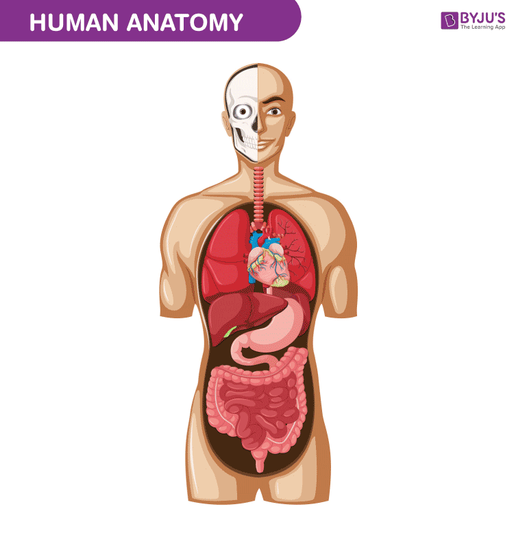
See human anatomy stock video clips. Boy body anatomy with internal organs. Image of free textbook below.
Anatomy and physiology presented in 3D model sets 3D animations and illustrations. Each unit presents a body system in a series of chapters with bite-sized visual interactivities and quizzes. Video lessons from awesome teachers 1 a day.
Human body side vector human heart hands anatomy 3d imaging human body body anatomy system prostate infection human heart with blood human painful joints pain concepts posture back view. Human heart and vascular system - anatomy of the human body stock pictures royalty-free photos images. Advertentie Anatomy And Physiology Resources.
Anatomy Books Made For Artists. Understanding the Human Figure. Browse 26983 anatomy of the human body stock photos and images available or start a new search to explore more stock photos and images.
Health medical icon human body physiology isolated on white background vector illustration. Anatomy and Physiology of Human Body - YouTube. See human body anatomy stock video clips.
About Press Copyright Contact us Creators Advertise Developers Terms Privacy Policy Safety How. Cells organs systems Pathology study of anatomy and physiology. Search from Human Anatomy Physiology stock photos pictures and royalty-free images from iStock.
Some sites also provide study aids such as flash cards and quizzes. Anatomy And Physiology of Human body 1. Human anatomy icons - anatomy of the human body stock illustrations.
DEFINITION OF ANATOMY PHYSIOLOGY Anatomy study of structures by dissection imaging and microscopy macro - gross systemic embryology development micro - cytology histology Physiology study of functions by chemical and physical means. Model of the intestine and the doctor on blurred background. An Orientation-Flashcards Anatomical Terminology-Abdominopelvic regions and quadrants Holes Human Anatomy online text book Companion site.
Learning Outcome Questions Chapter 1 and 2. 690104 human anatomy stock photos vectors and illustrations are available royalty-free. The skeleton also protects several vital organs such as the heart lungs and the liver.
Trackable unit objectives with multiple-choice and dissection quizzes for. Joints are points at which two bones meet. Advertentie 3D And Live Models From Various Angles And Body Postures And Color-Coded Muscle Diagrams.
Body naked anatomy face and back human musculature front and side female profile running human body human body blood flow the human body with organs anatomy body organs human anatomy blue girl underwear. Video lessons from awesome teachers 1 a day. Bones are attached to other bones through ligaments a fibrous connective tissue.
Liquid enters to the glomerulus in Browmans capsule goes down by.
See human eye diagram stock video clips. Diagram of the eye diagram of eyeball cross section of human eye section of human eye cross section eye cross section of eye anatomy of the eye eye parts human eye diagram of eye.
Main Parts Of Human Eye Free Transparent Png Download Pngkey
The pupil is a small opening in the iris.
/GettyImages-695204442-b9320f82932c49bcac765167b95f4af6.jpg)
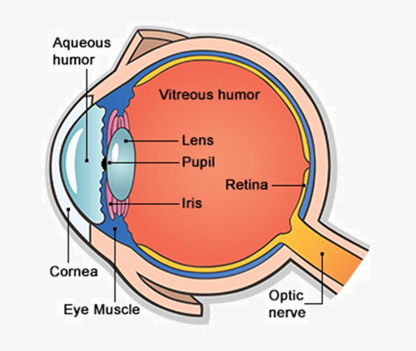
Picture of parts of the human eye. Light enters the eye. Human Body Parts List. When you look closely at an eye the cornea is the clear bulging surface that forms the shape for the front of the eye.
The iris controls the size of the pupil. The spaces within the eye are filled with fluids that help maintain its shape. Choose from Parts Of The Human Eye Pictures stock illustrations from iStock.
The lens can change shape by getting thicker or thinner to optimize the clarity of the picture. External components include structures which can be seen on the exterior of the eye. The eye is a hollow spherical structure about 25 centimeters in diameter.
The front transparent part of the sclera is called cornea. There are 6 sets of muscles attached to outer surface of eye ball which helps to rotate it in different direction. The pupils function is to adjust the amount of light entering the eye.
6513 human eye diagram stock photos vectors and illustrations are available royalty-free. 56779 human eye anatomy stock photos vectors and illustrations are available royalty-free. A human eye is roughly 23 cm in diameter and is almost a spherical ball filled with some fluid.
Anatomically the eye comprises two components fused into one. Human retinas - left and right eye - anatomy of the eye stock pictures royalty-free photos images. Find high-quality royalty-free vector images that you wont find anywhere else.
Floating eyeball - anatomy of the eye stock pictures royalty-free photos images. The eye is the photo-receptor organ. This part of the eye works along with the cornea to focus the light on the retina located in the back of the eye.
Choose from Parts Of The Human Eye stock illustrations from iStock. Search from Parts Of The Human Eye stock photos pictures and royalty-free images from iStock. See human eye anatomy stock video clips.
It is a protective tough white layer white part of the eye. A schematic section through the human eye with a schematic enlargement of the retina The retina is approximately 05 mm thick and lines the back of the eye. Picture of Internal Organs.
The outer covering of the eye is called sclera. It is the outer covering a protective tough white layer called the sclera white part of the eye. Anatomy parts and structure.
Try these curated collections. Learn about their function and problems that can affect the eyes. Structure of the human eye.
Studio close up of mid adult womans gazing blue eye - anatomy of the eye stock pictures royalty-free photos images. Structure of the Human Eye. The cornea is the outer.
WebMDs Eyes Anatomy Pages provide a detailed picture and definition of the human eyes. Find high-quality stock photos that you wont find anywhere else. The transparent part in front of the sclera is called the cornea.
Search from Illustration Of Parts Of The Human Eye Pictures stock photos pictures and royalty-free images from iStock. Find high-quality stock photos that you wont find anywhere else. The structure of the human eye is shown above in the image Parts of the Human Eye.
It is enclosed within the eye sockets in the skull and is anchored down by muscles within the sockets. Hence it does not possess a perfect spherical shape. The optic nerve contains the ganglion cell axons running to the brain and additionally incoming blood vessels that open into the retina to vascularize the retinal layers and neurons Fig.
This clear part of the eye focuses the light so the image can reach the back of the eye. Find high-quality royalty-free vector images that you wont find anywhere else. The human eye is a roughly spherical organ responsible for perceiving visual stimuli.
Its wall has three distinct layersan outer fibrous layer a middle vascular layer and an inner nervous layer. It consists of the following parts. Structure of Human Eye.
Human eye is spherical about 25 cm in diameter. Parts of the Body. It is situated on an orbit of skull and is supplied by optic nerve.
13 Zeilen Eye Parts.
Leaf-blade or Lamina. Download and use 70000 leaf stock photos for free.
Leaf Labelled Stock Photo Download Image Now Istock

They help plants in a variety of ways including producing food and oxygen through photosynthesis balancing water loss regulating gas exchange and transporting the products of photosynthesis.

Labelled picture of a leaf. There are different types of leaves which let us to distinguish the different kinds of plants but essentially each leaf is formed by the following parts. ᐈ Labeled Diagram Of The Ribs Stock Pictures Royalty Free Midrib Vectors On Depositphotos Draw A Labelled Diagram Of The External Structure Leaf Brainly In Solved Section 1 Examination Of The Internal Structure O Chegg Com. See leaf label stock video clips.
They are arranged closely together so that a lot of light. It is normally green in colour and manufactures food for the whole plant. They can be in many different forms ie.
Leaf anatomy Both Internal and External with Labelled Diagram. On the basis of number to leaflet a palmately compound leaf may be of the following types. Leaf apex - the outer end of a leaf.
Leaves are the main photosynthetic organs of the plant. Lamina - the blade of a leaf. Palisade cells are column-shaped and packed with many chloroplasts.
Under a powerful microscope we can see three main internal parts of a leaf ie. It is the thin flat part of the leaf that is typically green in color. Anatomy of the leaf is the detailed study of the internal structure of a leaf usually revealed by its dissectionLeaves are responsible for converting sunlight and carbon dioxide.
Generally leaf base petiole and lamina together form the main parts of a leaf. The major function of the leaf is to absorb sunlight energy to make food. November 1 2019 by Ranganr.
Definition of a Leaf. Conifer needle-like leaves Apple Leaf Ash Green Leaf Aspen Quaking Leaf Buckeye Ohio Leaf Catalpa Western Leaf Chokecherry Leaf Coffeetree Kentucky Leaf. Hi In this post we bring you various inspiring images that we collected for people like you this time we are pay more attention concerning Parts of a Leaf Worksheet.
Label Simple Leaf External Anatomy. Axil - the angle between the upper side of the stem and a leaf or petiole. External Parts of a Leaf.
33477456 leaf stock photos vectors and illustrations are available royalty-free. This is the tip of the leaf. Click on leaf images to enlarge.
1 Petiole- the stalk that supports a leaf in a plant and attaches the leaf. Epidermis mesophyll and vascular bundle. Thousands of new images every day Completely Free to Use High-quality videos and images from Pexels.
Draw A Labelled Diagram Of Cross Section Leaf Lamina To Show Chloroplasts From Science Life Processes Class 10 Cbse. See leaf stock video clips. In the leaves of these plants leaflets are present in primary stage so palmate habit appears in.
A leaf has two main parts. Leaf base has two small leaf-like structure called stipules. This is the middle vein of the leaf.
Tropical tropical leaf on white fall seniors out of focus spring trees sun abstract green leaves green foliage white background macro green leaves background texture material tropical leaves plant leaf close up. 741024 leaf label stock photos vectors and illustrations are available royalty-free. In plants like paddy wheat and other monocotyledons this leaf.
Light absorption happens in the palisade mesophyll tissue of the leaf. This is the blade of the leaf. It is further divided into three parts.
Structure Of A Leaf - Internal External. Leaves are vital to the survival of plants. The internal structure of a leaf.
Bio tag soap label templates lxuury frames invite black and white wedding border circle eco logo bio tags invitation gold leaves gold and bronze design style hand drawn menu badges elegant white and gold invitations. The leaves take up water and carbon dioxide and convert them into carbohydrates in the presence of sunlight and chlorophyll. By the way concerning Parts of a Leaf Worksheet we already collected various variation of images to inform you more.
Leaf Identification Identify Trees by Their Leaves. The leaf is one of the most important part of the plants because it is the one which is in charge of accomplishing the photosynthesis the respiration and transpiration. Leaves are very important in structuresThey are the plants food factories.
Leaf parts coloring page leaves worksheets activities and parts of a plant printable worksheet are three. I leaf apex the tip of the leaf blade ii leaf margin the edge of the leaf and iii leaf veins the small channels or capillaries which. They are the thin and flat organs of plants that are responsible for photosynthesis.
This is the outer edging of the leaf. Labeled diagram a leaf midrib this just means central vein within the leaf all the other veins sprout off of it blades these generally are long flat and have a good surface area to capture the maximum amount of sunlight nodes this is the exact spot where the leaf connects itself ot the stem bbc gcse bitesize inside the leaf inside the leaf higher tier you should be able to explain how the cellular structure of a leaf is. The leaf is a flattened lateral outgrowth of the stem in the branch developing from a node and having a bud in its axil.
In this type only one leaflet is attached on the apex of lamina eg Citrus lemon. This is the part where a leaf attaches to the stem. Read the plant definitions below then label the simple leaf morphology diagram below.
Find high-quality stock photos that you wont find anywhere else. Motte and Bailey Castles - Labelled diagram.
What Is A Motte And Bailey Castle Twinkl Teaching Wiki
Relatively easy to build with unskilled labour but still militarily formidable these castles were built across northern Europe from the 10th century onwards spreading from Normandy.
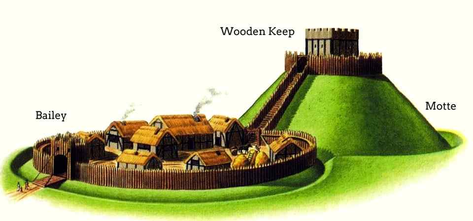
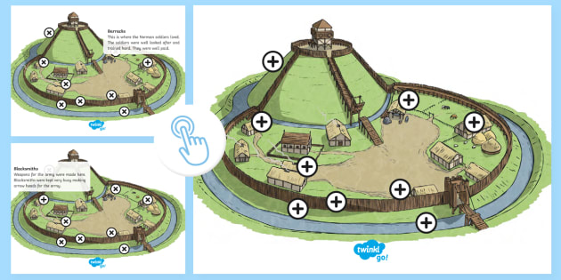
Labelled picture of a motte and bailey castle. To make a motte and bailey castle start by getting a base for your castle like a piece of styrofoam or plywood and drawing 2 overlapping circles on it. Keep Motte Drawbridge Ditch Bailey Fence. Windsor Castle with motte in the center and baileys on both sides.
The huge motte with its timber tower on top gave the defenders an advantage. Find the perfect motte and bailey castle stock photo. 47 An alternative approach focuses on the links between this form of castle and what can be termed a feudal mode of society.
Motte Bailey Pallisade Ditch Keep Drawbridge. Huge collection amazing choice 100 million high quality affordable RF and RM images. David J Morgan CC-BY-SA-20.
It is predominantly rounded in plan but square or rectangular mottes are known especially in Scotland. A stone castle was built on the Motte but this was dismantled in about 1629. The Motte was around 15 metres high and surrounded by a moat.
Then glue or tape an upside-down bowl over one of the circles which will be the motte that the castle sits on. Motte and bailey castles Norman castles were designed for a different purpose they were not defensive structures like the burhs they were designed to intimidate the conquered Anglo-Saxons and. Motte and bailey castle labelled.
Tonbridge Castle Kent Victorian engraving Victorian engraving of the gatehouse of Tonbridge Castle in Kent England. Browse 135 motte and bailey castlestock photos and images available or start a new search to explore more stock photos and images. The remains of Pleshey Castle a Motte and Bailey castle in Essex England.
You can still clearly see the Motte though. Motte and bailey castle labelled. No need to register buy now.
Motte-and-bailey castle raised earth mound and enclosed courtyard surrounded by water ditch. Label Motte and Bailey Castle - Labelled diagram. A motte-and-bailey castle is a European fortification with a wooden or stone keep situated on a raised area of ground called a motte accompanied by a walled courtyard or bailey surrounded by a protective ditch and palisade.
It doesnt burn like wood does and it is more difficult to knock down a stone wall than a wooden wall. Next use popsicle sticks to build a wall around the motte. Relatively easy to build with unskilled labour but still militarily formidable these castles were built across northern Europe from the 10th century onwards spreading from Normandy and Anjou in France into the Holy Roman Empire in the 11th century.
Jan 25 2016 - View 26 Best labelled diagram of a motte and bailey castle images. 135 Motte And Bailey Castle Premium High Res Photos. Jan 25 2016 - View 26 Best labelled diagram of a motte and bailey castle images.
Cut out and stick in picture of Motte and BaileyCut out and stick in pict. Search from Motte And Bailey Castle stock photos pictures and royalty-free images from iStock. Motte and bailey castle motte and bailey castle yelden bedfordshire home counties england uk europe motte and bailey stock pictures royalty-free photos images.
The bailey was designed so that any point on its circumference outer edge would be within bowshot of the tower. Motte and bailey castles were quick and cheap to erect - - some only took a couple of weeks. - motte and bailey castle stock illustrations.
Amotte is an enditched mound usually artificial which supported the strongpoint of the motte-and-bailey castle overshadowing the bailey or enclosed courtyard below.
Old medical illustration head engraved head vintage brain lobes head etching anatomy brain vintage face illustration head engraving anatomy of the brain science old draw. Signals from the brain tell muscles to contract.
Solved Label The Parts Of The Human Brain And Describe Their Functions Course Hero
Connecting the brain to the spinal cord the brainstem is the most inferior portion of our brain.
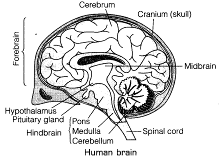
Label picture of human brain. 300 Free Human Brain Brain Images. The brain controls voluntary and involuntary movements. Brain diagram with labels hypothalamus vector brain diagram pons cerebrum and cerebellum brain pons brain anatomy amygdala brain labelled amygdala brain human midbrain diagram pons.
Your brain may look like an ugly wrinkled gray sponge but it is actually the lord and master of your body. Labels are usually small in size so you should carefully choose the font of the texts to make sure it is readable. In short it is the brain that embodies the essence of mind and soul and death of this master organ literally means the death of entire body and the end of life.
The average dimension of the adult human brain is 55 inches in width and 65 inches in length. Using radio waves in a magnetic field an MRI scanner creates highly detailed images of the brain and other parts of the head. Browse 849 human brain diagram stock photos and images available or start a new search to explore more stock photos and images.
Free for commercial use No attribution required High quality images. Human brain diagram blank label the brain anatomy diagram answers and plant cell diagram with labels are three of main things we want to show you based on the post title. Magnetic resonance imaging.
The total surface area of the cerebral cortex is about 2500 cm2 and when stretched it. Daniel Nelson on June 10 2019 Leave a Comment. It is this part of the human body that serves as a control center for every activity in the body.
Mental power concept - human brain stock illustrations. You can also put your logo at the top or bottom corner of the label. The human brain controls nearly every aspect of the human body ranging from physiological functions to cognitive abilities.
The height of the human brain is about 36 inches and it weighs about 4 to 5 lbs at birth and 3 lbs in adults. The Center of CNS. Divided into different sections it constitutes the central and superior most part of the central.
2653 labeled brain anatomy stock photos vectors and illustrations are available royalty-free. Labels are a means of identifying a product or container through a piece of fabric paper metal or plastic film onto which information about them is printed. It functions by receiving and sending signals via neurons to different parts of the body.
Brain images with labels. Now lets begin exploring the main structures of our awesome human brain. Talking related with Brain Label Worksheet below we will see particular related photos to inform you more.
Find images of Human Brain. Wednesday July 11th 2018. 111562 human brain anatomy stock photos vectors and illustrations are available royalty-free.
Browse 25645 human brain stock photos and images available or search for human brain anatomy or human brain illustration to find more great stock photos and pictures. Input from the brain controls the function of other organs in the body. Thalamus Intermediate mass Hypothalamus BRAIN STEM.
These are called the functions of brain. The brain stem begins inferior to the thalamus and runs approximately 7 cm before merging into the spinal cord. The brain is a fascinatingly complex organ responsible for all that we do.
Utilize the model of the human brain to locate the following structures landmarks for the diencephalon. T he human brain just like most other mammals has the same basic structure but it is better developed than any other mammalian brain. Human brain icons - vector illustration - human brain stock illustrations.
When we talk about Brain Parts Worksheet we have collected various similar pictures to inform you more. Labeled Model Of The Human Brain. Many of the most basic survival functions of the brain are controlled by.
Johannes Sobott via Wikimedia Commons Public Domain. See labeled brain anatomy stock video clips. The brain is the complex organ responsible for processing sensory information sound touch taste sight and smell.
Make your work easier by using a label. Wide collections of all kinds of labels pictures online. Label the Human Brain Label the Human Brain.
Labeled Brain Model Diagram. Human brain parts worksheet label heart diagram worksheet for kids and brain label worksheet are three main things we want to present to you based on the gallery title. See human brain anatomy stock video clips.