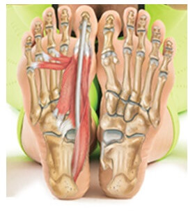
The tendons are thick bands that connect muscles to bones. The muscles at the top of the foot fan out to supply the individual toes.

Foot Anatomy Eorthopod Com
Both tendons run down across the front of the ankle across the top of the foot and then fan out attaching to the tips of the toes.
Parts of foot tendon. The TMT joint is made stable not only by strong ligaments connecting these bones but also because the second metatarsal. The Achilles tendon is the large tendon that attaches the back of your heel to your calf muscle. Your flexor foot tendons are located on the bottom of your feet.
Flexor Foot Tendons. Symptoms Achilles tendonitis causes an aching or burning pain with activity or stretching and the affected tendon is usually painful to the touch. The calf muscles through the Achilles tendon are the main plantarflexors of the ankle which pulls the foot down.
These parts are also responsible for different actions such as jumping climbing walking and running. The calcaneus which is. If you would like to learn all the parts of the foot structure you have come to the right place.
The cuboid is on the lateral side of the foot outer foot and sits in front of the calcaneus. Flexor hallucis longus flexor hallucis brevis flexor digitorum longus and posterior tibialis. Fourth layer-Interossei Dorsal 4 Planter 3 Paroneus longus tendon Tibialis posterior tendon Muscles of the dorsum of the foot This group has two muscles.
On the top of the foot are extensor tendons that help move your foot towards the front of the leg dorsiflexion There include. The Achilles tendon is the strongest and thickest tendon in the body and can withstand strains of up to 10 tons. On the side of the foot are.
When the muscles tighten contract they pull on the tendons which in turn move the bones. The peroneal brevis tendon the tendon of the tibialis anterior muscle the tendon of the tibialis posterior muscle the tendon of the peroneus longus and the tendons of the flexor hallucis longus and flexor digitorum longus muscles are some of the tendons in feet which are prone to injuries. 16 There are four flexor tendons.
There are 33 different joints in the foot. The extensor tendons sit between the skin and the bones and there is little padding around them making them prone to injury resulting in top of foot pain. Functionally the Achilles ankle tendon provides the large propulsive force needed for walking running and jumping.
Each foot contains five metatarsals numbered 1-5 medial great toe to lateral. Ballet dancers often have cases of tendinitis in their flexor hallucis longus because of the repeated movements associated. Achilles tendonitis refers to irritation or inflammation of the Achilles tendon which connects the calf and lower leg muscles to the heel bone of the foot.
The tendons are thick bands that connect muscles to bones. It is made up of bones muscles tendons ligaments and 100 other which are designed o allow the foot to balance the body on two legs. The first three metatarsals medially are more rigidly held in place than the lateral two.
This is the weakest part of the tendon and. Achilles tendonitis usually occurs one to four inches above the area where your Achilles attaches to your heel bone. The talus and the three cuneiforms.
The muscles are located mainly in the sole of the foot and divided into a central medial group and a group on either side lateral. If you talk about human body foot it is one of the most difficult and complex structure to understand. The Achilles tendon is a tough band of fibrous tissue that connects the calf muscles to the heel bone calcaneus.
Human feet are mostly subjected to injury due to their. The phalanges which are the bones in your toes The metatarsals which run through the flat part of your foot The cuneiform bones the navicularis and the cuboid all of which function to give your foot a solid yet somewhat. The posterior tibial tibia tendon located at the inner side of your foot.
Tendon of the tibialis anterior. The navicular is on the medial inner side of the foot between the talus and the cuneiform bones in front. The Achilles tendon is also called the calcaneal tendon.
Arguably the most important tendon is the achilles tendon which allows the calf muscles. A foot pain diagram is a great tool to help you work out what is causing your ankle and foot pain. Extensor hallucis longus tendon.
The metatarsals articulate with the mid-foot at their base a joint called the tarsal-metatarsal TMT joint or Lisfranc joint. These tendons help the flexor muscles to stabilize your toes. Arguably the most important tendon is the Achilles tendon.
The joints are made up of two bones coming together but they also involve tendons ligaments and muscles. The muscles tendons and ligaments. Extensor digitorum muscle tendon.
The navicular forms joints with four bones.