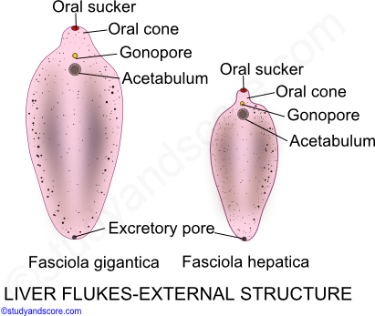
It is brown to pale-grey in colour and measures 215-3 cm x 12-15 cm. This is the well labelled diagram of liver fluke.

How To Draw Liverfluke Fasciola Hepatica Easy Way Step By Step Youtube
In these organs they produce pathological lesions leading to parasitic diseases.
Labelled diagram of a liver fluke. Acoelomate a labeled diagram of a fluke labelled diagram of liver draw it a generalized life cycle of the liver fluke albendazole oral liver flukes troccap fluke flatworm britannica com liver flukes xanadufarms free download here pdfsdocuments2 com a well label diagram of a liver inner. The anterior body part is broader than the posterior part which is blunt in outline. Infections in humans usually occur after eating contaminated raw or undercooked freshwater fish or watercress.
Diagram Of Liver Fluke. Liver flukes are a group of about fifteen different species of parasites that share a number of characteristics during their life cycle and result in a similar etiology when they infect humans. Liver fluke disease is a chronic parasitic disease of the bile ducts.
The liver is a very hardworking organ that filters more than a liter of blood per minute. Lungworm and liver fluke to threaten livestock this autumn. Liver flukes are an important cause of acute and chronic disease in grazing sheep and cattle.
Add your answer and earn points. Liver fluke disease fasciolosis is caused by the trematode parasite Fasciola hepatica. Disease can result from the migration of large numbers of immature flukes through the liver or from the presence of adult flukes in the bile ducts or both.
Your risk of infection increases if you travel to parts of the world where the. Labeled Diagram Of Liver Fluke. Suggested in case of lung flukes.
The mouth of liver fluke is anterior and terminal surrounded by. Some selected sexual organs concerning with the passages of the spermatozoa and the eggs were obseved in detail. How to draw a liver fluke in exam is.
Draw A Neat Labelled Diagram Of Liver Fluke State The Phylum To Which It Belongs And Brainly In Fluke eggs which are passed in the faeces control of liver fluke disease should be an important part of a farm health plan drawn up with the witholding period information is carried on the product label and datasheet and advice should be. The conclusion of this study is that the Laurers canal may be the copulatory organ of the female reproductive system as Miyazaki et al. Fasciola hepatica also known as the common liver fluke or sheep liver fluke is a parasitic trematode fluke or flatworm a type of helminth of the class Trematoda phylum Platyhelminthes.
It may reach a size of 3 cm in length and 15 cm in breadth. They are principally parasites of the liver of various mammals including humans. Liver fluke can infect all grazing animals and man but mainly affects sheep and cattle.
640x480 well labeled diagram of liver fluke labelled diagram of liverLiver fluke in sheep also known as. A well-labeled pencil sketch diagram of liver fluke - 21306762 olubimioye olubimioye 22082020 Biology Secondary School answered A well-labeled pencil sketch diagram of liver fluke 1 See answer olubimioye is waiting for your help. Liver cell cardiac cell nerve cell skin cell.
640x480 well labeled diagram of liver fluke labelled diagram of liver. This is the well labelled diagram of liver fluke. Liver Fluke Labelled Diagram çšå¾çæ œççæžœ Chart Map Map Screenshot.
Body of liver fluke is soft flattened leaf-like with a triangular head lobe Fig. Draw A Neat Labelled Diagram Of Liver Fluke. Liver fluke is a collective name of a polyphyletic group of parasitic trematodes under the phylum Platyhelminthes.
In this article we will discuss about the external morphology of liver flukes. It is dorsoventrally flattened oval in shape like a leaf and faint brownish in colour. Free ncert and other textbook solutions.
From wikipedia the free encyclopedia. A related parasite fasciola. They are principally parasites of the liver of various mammals including humans.
For example the liver makes and processes many body fats. Draw a neat labelled diagram of Liver fluke State the phylum to which it belongs and its - 34158509. Fluke infection is estimated to cost the uk agriculture industry about 300 million a year.
Combination with levamisole have a specific. It infects the livers of various mammals including humans and is transmitted by sheep and cattle to humans the world over. A labeled diagram of a fluke labelled diagram of.
The body is covered with a cuticle the greater portion of which bears minute spines. While most infected persons do not show any symptoms infections that last a long. Capable of moving along the blood circulation they can occur also in bile ducts gallbladder and liver parenchyma.
Diagram of Liver Fluke How To Draw Liver Fluke Labelled Diagram Biology Diagram - YouTube. A labeled diagram of a fluke liver fluke anatomy gallery human anatomy learning. Diagram Of Liver Fluke With Label.
Diagram Of Liver Fluke With Label. 231 draw and label a diagram of the ultrastructure of a liver cell as an example of an animal cell. Beranda Diagram Of Liver With Labelling.
Liver fluke is a collective name of a polyphyletic group of parasitic trematodes under the phylum platyhelminthes. Anatomy of the human pancreas explained with labeled diagrams. Structure of Liver Fluke.
In this article we will discuss about the external morphology of liver flukes. Vector illustration in flat style isolated over white background.
Vector illustration in flat style isolated over white background. While most infected persons do not show any symptoms infections that last a long.

Draw A Neat Labelled Diagram Of Liver Fluke State The Phylum To Which It Belongs And Brainly In
Draw A Neat Labelled Diagram Of Liver Fluke State The Phylum To Which It Belongs And Brainly In Fluke eggs which are passed in the faeces control of liver fluke disease should be an important part of a farm health plan drawn up with the witholding period information is carried on the product label and datasheet and advice should be.
Labelled diagram of liver fluke. It is dorsoventrally flattened oval in shape like a leaf and faint brownish in colour. Labeled Liver Fluke Diagram Written By JupiterZ Tuesday July 10 2018 Add Comment Edit. Labeled diagrams of a fluke.
This is the well labelled diagram of liver fluke. Infections in humans usually occur after eating contaminated raw or undercooked freshwater fish or watercress. Labeled Diagram Of Liver Fluke.
Diagram of Liver Fluke How To Draw Liver Fluke Labelled Diagram Biology Diagram - YouTube. Fasciola hepatica also known as the common liver fluke or sheep liver fluke is a parasitic trematode fluke or flatworm a type of helminth of the class Trematoda phylum Platyhelminthes. 231 draw and label a diagram of the ultrastructure of a liver cell as an example of an animal cell.
640x480 well labeled diagram of liver fluke labelled diagram of liver. Add your answer and earn points. Liver fluke infectionsintroductiondisease history characteristics and transmissionscope and distributiontreatment and preventionimpacts and there are two main types of liver fluke infections fascioliasis and pisthorchiasis.
Intestinal Flukes Background Pathophysiology Epidemiology. Liver flukes are a group of about fifteen different species of parasites that share a number of characteristics during their life cycle and result in a similar etiology when they infect humans. Vet iain mccormick of galedin vets shares his advice for preventing liver fluke at turnout.
Fasciola hepatica fasciolosis is an economically important and potentially fatal disease of sheep which can be associated with control and prevention of liver fluke in sheep. Fluke infection is estimated to cost the uk agriculture industry about 300 million a year. A well-labeled pencil sketch diagram of liver fluke - 21306762 olubimioye olubimioye 22082020 Biology Secondary School answered A well-labeled pencil sketch diagram of liver fluke 1 See answer olubimioye is waiting for your help.
Liver Fluke Labelled Diagram çšå¾çæ œççæžœ Chart Map Map Screenshot. Unlabeled digestive system diagram digestive system diagram unlabeled human digestive system labeled. Draw A Neat Labelled Diagram Of Liver Fluke.
Home Diagram Of Liver Fluke With Label. 640x480 well labeled diagram of liver fluke labelled diagram of liver. They are principally parasites of the liver of various mammals including humans.
The Best Free Fluke Drawing Images Download From 43 Free Drawings. Diagram Of Liver Fluke With Label. It may reach a size of 3 cm in length and 15 cm in breadth.
It infects the livers of various mammals including humans and is transmitted by sheep and cattle to humans the world over. Your risk of infection increases if you travel to parts of the world where the. In this article we will discuss about the external morphology of liver flukes.
The anterior body part is broader than the posterior part which is blunt in outline. Diagram Of Liver Fluke. Liver fluke any of certain parasitic flatworms that invade the liver of the host animal.
Draw a neat labelled diagram of Liver fluke State the phylum to which it belongs and its - 34158509. Liver flukes are thought to invade the liver from the peritoneal cavity and migrate through the liver parenchyma to the biliary tree where an inflammatory reaction develops. A related parasite fasciola.
Beranda Diagram Of Liver With Labelling. Learn vocabulary terms and more with flashcards games and other study tools. A labeled diagram of a fluke labelled diagram of liver fluke.
640x480 well labeled diagram of liver fluke labelled diagram of liverLiver fluke in sheep also known as. Acute liver fluke disease is related to the damage caused by the migration of immature flukes which leads to liver inflammation hemorrhage necrosis and fibrosis. Labeled diagram of liver fluke liver fluke anatomy gallery.
Acoelomate a labeled diagram of a fluke labelled diagram of liver draw it a generalized life cycle of the liver fluke albendazole oral liver flukes troccap fluke flatworm britannica com liver flukes xanadufarms free download here pdfsdocuments2 com a well label diagram of a liver inner. Acute liver fluke disease is related to the damage caused by the migration of immature flukes which leads to liver inflammation hemorrhage necrosis and fibrosis. Diagram Of Tapeworm Liver Fluke Earthworm Hydra With Labelling.
Magna infections in sheep and goats can be fatal as the result of just one fluke tunneling through hepatic tissue. The above picture contains all the labelling of liver. Mode of transmission of liver fluke.
A liver fluke is a parasitic worm. What are the functions of the stomach spleen and pancreas.

Labelled Diagram Of Liver Liver Images Human Liver Diagram In 2021 Human Liver Liver Anatomy Medicine Images
A Draw a well labelled diagram of human alimentary canal and label the following parts.

Draw the labelled diagram of liver. Draw a simple diagram outlining the relationships. Embryologically it develops from the foregut and it spans the upper right and part of left abdominal quadrants. Select size format.
This is the well labelled diagram of Liver Fluke. This is the diagram of liver fluke. This is a well labelled.
14 minutes The liver is the largest internal organ of the human body weighing approximately 15 kg. Draw and label a diagram of where the gallbladder is on the liver. Plus get full access to a library of over 316 million images.
Illustration of the biliary system with the liver gallbladder duodenum pancreatic duct common bile duct pancreas cystic duct and hepatic ducts labeled View full-sized image Download Media Please credit each image as. Draw and label a diagram of the ultrastructure of Escherichia coli E. Cell Drawing Liver F.
Anatomically the liver is a meaty organ that consists of two large sections called the right and the left lobe. How to draw a liver fluke in exam is the topic. Draw And Label A Dia.
Liver damage occurs when the juvenile immature fluke migrate through the liver of the animal. Select size format. I Absorption of digested food ii Absorption of water.
2 liver converts it to calcidiol 3 kidney converts that to. What are the functions of the liver and the gallbladder. C Name the organ which performs the following functions in humans.
Eukaryotic Cells A B. B State the roles of Liver and Pancreas. Draw and label a diagram of the ultrastructure of a liver cell as an example of an animal cell.
The liver is one of the most important organs in the human body. Adrian Rad BSc Hons Reviewer. Alimentary System 1 Label a diagram of the alimentary system.
Anatomically the liver consists of four lobes. Uruj Zehra MBBS MPhil PhD Last reviewed. June 17 2021 Reading time.
In this video Im going to draw diagram of Liver Stomach and Pancreas labelled diagram from chapter Human Nutrition of class 11 biologyHow to draw Liver. Draw and label a picture of a cross section of the spinal chord. National Institute of Diabetes and Digestive and Kidney Diseases National Institutes of Health.
Coli as an example of a prokaryote. OTHER SETS BY THIS CREATOR. 167 137 in 300 DPI JPEG.
How can you tell the difference between the small and large intestine. 4 821 Draw and label a diagram showing the structure of a chloroplast as seen in electron micrographs. Liver Anatomy Labeled Diagram Stock Illustration 220894729.
813 Draw and label a diagram showing the structure of a mitochondrion as seen in electron micrographs. The rib cage partly protects the liver and cannot be felt if you were to touch it. Diagram Of Liver Fluke - This is the well labelled diagram of liver fluke.
However it can be felt ascending and descending if you. The liver fluke is a parasite found in the bile ducts and the liver. In this video I will draw human liverThe liver is an organ only found in vertebrates which detoxifies various metabolitessynthesizes protines and produ.
I Liver ii Pancreas iii Small intestine iv Large intestine. A Draw a diagram depicting Human Alimentary Canal and label on it. Get this image for FREE.
Download this image now with a free trial. Draw and label a diagram of where the gallbladder is on the. Liver in section appears to consist of lobules.
Maret 13 2021. This is a Request videoThis videofor Yasir Rishi and he feel deficulty in liver diagramso i make again siimple trick video about liver diagramso watch all f. The Cytoplasm And Ce.
Gall bladder Liver and Pancreas. 4 911 Draw and label plan diagrams to show the distribution of tissues in the stem and leaf of a dicotyledonous plant. Liver histology Author.
Large 5007 4116 pixels.
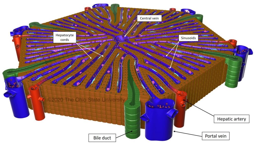
A small subunit of the liver composed of cells hepatocytes that process blood from an incoming portal venule and send the resulting blood to an outgoing hepatic venule. The central veins of different liver lobules converge to form the hepatic vein which carries blood from the liver to the inferior vena cava.
Hepatic Blood Supply Veterinary Histology
This bile is transported via bile ducts that eventually join and form the common bile duct that.

What does the liver lobule contain. Blood enters into the liver lobules via the hepatic artery which then forms hepatic sinusoids that drain into the central vein in the centrilobular region of the liver also called zone 3. Arterial blood and portal venous blood containing molecules absorbed in the GI tract thus mix as the blood flows within the sinusoids from the periphery of the lobule to the central vein. The mammalian liver also containing Kupffer cells which are one type of phagocytic cell and Fat storage cells.
They contain two blood vessels and a. The hepatic lobule is a building block of the liver tissue consisting of a portal triad hepatocytes arranged in linear cords between a capillary network and a central veinLobules are different from the lobes of the liver. The liver also produces an estimated 800 to 1000 milliliters ml Trusted Source.
The hepatic lobule is a building block of the liver tissue consisting of a portal triad hepatocytes arranged in linear cords between a capillary network and a central vein. The portal vein is an additional source of blood bringing nutrient- and antigen-rich blood from the gastrointestinal system to. A subdivision of the four main liver lobes the basic functional unit of the liver.
In the anatomic model liver lobules are organized into irregular polygons demarcated by connective tissue and composed of plates of hepatocytes radiating outward from the central vein to the portal triads Figure 61-1. Blood enters the lobules through branches of the portal vein and hepatic artery proper then flows through sinusoids. What is a lobule in the liver.
Of clusters of cells called lobules where the vital functions of the liver are carried out. Lobules are different from the lobes of the liver. The classic liver lobule is enveloped by a limiting plate of hepatocytes surrounded by various vessels embedded in connective tissue.
These fibres enter the liver at the porta hepatis and follow the course of branches of the hepatic artery and portal vein. Liver Physiology Functions Lobule Hepatocytes. Structure of a liver lobule.
Hepatic macrophages Kupffer cells are also present in the sinusoids which remove bacteria and cell debris. Read remaining answer hereAlso question is what do liver lobules do. Finally the liver has immunologic functions as the liver contain cells involved in adaptive and innate immunity.
The structural and functional units of liver are Hepatic lobules containing hepatic cells. The liver also stores vitamins E and K. The lobules of liver lobuli hepatis form the chief mass of the hepatic substance.
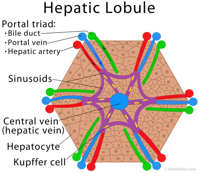
There are two types of liver lobule which look at the same cluster of liver cells from opposite ends. The bile flows in the opposite direction green arrow. Hepatic triads are areas present at each corner of the lobule.
The liver is made of very soft pinkish-brown tissues encapsulated by a connective tissue capsule. They are the smaller divisions of the lobes. Focusing on the outflow of blood the classical lobule is.
The hepatic lobule is a building block of the liver tissue consisting of a portal triad hepatocytes arranged in linear cords between a capillary network and a central vein. The parenchyma of the liver is innervated by the hepatic plexus which contains sympathetic coeliac plexus and parasympathetic vagus nerve nerve fibres. This allows the liver to remove glucose amino acids iron vitamins and other nutrients.
Each lobule measuring about one millimetre in diameter consists of numerous cords of rectangular liver cells or hepatocytes that radiate from central veins or terminal hepatic venules toward a. Iron is stored in the liver as ferritin an iron-protein complex and is released when needed for red blood cell production. Each lobule is made up of millions of hepatic cells that are the basic metabolic cells of the liver.
They may be seen either on the surface of the organ or by making a section through the gland as small granular bodies about the size of a millet-seed measuring from 1 to 25 mm. The blood flows from the periphery via the sinusoids into the central vein violet arrow. Of bile each day.
The hepatic acinus is the functional unit of the liver. This capsule is further covered and reinforced by the peritoneum of the abdominal cavity which protects the liver and holds it in place within the abdomen. The lobes are further divided into lobules the functional units of the liver.
Blood enters into the liver lobules via the hepatic artery which then forms hepatic sinusoids that drain into the central vein in the centrilobular region of the liver also called zone 3. The structure of the livers functional units or lobules.
Lobule Liberal Dictionary
The liver consists of four lobes.

What is lobules of liver. Liver lobules are the basic functional units of the liver. There are two types of liver lobule which look at the same cluster of liver cells from opposite ends. The basis of this page is on Wikipedia.
The right lobe and the left lobe. The lobules of liver or hepatic lobules are small divisions of the liver defined at the microscopic histological scale. Two larger ones right and left and two smaller ones quadrate and caudate.
What is WikiRIP There is a free information resource on the Internet. The hepatic acinus is. In the anatomic model liver lobules are organized into irregular polygons demarcated by connective tissue and composed of plates of hepatocytes radiating outward from the central vein to the portal triads Figure 61-1.
Histologically speaking it has a complex microscopic structure that can be viewed from several different angles. A small subunit of the liver composed of cells hepatocytes that process blood from an incoming portal venule and send the resulting blood to an outgoing hepatic venule. Discussion of the hepatic lobule including histology.
The lobules of liver or hepatic lobules are small divisions of the liver defined at the microscopic histological scale. Seen from the front the diaphragmatic surface - the liver is divided into two lobes. Microscopically the cells of the liver known as hepatocytes are arranged into lobules.
Focusing on the outflow of blood the classical lobule is. The hepatic lobule is the anatomic unit of the liver. The hepatic lobule is a building block of the liver tissue consisting of a portal triad hepatocytes arranged in linear cords between a capillary network and a central vein.
Lobules are different from the lobes of the liver. In the human subject their outlines are very irregular. They are classically described classic lobules as hexagonal structures made of six vertically aligned portal canals with a central vein.
Each anatomical lobule is hexagonal-shaped and is drained by a central vein. Blood enters the lobules through branches of the portal vein and hepatic artery proper then flows through sinusoids. They may be seen either on the surface of the organ or by making a section through the gland as small granular bodies about the size of a millet-seed measuring from 1 to 25 mm.
The left and right lobe are divided by the falciform sickle-shaped in Latin ligament which connects the liver to the abdominal wall. These are the structural units of the liver. The hepatic lobule is a building block of the liver tissue consisting of a portal triad hepatocytes arranged in linear cords between a capillary network and a central vein.
The larger right lobe and left lobe and the smaller caudate lobe and quadrate lobe. The portal vein is an additional source of blood bringing nutrient- and antigen-rich blood from the gastrointestinal system to. The four lobes are the right lobe the left lobe the caudate lobe and the quadrate lobe.
Biology and Engineering of Stem Cell Niches 2017. From Wikipedia the free encyclopedia. The lobules of liver or hepatic lobules are small divisions of the liver defined at the microscopic histological scale.
Viewed from the underside the visceral surface the other two smaller lobes the caudate lobe and the quadrate lobe are also visible. Structure of a liver lobule. Anatomically the liver consists of four lobes.
What is a lobule in the liver. The lobules of liver lobuli hepatis form the chief mass of the hepatic substance. At the periphery of the hexagon are three structures collectively known as the portal triad.
However microscopic evaluation of the liver usually shows a lack of classic liver lobule as a well-defined connective tissue septum is. The human liver is divided grossly into four parts or lobes. Liver lobules are hexagonal and at each of six corners there is a portal triad of vessels consisting of a portal vein hepatic artery and bile duct.
The hepatic lobule is a building block of the liver tissue consisting of a portal triad hepatocytes arranged in linear cords between a capillary network and a central vein. It is open to any user. Wiki is a library that is public and multilingual.