Nucleus cell wall cell membrane cytoplasm chloroplast mitochondrion endoplasmic reticulum vacuoles mitochondrion Golgi body To. The set includes two versions of a plant cell to color and label and two versions of an animal cell to color and label.

Animal Cell Color Page Worksheet And Quiz Ce 3 In 2021 Animal Cells Worksheet Animal Cell Cells Worksheet
Include descriptions of what each part does.
Color and label the parts of an animal cell. Function of Cell Part. When autocomplete results are available. Using arrows and Textables label each part of the cell and describe its function.
Use the animal cell reference chart as a guide. Animal Cell Label Color Directions. Color the organelle orange that controls.
Cell Membrane Coloring Worksheet - Kenton County School District Cell Membrane Coloring Worksheet Composition of the Cell Membrane u0026amp. Use the colors indicated in the box. Color the animal cell drawn below.
The worksheets recommended for students of grade 4 through grade 8 feature labeled animal and plant cell structure charts and cross-section charts cell vocabulary with descriptions and functions and exercises like identify and label the parts of the animal and plant cells color the cell organelles match the part to its description fill in the blanks crosswords and more. Learn the parts of animal and plant cells by labeling the diagrams. Use the organelles listed in the word bank.
Students are ask to label the following organelles. 5th grade science and biology. Dec 17 2019 - Label an animal cell and match the parts with its definition.
Color the organelle the color listed on the cells. The Animal Cell to Color Name. You may also like.
Can you label and color these important parts of the animal cell. A cell is the smallest unit of life. If time permits have a paint program open for them to practice their mouse skills.
Cellcolorpdf - Read File Online - Report Abuse. RER Rough Endoplasmic Reticulum synthesizes proteins. The lysosomes are oval and the vacuoles are more rounded 1.
Color Coded Cells Read the descriptions and write in the name of the cell organelle. A digital version of this resource is also included and is compatible with Google Classroom. NUCLEUS control center for cell cell growth cell metabolism cell reproduction.
Color the cell part to match. Color all organelles different colors cut them out glue them into your cell and label them with the correct organelle name. Jun 22 2016 - The set includes two versions of a plant cell to color and label and two versions of an animal cell to color and label.
Typical plant and animal cells diagram and coloring activity cell part color function 1. Choose a color for each of the parts below and fill in the square with the color of your choice. Label and color the animal and plant cells answers.
Color the cell part to match. 650x703 px Animal And Plant Cell Labeling - Click the buttons to print each worksheet below you will see a microscope label the following parts. Color the text boxes to group them into organelles.
Parts of a Plant Cell - Labeling MatchingParts of an Animal Cell - Organelle. Color and label the. Asked in genetics diy projects draw and label a plant cell.
Choose a color for each of the parts below and fill in the square with the color of your choice. Parts Animal Cell Labeling Matching Science Cells Posted in Worksheet November 6 2020 by Kimberly R. Answer keys are included for easy grading.
Color as per directions. RIBOSOMES the site of protein building this is where translation takes place mRNA in language of nucleic acids is translated into the language of amino acids. Parts Of An Animal Cell Labeling Matching Distance Learning Animal Cell Animal Cell Parts Cell Parts - A set of diagrams of an animal cell.
Both plant and animal cells have double membranes and their own dna. Dec 7 2014 - Printable animal cell diagram to help you learn the organelles in an animal cell in preparation for your test or quiz. Diffuse into a.
Nucleus cell wall cell membrane cytoplasm chloroplast mitochondrion endoplasmic reticulum vacuoles mitochondrion Golgi. Label both a plant and animal cell on a poster layout. Older students can be challenged to identify and label the animal cell parts.
Students are ask to label the following organelles. Animal Cell Coloring Directions. Cell membrane nucleus nucleolus nuclear membrane cytoplasm endoplasmic reticulum Golgi apparatus ribosomes mitochondria centrioles cytoskeleton vacuoles and vesicles.
Download a free printable outline of this video and draw along with us. Foreman Be assessed on your knowledge of different types of hardware andSep discuss directions and have students complete the computer parts worksheet. There are 13 main parts of an animal cell.
Identify if the description is the organelles structure or function by circling. Label the animal cell drawn below and then give the function of each cell part. Cells tend to be 1 100 micrometers μm in diameter and each cell while typically specialized in function carries out the.
Animal Cell Picture with Labels Younger students can use the animal cell worksheets as coloring pages. 5th grade science and biology. Dec 7 2014 - Printable animal cell diagram to help you learn the organelles in an animal cell in preparation for your test or quiz.
Cell Membrane Cell Wall Nucleus Cytoplasm Mitochondrion Chloroplast Vacuole Lysosome 1. Httpsartforallmevideohow-to-draw-an-animal-cellThank you for watching. Find diagrams of a plant and an animal cell in the Science tab.
Head Base and Arm. Light Microscope Definition Uses Amp Parts Cell Parts Ask A Biologist.

Label And Color The Parts Of Both Microscopes Trovoadasonhos
Microscope world has pdf printable versions of both microscope activities shown below that you can print and use for free here.
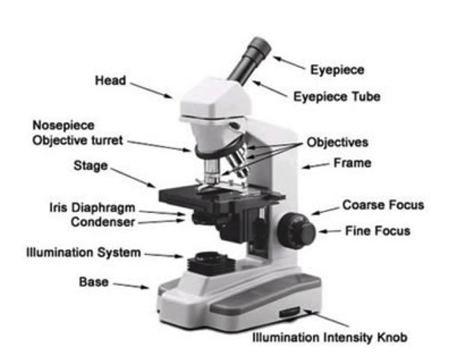
Label and color parts of both microscopes. Related for label and color the parts of both microscopes blue label fiyat creative labels label gallery get some ideas to make labels for bottles jars packages products boxes or classroom activities for free. Label the parts of the microscope. Label and color the ocular lens light blue.
There are three structural parts of the microscope ie. 32 Label And Color The Parts Of Both Microscopes Written By Paul A Cho Saturday May 15 2021 Add Comment Edit. Label and color the parts of both microscopes.
You can view a more in-depth review of each part of the microscope here. Labels are usually small in size so you should carefully choose the font of the texts to make sure it is readable. A compound light microscope.
This activity has been designed for use in homes and schools. An easy and convenient way to make label is to generate some ideas first. Color the stage blue.
You can also put your logo at the top or bottom corner of the label. Stage clips J on the stage can hold the slides in place. Label And Color The Parts Of Both Microscopes.
Related for label and color the parts of both microscopes. Diagram of parts of a microscope. Always lift a microscope by holding both the arm and base with two hands.
Color the light source yellow. Revolving nosepiece objective lens stage condenser illuminator base label parts of the microscope. Label and color the parts of both microscopes.
Label and color the parts of both microscopes microscopeparts label gallery get some ideas to make labels for bottles jars packages products boxes or classroom activities for free. The upper lens is called the ocular lens or eyepiece and the lower lens or lenses as there may be a choice of sizes is called the objective lens. Each microscope layout both blank and the version with answers are available as pdf downloads.
Parts of a microscope a compound light microscopes use two lenses for greater magnification. Label And Color The Parts Of Both Microscopes Ythoreccio Label And Color The Parts Of A Plant A Free Printable First Grade Label And Color The Parts Of A Snail Label And Color The Parts Of A Seed By Heather Ward Tpt Parts Of A Plant Cut Label Color Activity And Or Define Tpt. Light microscopes use either a bulb or a mirror M as their light source.
Base It acts as microscopes support. The eyepiece is usually 10x. There are three major structural parts of a microscope.
Labeling the parts of the microscope is a common activity in schools. The upper lens is called the ocular lens or eyepiece and the lower lens or lenses as there may be a choice of sizes is called the objective lens. Label and color the parts of both microscopes answers.
Label and color the parts of both microscopes. Correlated Light And Electron Microscopy Ultrastructure Lights Up Nature Methods. Label Gallery Get some ideas to make labels for bottles jars packages products boxes or classroom activities for free.
Color the parts of the microscope. Buki France Microscope 30. It also carriers the microscopic illuminators.
Label and color the ocular lens light blue. Labeling the parts of the microscope. Color the microscope parts label and color the parts of both microscopes design styles light microscopy clipart labeled pencil and in color parts of the microscope color by number by science label and color the parts of both microscopes design styles label and color the parts of both microscopes design styles.
Color the stage clips gray. Head base and arm. Below you will find both the label the microscope activity worksheet as well as one with answers.
Label and color the parts of both microscopes image002 label gallery get some ideas to make labels for bottles jars packages products boxes or classroom activities for free. The objective lenses are located on a revolving nosepiece at the bottom of the body tube. The switch for this light is usually found on the base of the microscope.
Parts of a microscope a compound light microscopes use two lenses for greater magnification. Label the parts of the microscope. Label and color the low power objective pink and the high power objective red.
Color the nosepiece blue green. Label and color the parts of both microscopes microcope coloring key label gallery get some ideas to make labels for bottles jars packages products boxes or classroom activities for free. The total magnification obtained is.
Head This is also known as the body it carries the optical parts in the upper part of the microscope. Most eyepiece lenses are 10x magnification. Each microscope layout both blank and the version with answers are available as PDF downloads.
Compound microscopes have more than one lens to generate high magnification images of flat thin specimens. Label and color the parts of both microscopes image002 label gallery get some ideas to make labels for bottles jars packages products boxes or classroom activities for free. Each microscope layout both blank and the version with answers are available as pdf downloads.
The diaphragm dark purple. Labeling the Parts of the Microscope. Color the microscope parts label and color the parts of both microscopes design styles light microscopy clipart labeled pencil and in color parts of the microscope color by number by science label and color the parts of both microscopes design styles label and color the parts of both microscopes design styles.