The development of the middle ear mechanism which began in the amphibians greatly improved the transmission of energy from air to the fluid of the inner ear by acting as an impedance transformer that matched the high impedance of the fluid to the much lower impedance of the air. It is classical to ascribe three functions to the middle ear.
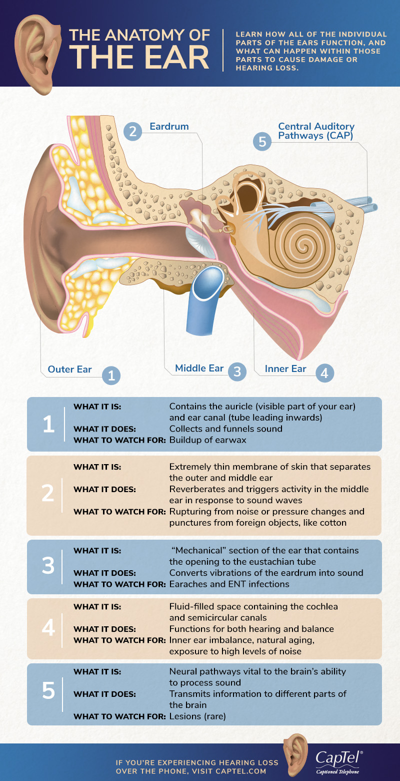
The Anatomy Of The Ear Infographic
Lorenzo Crumbie MBBS BSc Reviewer.

What are the function of middle ear. 1 The area ratio pressure transformer 2 The ossicular lever 3 The catenary lever buckling effect of TM. The middle ear comprises the following parts. The middle ear communicates anteriorly with the nasopharynx through the auditory tube and posteriorly with the mastoid antrum and mastoid air cells above the aditus to the mastoid antrum.
The transmission of acoustic vibrations from the tympanic membrane to the cochlea impedance matching between the air in the external auditary meatus and the labyrinthine fluids and protection of the inner ear by means of the acoustic reflex. Also known as the tympanic cavity the middle ear is an air-filled membrane-lined space located between the ear canal and the Eustachian tube cochlea and auditory nerve. Transmits the vibrations of the tympanum caused by sound waves to the inner ear 2.
Then what are the 3 functions of the middle ear. What is the function of the middle ear. This membrane separates the middle ear and the external ear.
Functions of middle ear 1. What are the three mechanisms of the middle ear pressure transformer that amplifies and transmits sound energy from TM to oval window air to fluid. 12 minutes The human ear is one of the most intricate pieces of machinery found in the human body.
Ryan Sixtus MPhEd Last reviewed. The primary function of the middle ear is to offset the decrease in acoustic energy that would occur if the low impedance ear canal air directly contacted the high-impedance cochlear fluid. Behind the ear drum is the middle ear space which is normally filled with air.
July 21 2021 Reading time. The opening of the eustachian you STAY shun tube is in the middle ear space. It is responsible for transmitting and converting vibrational energy so that sound can be appreciated.
Your middle ear turns sound waves from the world around you into vibrations which can be used to make nerve signals for your brain. The ear drum is where the middle ear starts. The primary function of the middle ear is to offset the decrease in acoustic energy that would occur if the low impedance ear canal air directly contacted the high-impedance cochlear fluid.
Its central part is known as the umbo. The middle ear is likened to a pistol in the sloping course of the aditus to the epitympanic recess and the auditory tube. The role of the eardrum also called as the tympanic membrane is always to take sound waves to bone fragments which have been found in the middle ear.
Middle ear Author. The transmission of acoustic vibrations from the tympanic membrane to the cochlea impedance matching between the air in the external auditary meatus and the labyrinthine fluids and protection of the inner ear by means of the acoustic reflex. This part receives and amplifies the sound waves.
It is classical to ascribe three functions to the middle ear. Make a clarity in the voice by passing through a series of cellular layers. All these bone tissues are referred to ossicles.
This is a big job for the tiniest bones of your body. The outer ear acts like a funnel to quickly send sounds to the ear drum.
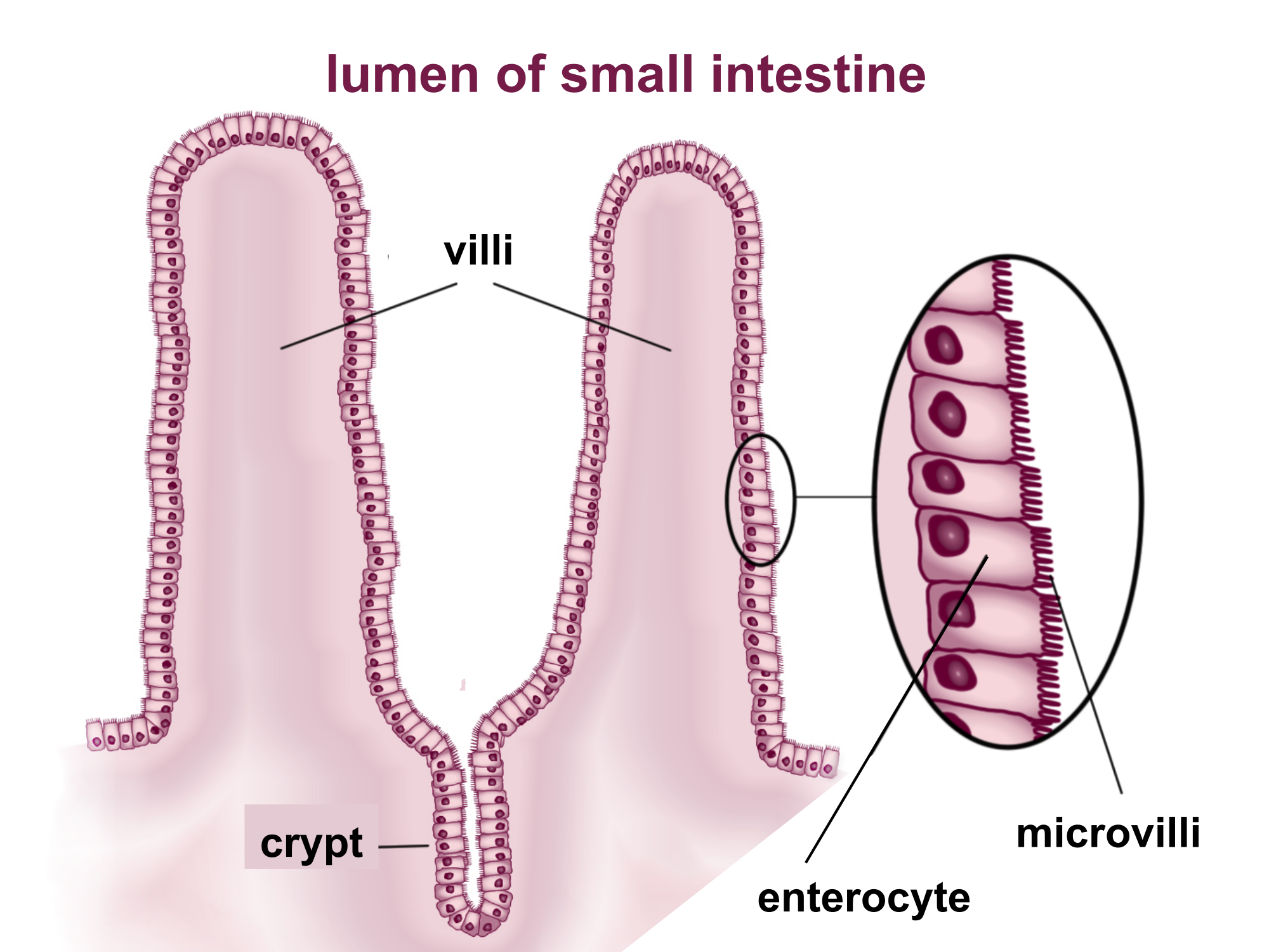
Villi numbers vary between 10 to 40 per square metre of tissue and are typically 05 to 1 mm long. There the villi and the microvilli increase intestinal absorptive surface area approximately 30-fold and 600-fold respectively providing exceptionally efficient absorption of nutrients in the lumen.
How Is The Structure Of A Villi In The Small Intestine Related To Its Function Socratic
Their function is to increase the surface area of the small intestinal wall for absorption of the digested food.
What is the function of a villus. The nucleic acid may be single- or double-stranded. Intestinal Villus Roles of Substance P and ATP in the Subepithelial Fibroblasts of Rat Intestinal Villi. What Is the Function of Spikes on a Virus.
The villi are further covered by even smaller finger-like projections of the plasma membranes of the epithelial cells lining the vili which are known as microvilli. Absorb fatty acids and glycerol. Intestinal Regeneration and Adaptation Models.
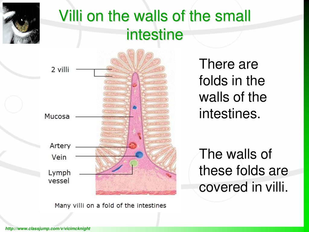
Intestinal villi are small thread-like objects that line the intestines in order to effectively increase the surface area of the intestinal wall. Viruses possess unique infective properties and thus often cause disease in host organisms. The diagram shows a section through a villus.
What are the two functions of villi. Inside each villus are. The villi increase the surface area of the small intestine over which food may be absorbed.
The entire infectious virus particle called a virion consists of the nucleic acid and an outer shell of protein. Virus infectious agent of small size and simple composition that can multiply only in living cells of animals plants or bacteria. Absorb amino acids and glucose.
Spikes made of the glycoprotein hemagglutinin or H spikes enable viruses to latch onto their host cells while N spikes those made of the glycoprotein neuraminidase enable viruses to escape their host cells upon reaching maturity explains Midlands Technical College. Functions of the Villi. From the stomach to the small intestine villi lines the intestine and the villi absorbs the nutrients and vitamins which will be used by the body.
The villi of the small intestine project into the intestinal cavity greatly increasing the surface area for food. I To absorb amino acids ii To carry blood. The primary function of the villi in the small intestine is to increase the absorption of nutrients from food passing through the small intestine.
A virus is a small parasite that cannot reproduce by itself. Most viruses have either RNA or DNA as their genetic material. Important Structural Characteristics of the Villus which Aid its Function.
Villus plural villi in anatomy any of the small slender vascular projections that increase the surface area of a membrane. Iii To transport fats. Their function is to increase the surface area of the small intestinal wall for absorption of the digested food.
Their function is to increase the surface area in order to maximise the absorption of digested food. These projections absorb the protein molecules and help in the transfer of the proteins to all cells and tissues. If a section of small intestine was turned inside out its surface would be kike a carpet.
Weve already stated that the small intestines main job is to absorb nutrients from the food you eat and that your villi help by increasing the surface area the intestine has for absorption. Important villous membranes include the placenta and the mucous-membrane coating of the small intestine. There are also enzymes enterocyte digestive enzyme on the surface for digestion.
Functions of the Villi Weve already stated that the small intestines main job is to absorb nutrients from the food you eat and that your villi help by increasing the surface area the intestine. Each villus consists of arteries veins a complex capillary system and a lymphatic vessel called lacteal. When the food has been broken down into nutrients vitamins.
Learn about the history types and features of viruses. Once it infects a susceptible cell however a virus can direct the cell machinery to produce more viruses. These microvilli further increase the surface area for absorption on the wall of the.
The intestinal villus microcirculation is supplied by a central arteriole. What is the function of structure X. Intestinal villi are a unique.
These projections absorb the protein molecules and help in the transfer of the proteins to all cells and tissues. Many blood vessels are present within these villi that help in the absorption of digested food and carry it to. Villi are finger like projections that increase the surface area for absorption.
It is flexible and allows unicellular organisms to move. Cholesterol is found in high quantities in bacon egg cheese and many other comfort foods.

Cell Membrane Flashcards Quizlet
First to be a barrier keeping the constituents of the cell in and unwanted substances out and second to be a gate allowing transport into the cell of essential nutrients and movement from the cell of waste products.

What is the function of cholesterol in the cell membrane quizlet. Cholesterol molecules regulate the fluidity of the cell membrane which is necessary for the cell to retain its shape. -Synthesis of Cholesterol esters by acyl-CoA-cholesterol acyl transferase ACAT a more hydrophobic form of cholesterol for transport and storage lipid droplets Transport to extrahepatic tissues in VLDL and LDLs-Synthesis of Bile salts to aid lipid digestion -Direct excretion of cholesterol into bile. The presence of cholesterol has been shown to provide stability and in general to decrease the permeability of biological membranes to various solutes.
Cholesterol is an essential component of eukaryotic membranes and plays a crucial role in membrane organization dynamics and function. What is the function of the cell membrane quizlet. The cell membrane therefore has two functions.
Such stability would be beneficial to the cell only in so far as it does not interfere with specific transport properties. The primary function of the. The modulatory role of cholesterol in the function of a number of membrane proteins is well established.
If cholesterol is absent in the cell membrane then because of excess fluidity there may be chances of cell lysis - breaking down of the cell. While too much cholesterol can harm the body regulated amounts of cholesterol in cell membranes are. Cholesterol and membrane rafts Cholesterol displays a very important function as a component of cellular membranes specially the cell plasma membrane where it is found in higher concentrations.
Specifically cholesterol allows the cell membrane to stay flexible and allow lipids to pass through. Cholesterol plays a role in membrane fluidity but its most important function is in reducing the permeability of the cell membrane. Also Know what does the plasma membrane do quizlet.
The most unique feature of the eye lens fiber-cell plasma membrane is its extremely high cholesterol content. 1 Excretion in feces represents significant mechanism for elimination of excess cholesterol 2 Solubilize Cholesterol in Bile preventing precipitation of cholesterol in Gall Bladder 3 Faciliate digestion of TAG by emulsification 4 Facilitate intestinal absorption of fat-soluble vitamins. Cholesterol functions to immobilise the outer surface of the membrane reducing fluidity.
Since human cell membranes are made with cholesterol its no surprise that cholesterol is needed for cell maintenance and creation. Cholesterol plays a critical role in the function of the cell membrane which has the highest concentration of cholesterol with around 25-30 of lipid in the cell membrane being cholesterol. Cholesterol saturates the bulk phospholipid bilayer and induces formation of immiscible cholesterol bilayer domains CBDs within the membrane.
Cholesterol is also a key determinant of membrane fluidity. Cholesterol helps to restrict the passage of molecules by. Cholesterol chemical structure.
Gives shape to the cell. In this lesson you learned that the role of cholesterol in the cell membrane is to maintain stability anchor other molecules and keep the membrane fluid in cold temperatures. While at low temperatures it inserts into phospholipids and prevents them from interfering with each other to avoid aggregation 39.
Cholesterol helps to restrict the passage of molecules by increasing the density of the packing of phospholipids. Cholesterol interacts with the fatty acid tails of phospholipids to moderate the properties of the membrane. It regulates what enters and leaves the cell.
It also provides protection and support. Maintains homeostasis or cell equillibrrium it does this by controlling what enters and leaves the cell. Cholesterol acts as membrane stabilizer.
It makes the membrane less permeable to very small water-soluble molecules that would otherwise freely cross. Cholesterol plays has a role in membrane fluidity but its most important function is in reducing the permeability of the cell membrane. Sterols such as cholesterol in mammals ergosterol in fungi and phytosterols in plants buffer membrane fluidity.
So cholesterol is essential for the stability of the cell membrane. Protects and encloses the cell. At high temperatures cholesterol acts to stabilize the cell membrane and increase its melting point.
The plasma membrane regulates the entry.
Anatomy and Physiology of the Ear. This structure helps to give each of us our unique appearance.

Hearing Mechanism Anatomy Of Ear Hearing Loss Flashcards Quizlet
The malleus is the largest and the outermost of the bones which are part of the auditory system.
Ear anatomy and function quizlet. It is made up of. Middle Structures and Hearing Functions. Learn vocabulary terms and more with flashcards games and other study tools.
Inner_ear_anatomy_quizlet 44 Inner Ear Anatomy Quizlet Inner Ear Anatomy Quizlet This is likewise one of the factors by obtaining the soft documents of this inner ear anatomy quizlet by online. The middle ear contains three bones the malleus. Start studying Ear anatomy and functions.
Terms in this set 42 what are the functions of the ear. Memorizing the parts of the ear isnt difficult. The ear is made up of three parts.
What are the subdivisions of the ear. There are three different parts to the outer ear. Middle Ear Muscle Function.
Start studying Ear Anatomy. The tragus helix and the lobule. Start studying CHAPTER 6 term test--- GENERAL ANATOMY AND PHYSIOLOGY.
Where is the ear located. The medical term for the outer ear is the auricle or pinna. This is the smallest bone in the human body it is 025 to 033 cm long.
Ear Anatomy Outer Ear. In some cases you likewise attain not discover. Transfers the vibrations from the tympanic membrane to the middle of the ear to the oval window.
Each part of the inner ear has a specific function. Nerve that affects the external ear and skin above the temple up to the top of the skull. The cochlea is responsible for hearing.
Transmits vibrations from the ossicles to the outer ear. Start studying ear anatomy functions. All three parts of the ear are important for detecting sound by working together to move sound from the outer part through the middle and into the inner part of the ear.
The end part of the Ear is Inner Ear. The Ear - Science Quiz. Its main function is to refer information on equilibrium and skull position to the Brain.
Together the three bones make up an area no larger than the seed of an orange. It contains three parts. Function is to transport blood to and from the heart and then to.
The External Ear emanates in Shape and Size. Start studying Ear Anatomy - Functions. Capillaries venules and veins.
Each part plays a vital role in our hearing. Learn vocabulary terms and more with flashcards games and other study tools. The human ear is made up of three main parts the outer middle and inner ear.
The outer ear comes in all types of shapes and sizes. Not when you use this quiz game that is. Our online ear anatomy trivia quizzes can be adapted to suit your requirements for taking some of the top ear anatomy quizzes.
Ears also help to maintain balance. The outer ear is made up of cartilage and skin. Connects to the malleus.
External ear middle ear and inner internal ear. Youll be tested on vocabulary terms for the various parts composing the external ear as well as some of their functions. Start studying Ear Anatomy.
Get your study survival kit for 50 off. The outer ear gathers sound and allows it to pass through the ear canal to the eardrum. Anatomy of the Ear.
The malleus hammer incus anvil and stapes stirrup are the three bones also known as ossicles of the inner ear. Stirrup - one of the ear ossicles a tiny U-shaped bone that passes vibrations from the stirrup to the cochlea. Help maintain ossicles in proper position Protect inner ear from excessive sound levels This protective reflex termed acoustic reflex 020708 Tinnitus Support Group 1010 Eustachian Tube.
Connects to the incus. The outer middle and inner ear. You might not require more times to spend to go to the book initiation as capably as search for them.
Learn vocabulary terms and more with flashcards games and other study tools. Anatomy of the Ears External Structures - Quiz Worksheet Chapter 11. Learn vocabulary terms and more with flashcards games and other study tools.
Learn vocabulary terms and more with flashcards games and other study tools. A comprehensive database of ear anatomy quizzes online test your knowledge with ear anatomy quiz questions. Learn vocabulary terms and more with flashcards games and other study tools.
Oval Window is a specialized part that joins the Middle Ear to the Inner Ear. The purpose of the inner ear is to sense and process information about sound and balance and send that information to the brain. The ear is located in the temporal bone41 of.
The ear contains the organs for both balance and hearing.
The review of anatomy and structure indicates a lack of quantitative data. Following are the important function of the ear.
Human Ear The Physiology Of Hearing Britannica
The eardrum represents the boundary between the outer ear and the middle ear.

Describe the structure and function of the eardrum. The tympanic membrane is a vital component of the human ear and is more commonly known as the eardrum. The ossicles will. At the bottom of the stapes sits the oval window followed by the semicircular canals also called the labrynthine.
The eardrum is circular and flexible and begins to vibrate as the incoming sound waves strike it. Is the long narrow canal leading to the eardrum or tympanic membrane. Includes the ossicles three minuscule bones called the malleus incus and stapes the latter being the smallest bone in the human body.
The footplate of the stapes fits tightly into the oval window of the bony cochlea. It is a thin circular layer of tissue. When sound waves enter your ear they run into your eardrum which is also called the tympanic membrane.
Structure and Function of Human ear. The vibrations produced pass through the tympanic membrane to the tympanic cavity. The mechanism of hearing involves the following steps.
The sound waves produce pressure changes over the surface of the tympanic membrane. The semicircular canals are filled with a fluid called endolymph and function to provide the body with a proper sense of balance. The eardrum vibrates according to the frequency and the amplitude of sounds that strike it.
The sound waves pass through the auditory canal and reach the eardrum. Behind the eardrum are the ossicles tiny bones that play a vital role in hearing. Tympanic membrane also called eardrum thin layer of tissue in the human ear that receives sound vibrations from the outer air and transmits them to the auditory ossicles which are tiny bones in the tympanic middle-ear cavity.
You might want to. The entrance to this canal is called the external auditory meatus. The inner ear is filled with fluid.
Eardrum structure properties and behaviour in order to provide a basis for quantitative modelling and to identify areas where further information is required. The deep bowl-like portion of the pinna adjacent to the external auditory meatus is known as the concha. It also serves as the lateral wall of the tympanic cavity separating it from the external auditory canal.
The eardrum is exceedingly sensitive as well as pressure from sound waves tends to make the eardrum vibrate. The middle ear function of human ear is to transmit and amplify the sounds vibrated from the eardrum towards the oval window. The resulting movements of the eardrum are transmitted through the three middle-ear ossicles malleus incus and stapes to the fluid of the inner ear.
The ear is divided into three interconnecting sections. The middle ear. It also acts as a dampener to loud sounds that may damage the cochlea.
Functions of External Ear. Anatomically Human ear mainly consists of 3 parts. The cerumen ear wax prevents the entry of the foreign bodies into the ear.
This is a thin membrane that separates the outer ear from the middle ear. While the external and middle ears are mainly concerned with the transmission of sound the inner ear contains the cochlea often called the organ of hearing and also houses the bodys organ of balance. The function of the tympanic membrane eardrum is to transmit sound waves from the environment into sound vibrations that are picked up by the middle ear auditory ossicles.
The pinna and external auditory meatus collect these waves change them slightly and direct them to the tympanic membrane. External middle and inner ears Fig 1. Human ear organ of hearing and equilibrium that detects and analyzes noises by transduction or the conversion of sound waves into electrochemical impulses and maintains the sense of balance equilibrium.
The eardrum tympanic membrane is a membrane towards the end of the auditory canal and also scars the opening of the middle ear. It directs sound waves towards the tympanic membrane. Hence called as the Stato acoustic organ of the body.