Finden Sie perfekte Stock-Fotos zum Thema Digestive System Drawing sowie redaktionelle Newsbilder von Getty Images. Digestive system zoom.

How To Draw A Model Of The Digestive System 15 Steps
Wählen Sie aus erstklassigen Inhalten zum Thema Digestive System Drawing in höchster Qualität.

Human digestive system drawing image. Human Internal Digestive Organ Pancreas Anatomy. 156951315 stock photos online. Human Digestive System Extraction.
Human digestive system - human digestive system stock illustrations. Find the perfect Digestive System Drawing stock illustrations from Getty Images. 4502 digestive system drawing stock photos vectors and illustrations are available royalty-free.
Choose from Diagram Of The Human Digestive System Drawing stock illustrations from iStock. Choose from Human Digestive System Drawing stock illustrations from iStock. Select from premium Digestive System Drawing of the highest quality.
Posted on 29 Mar 0131. No need to register buy now. Select from premium Digestive System Drawing images of the highest quality.
See more ideas about digestive system system anatomy and physiology. Find human digestive system drawing stock images in HD and millions of other royalty-free stock photos illustrations and vectors in the Shutterstock collection. Diagram of the human digestive system drawn on a whiteboard together with a stethoscope conceptual image.
Treatment with probiotics bowel irritation. Select from premium Digestive System Drawing images of the highest quality. 1297 human digestive system stock photos are available royalty-free.
Browse 7994 human digestive system stock illustrations and vector graphics available royalty-free or search for human digestive system anatomy or human digestive system illustration to find more great stock images and vector art. Huge collection amazing choice 100 million high quality affordable RF and RM images. Download 1398 Digestive System Drawing Stock Illustrations Vectors Clipart for FREE or amazingly low rates.
New users enjoy 60 OFF. Find the perfect Digestive System Drawing stock photos and editorial news pictures from Getty Images. Organs of the digestive system human digestive system black and white anatomy for children intestine digestive system in a body cartoon child body system digestive system organs esophagus.
Find high-quality royalty-free vector images that you wont find anywhere else. 1211 Human Digestive System Drawing. Find high-quality stock photos that you wont find anywhere else.
Find high-quality royalty-free vector images that you wont find anywhere else. Thousands of new high-quality pictures added every day. Digestive system - human digestive system.
49685 human digestive system stock photos vectors and illustrations are available royalty-free. Find the perfect Digestive System Drawing stock illustrations from Getty Images. Aug 13 2016 - Explore Sahu Anu Anubiss board DIGESTIVE SYSTEM IMAGES AND HOW IT WORKS followed by 2380 people on Pinterest.
Human intestines on a blue background constipation and digestive problems. Organs in body anatomical body vector human digestion human body intestine body-organs spleen liver systems human body in body anatomy of the digestive system human body a. See digestive system drawing stock video clips.
See human digestive system stock video clips. Find the perfect digestive system diagram stock photo. Learn step by step drawing tutorialPrintable Link.
Human digestive system extracted against a white background. Cartoon Character of stomach with Circular arrow diagram. 1211 Human Digestive System Drawing.
Search from Diagram Of The Human Digestive System Drawing stock photos pictures and royalty-free images from iStock. Three pairs of glands make saliva.
Spirogyra Diagram Cell Wall Cell Membrane Life Science Labeled diagram of spirogyra. Cell wall consists of three layers of which inner two layers are made of pectose and the outer layer is composed of cellulose.
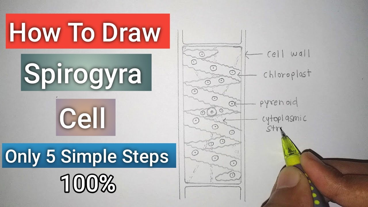
How To Draw Spirogyra Cell Only In 5 Simple Steps For Beginners Youtube
Chloroplasts conjugation tubes male gametes female gametes and zygospores.

Drawing of spirogyra with label. Spirogyra is a filamentous green alga which is common in freshwater habitats. Diagram of spirogyra with label. Spirogyra is a filamentous algae commonly found in freshwater ditches and ponds.
The vegetative structure or plant body of Spirogyra is known as thallus. Ine Image Spirogyra Conjugation- acies. It is very unusual among the plant-like protists.
Go through this article. Draw a neat diagram of spirogyra and label the following parts. Outer layer of the cell.
The green filamentous alga Spirogyra has a multicellular body with comparable cells. Be sure to CLEARLY label. Outermost layer of the cell.
Seen under the microscope each filament consists of an extensive chain of identical cells. Draw a colony of Volvox at 10x and label cells daughter colonies and flagella. Darkly coloured and dot like.
Image result for labelled diagram of a spirogyra cell. It has the appearance of very fine bright dark-green filaments moving gently with the currents in the water and is slimy to the touch when attempts are made to collect it. Each cell of Spirogyra filament is cylindrical and consists of 2 parts.
Upload your drawing to this assignment. Spirogyra Labelled Diagram. Figure 71 With a good microscope and a little fine tuning you can even.
Follow the guidelines for scien-tific drawings 011 Student Sheet 23A which you used in Lesson 2. The thallus is un-branched and filamentous shaped measuring approximately 10 to 100 μm in width and may grow up to several centimeters in length. The slime serves to deter creatures which otherwise attatch themselves to underwater.
Hello everyoneHere I have drawn the diagram of SPIROGYRAThis diagram is very important in biologylearn how to draw spirogyra diagram easilythanks fo. Organelle that performs the function of photosynthesis. Title your drawing Spirogyra Cell Label at least two organelles.
Observe the spiral-shaped chloroplasts within each cell. Male having a positive strain and female having a negative strain or fusion process of two isogametes called conjugation. How to draw spirogyra is the main topic of this video.
Under a light microscope Spirogyra is seen as long threadlike green colonies called filaments that are joined end to end. Jelly like substance in the cell where all organelles are suspended. Well Label Diagram Of Spirogyra And Volvox Brainly In Spirogyra filaments are slippery and float in large masses.
Draw a neat diagram of spirogyra and label the following parts i outermost layer of the cell ii organelle that performs the function of photosynthesis. Articles and drawings on Protoctista Protista Amoeba Paramecium Spirogyra Chlamydomonas Euglena Malaria Resources for Biology Education by D G Mackean. Next Drawing Spirogya is a filamentous alga.
Draw a neat diagram of Spirogyra and label the following parts. Outermost layer of the cell. Image result for labelled diagram of a spirogyra cell.
Diagram of spirogyra with label. Spirogyra a filamentous green alga 1. Draw a neat diagram of spirogyra and label the following parts.
Organelle that performs the function of. The fusion process of two opposite strains positive negative cell s is called conjugation. In this video i have explained hoe to draw a spirogyra for biology.
Cell wall and protoplast. Spirogyra is a filamentous algae commonly found in freshwater ditches and ponds. Outermost layer of the cell.
Describe at least two visible differences between Anabaena and Spirogyra. Its cells form long thin strands that in vast numbers contribute to the familiar green slimy blanket weed in ponds. Refer to Plant and Animal Cells.
Observe the row of cells that comprises each filament. Draw a neat diagram of Spirogyra and label the following parts. Place a few filaments of Spirogyra on a slide and make a wet mount.
In spirogyra sexual reproduction takes place by conjugation. Use photo below to complete the drawings of spirogyra as described in your lab manual. Draw 3 or 4 onion cells and label.
The Same But Different to help you identify the structures. Draw a neat diagram of spirogyra and label on it. Spirogyra has a cell wall nucleus pyrenoid and spiral chloroplasts.
Biological drawing showing Spirogyra Single Cell Biology Teaching Resources by D G Mackean. How does Spirogyra get nutrition. Spirogyra common names include water silk mermaids tresses and blanket weed is a filamentous chlorophyte green algae of the order zygnematales named for the helical or spiral arrangement of the chloroplasts that is characteristic of the genus.
Ii Give the functions of two labelled organelles b Describe briefly two levels of cell organization in living organisms giving an example of each type. Many different substances have to pass in and out of a cell in order for it to function.
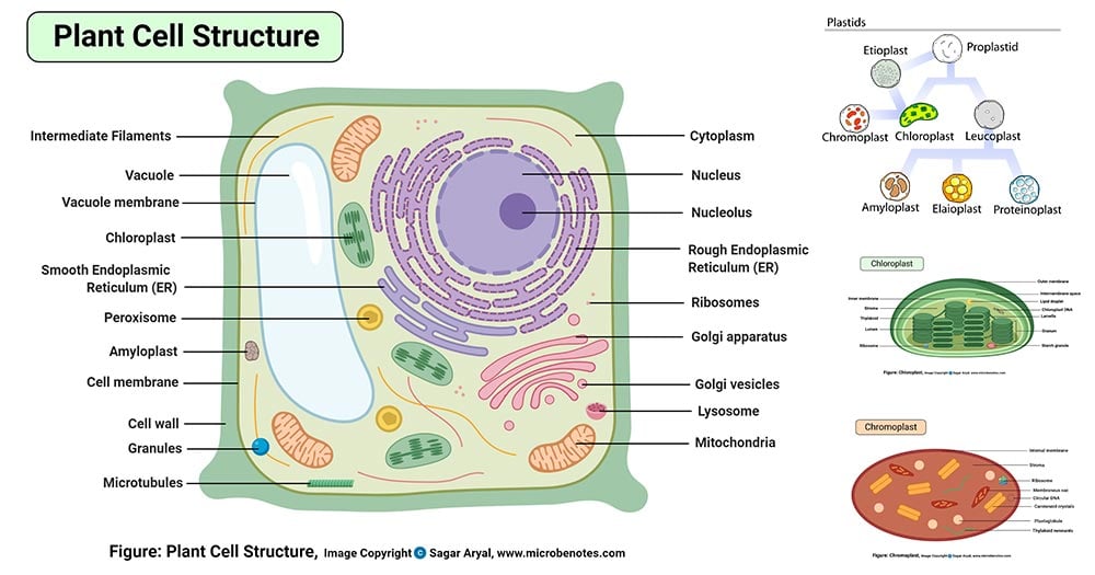
Plant Cell Definition Labeled Diagram Structure Parts Organelles
The typical characteristics that define the plant cell include cellulose hemicellulose and pectin plastids which play a major role in photosynthesis and storage of starch large vacuoles responsible for regulating the cell.
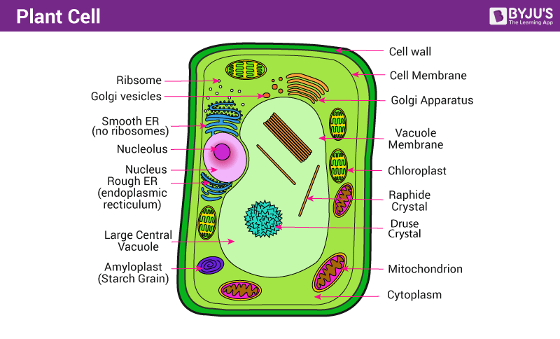
A labelled drawing of a typical plant cell to show its essential features. 1ai Make a labelled drawing of a typical plant cell to show its essential features httpstcoDilljGm3s6. Used in photosynthesis to convert sunlight carbon dioxide and water into food. A drawing of a typical animal cell.
All living organisms directly or indirectly depend on them for energy. A model of a typical plant cell is found to be rectangular in shape ranging in size from 10 to 100 µm. Grade Science Plant.
Cell membrane is made up of lipids and proteins and forms a barrier between the extracellular liquid bathing all cells on the exterior and the. Avail 25 off on study pack. Plants capture light from the sun and use it to build up chemical stores of energy.
As observed in the labeled animal cell diagram the cell membrane forms the confining factor of the cell that is it envelopes the cell constituents together and gives the cell its shape form and existence. A Picture Of A Plant. Cell Drawing Plantl.
The cell membrane is a thin layer that encloses the cells contents and separates the cell from its environment. All the best Plant Cell Drawing With Labels 37 collected on this page. Diagram Of Animal Ce.
A wall on the outside of the membrane which in combination with the vacuole as described below helps the plant cell maintain its shape and rigidity. In a plant cell chloroplasts are the most prominent forms of plastids that contain chlorophyll the green pigment. All the best Plant Cell Drawing 35 collected on this page.
Starch is the way plant cell store energy whereas animal cells store their energy as glycogen. The cell membrane controls which substances are allowed to enter and leave the cell. Contact us on below numbers.
We are aware that all life stems from a single cell and that the cell is the most basic unit of all living organisms. Each part known as an organelle works together to keep the cell functional. Plant Cells Are Eukaryotic Cells Of The Types Present In Green Plants Photosynthetic eukaryotes Of The Kingdom PlantaeDiagram Of Structure Of Plant CellHo.
All cells have a cell membrane around them. Starting early can help you score better. Plant Cell And Anima.
To begin with animal cells are typically substantially smaller than plant cells with plant cells ranging from 10 to 100 µm in length whereas animal cells are typically between 10 to 30 µm in length. Under the microscope it shows many different parts. Structures Unique to Plant Cells.
Most life on Earth depends upon plants for energy. Labelled diagram of plant and animal cell are as follow---Both plant and animal cells belong to eukaryotic cells. Plant and animal cells are different from each other as plant cell has cell wall plastids and a large central vacuole which is absent in animal cell.
Simple Plant Cell Di. Here lets study the plant cell. How to draw Plant Cell with easy method Step by Step GuidePlant cells are eukaryotic cells present in green plants photosynthetic eukaryotes of the kingdo.
Chloroplast in enclosed in two smooth membranes separated by a distinct periplastidial space. A Labeled Diagram of the Plant Cell and Functions of its Organelles. Eukaryotic cells are one which have organised nucleus with a nuclear membrane and genetic material is organised into chromosomes.
I Make a labelled drawing of a typical plant cell to show its essential features. We say the. The cell being the smallest unit of life is akin to a tiny room which houses several organs.
For Study plan details. Structure and Characteristics of a Plant Cell. C What role do the following processes play in the activities of a living cell i osmosis.
The most well-known plastids are chloroplasts which contain the chlorophyll that gives many plants their green hue. 1000 AM to 700. However the cell membrane in plant cells is quite rigid while the cell membrane in animal cells is quite flexible.
The chlorophyll enables the chloroplast to harness kinetic solar energy and trap it in the form of potential energy. The way the cells store energy is also different.
Science Animals including humans Human Body Heart. KS3 KS4 Science PE.

Simple Diagram Simple Human Heart Drawing Novocom Top
Once the thin border is perfect you can draw a thick border over it.
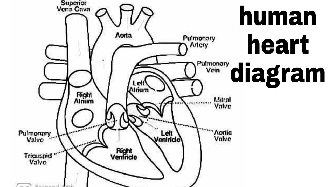
Heart diagram labeled drawing. This creates a border that gives them the confidence that their drawing will come out well. All the best Heart Diagram Drawing 36 collected on this page. Moreover the heart lies under the rib cage in the left of the breastbone sternum and the right behind the lungs and above the diaphragm.
The primitive streak is also a key component to the chick embryo. The lower two chambers of the heart are called ventricles. Well-Labelled Diagram of Heart.
Labeled Diagram Structures Function and Blood Flow. Heart diagram labeled drawing The heart is a muscle. Human Heart Diagram Labeled For Kids Random Heart Diagram Human Human Heart Diagram Human Body Pictures Science For Kids Free Human Heart Sketch Diagram Download Free Clip Art Free Clip Parts Of The Heart Diagram Worksheet By Gammaray Teaching Resources Easy Human Heart Drawing At Paintingvalleycom Explore Collection How Does The Heart Work Science Made Simple Evolution Of The Human Heart.
S04e04 How To Draw Human Heart Diagram Easy And Simple Biology. The heart wall is made up of three layers. The upper two chambers of the heart are called auricles.
Blank Heart Diagram Labeled Cashewapp Co Heart And Blood Flow Labeling Page Heart Labeling Worksheet Activities Human Worksheets For Step By Step Tutorials On Drawing Biology Diagrams Brains In 2019 Draw It Neat How To Draw Human Heart Labeled Free Printable Heart Diagram For Kids Labeled And Unlabeled Diagram Of Respiratory System Fetal Pig Heart Labeled Inspirational Drawing Of Heart Diagram. Label the Heart diagram L5 Labelled diagram. This is not only used to draw the human heart but for any drawing in general.
The wall of the heart has three different layers such as the Myocardium the Epicardium and the Endocardium. Heart diagram using Labelled diagram Labelled diagram. The human heart is about a closed fist s size weighs about 105 ounces and is somewhat cone-shaped.
The middle layer of the. Its situated a little to the left of your chest center and its around your fist size. The outer layer of the heart wall is called epicardium.
The heart is made up of two chambers. Label the Heart diagram Labelled diagram. How to draw Human Heart diagram Step by step मनव हदय क सरचन क चतर CBSE NCERT YouCanDrawIn this video youll watch how to draw human.
Labeled Heart Diagram Drawing Written By JupiterZ Tuesday September 17 2019 Add Comment Edit. A Schematic Diagram Of A Longitudinal Section Of The Human Heart. Anatomy of the heart and main cardiac structures including the heart valves chambers atria and ventricles and great vessels.
Draw A Heart Diagram Brainlyin How To Draw A Heart Drawing. A heart diagram labeled will provide plenty of information about the structure of your heart including the wall of your heart. We then simplified the anatomy of the heart even further with the below cartoon diagram and 2x2 table.
A heart diagram labeled will provide plenty of information about the structure of your heart including the wall of your heart. They draw around this using thin lines. The heart seen below the hind brain is in its first stages of growth.
Simple Easy Diagram Of Heart. Free Printable Heart Diagram For Kids Labeled And Unlabeled Heart Anatomy Blank Kind Of Letters Pertaining To Heart Diagram Blank Heart Diagram With Answers Bhd08 Ninja Heart Diagram The Parts Of The Heart Blank Printable Teachervision Free Unlabelled Diagram Of The Heart Download Free Clip Art Free Heart Diagram Sketch At Paintingvalleycom Explore Collection Of 19 Heart Diagram. The Heart Diagram Drawing Labeled A Perfect Cool Hands In Broken Transverse Section Of Human Heart 5 Download Scientific Diagram Label Heart Diagram Gcse Labeled Simple Cashewapp Co Diagram Of Nephron In Kidney Basic Cardiovascular Label Human Colored Diagram Of The Heart Lovely How To Draw Human Heart Anatomy Using Simple Heart Diagram Learning Medium For Kids Humandiagram Info Real Heart.
Heart diagram labeled Fattoria Estense Fattoria Estense Silver Label Farmacia Style Bottle Previously labeled as Aceto Balsamico di Modena Aged 10 years In July 2009 Aceto Balsamico di Modena Balsamic Vinegar of Modena obtained the Protected Geographical Indication status from the European Union. Anatomy of the heart and chart. This is how you can draw human heart in very easy way stay tuned for more videos like this.
Label The Diagram of The Heart Labelled diagram. This part of the embryo is responsible for creating bilateral symmetry and also lends support for a body axis. A labeled diagram of the human heart you really need to see the heart one of the most significant organs in the human body is.
A detailed explanation of the heart along with a well-labelled diagram is given for reference. Human Heart Diagram Drawing. At this point in development the form of the chick is growing.
Internal Human Heart Diagram Anatomy Poster Drawing By War Is. Function and anatomy of the heart made easy using labeled diagrams of cardiac structures and blood flow through the atria ventricles valves aorta pulmonary arteries veins superior inferior vena cava and chambers. This diagram labels the main aspects of the form at this stage of development.
Heart diagram labeled drawing. This is their start point. One secret all artists follow while drawing and sketching are they fix one point.
The wall of the heart has three different layers such as the myocardium the epicardium and the endocardium. Heres more about these three layers. A cloth called the pericardium.
Includes an exercise review worksheet quiz and model drawing of an anterior view frontal section of the heart in order to match the anatomy to the picture and test. How to draw Easy heart drawing for kids World Heart Day Drawing CompetitionSubscribe for More videos.