Like green plants use the process of photosynthesis to obtain food. Other questions on the subject.
Photosynthesis Definition Equation Steps Process Diagram
What is photosynthesis explain with diagram.

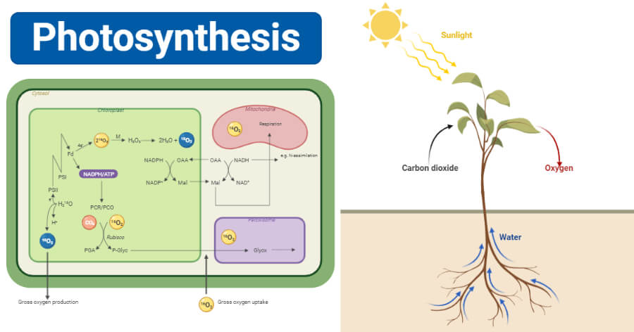
What is photosynthesis explain with diagram. Most life on Earth depends on photosynthesisThe process is carried out by plants algae and some types of. During photosynthesis in green plants light energy is captured and used to convert water carbon dioxide and minerals into oxygen and energy-rich organic compounds. Water soil and carbon dioxide.
Photosynthesis Photon Light Synthesis Putting together is an anabolic endergonic process by which green plant synthesize carbohydrates initially glucose requiring carbon dioxide water pigments and. Photosynthesis is the process by which plants and some microorganisms make substances like carbohydrates. The Process Of Photosynthesis Explained With Diagrams.
Broadly classifying organisms. This article will help you. The sugars are used by the cell as energy and to build other kinds of molecules.
Plants begin making their food which basically includes large quantities of sugars and carbohydrate when sunlight falls on their leaves. Plant mamtanegi00436 mamtanegi00436 21062021 Biology Primary School answered Q5 what is photosynthesis. Through a process called photosynthesis plants use energy in sunlight to turn a gas called carbon dioxide and water into sugar.
Explain with labeled diagram. What is photosynthesis explain with diagram. What is photosynthesis explain with diagram.
Through a process called photosynthesis. Although the process of photosynthesis is extremely complex when it comes to understanding it for research purposes the basic methodology can be easily explained with simple diagrams and the way the objects in it are labelled. Plants then use this sugar to grow.
KPG_Payless Shutterstock Photosynthesis is the process used by plants algae and certain bacteria to harness energy from sunlight and turn it into chemical energy. 2 See answers. Water soil and carbon dioxide.
What is photosynthesis diagram. Draw A Schematic Diagram And Explain The Photosynthesis Posted by Margaret Byrd Posted on March 6 2021 Draw a schematic diagram showing write photosynthesis representation of explained with the ofa stomatab definition equation to ilrate some what is site process in plants. According to the diagram of photosynthesis the process begins with three most important non-living elements.
Photosynthesis is essentially the only mechanism of energy input in the living world. Clearly Explained With Diagram For Kids. Photosynthesis in plants is necessary to maintain the oxygen levels in the atmosphere.
Click here to get an answer to your question What is photosynthesis. Explain with a diagram Answers. ExplanationAccording to the diagram of photosynthesis the process begins with three most important non-living elements.
This process goes on till the end of the. At the same time plants produce a gas called oxygen as a waste product which is lucky for us and other animals because we need oxygen to breathe. However the process by which an organism gets food differs to a great extent.
Photosynthesis is the primary source of energy in autotrophs where they make their food by utilizing carbon dioxide sunlight and photosynthetic pigments. Every organism in the world requires food to sustain life. In a nutshell how plants take in natural resources like light carbon dioxide and water and produce oxygen sugar and other elements that are important for life is what.
You might want to teach your kids what photosynthesis is. Click here to get an answer to your question what is photosynthesis explain with a diagram. The food is then stored aside by the plant and some of it is consumed during the day.
Photosynthesis is a process used by plants and other organisms to convert light energy into chemical energy that through cellular respiration can later. Photosynthesis photos-light synthesis-putting together is an anabolic process of manufacture of organic compounds inside the chlorophyll containing cells from carbon dioxide and water with the help of sunlight as a source of. The chemical energy is stored in the form of sugars which are created from water and carbon dioxide.
Why is photosynthesis important to. It is an endothermic takes in heat chemical process that uses sunlight to turn carbon dioxide into sugars. Science 20082019 0600 jack626259.
Photosynthesis is a process by which phototrophs convert light energy into chemical energy which is later used to fuel cellular activities. Photosynthesis Definition Photosynthesis is defined as the process utilized by green plants and photosynthetic bacteria where electromagnetic radiation is converted into chemical energy and uses light energy to convert carbon dioxide and water into carbohydrates and oxygen. Explain with a diagram _____ab points le re ho toh sab thanku boloxD.
The heart is roughly cone shaped hollow organ. They will be to the lower left of the Aorta.
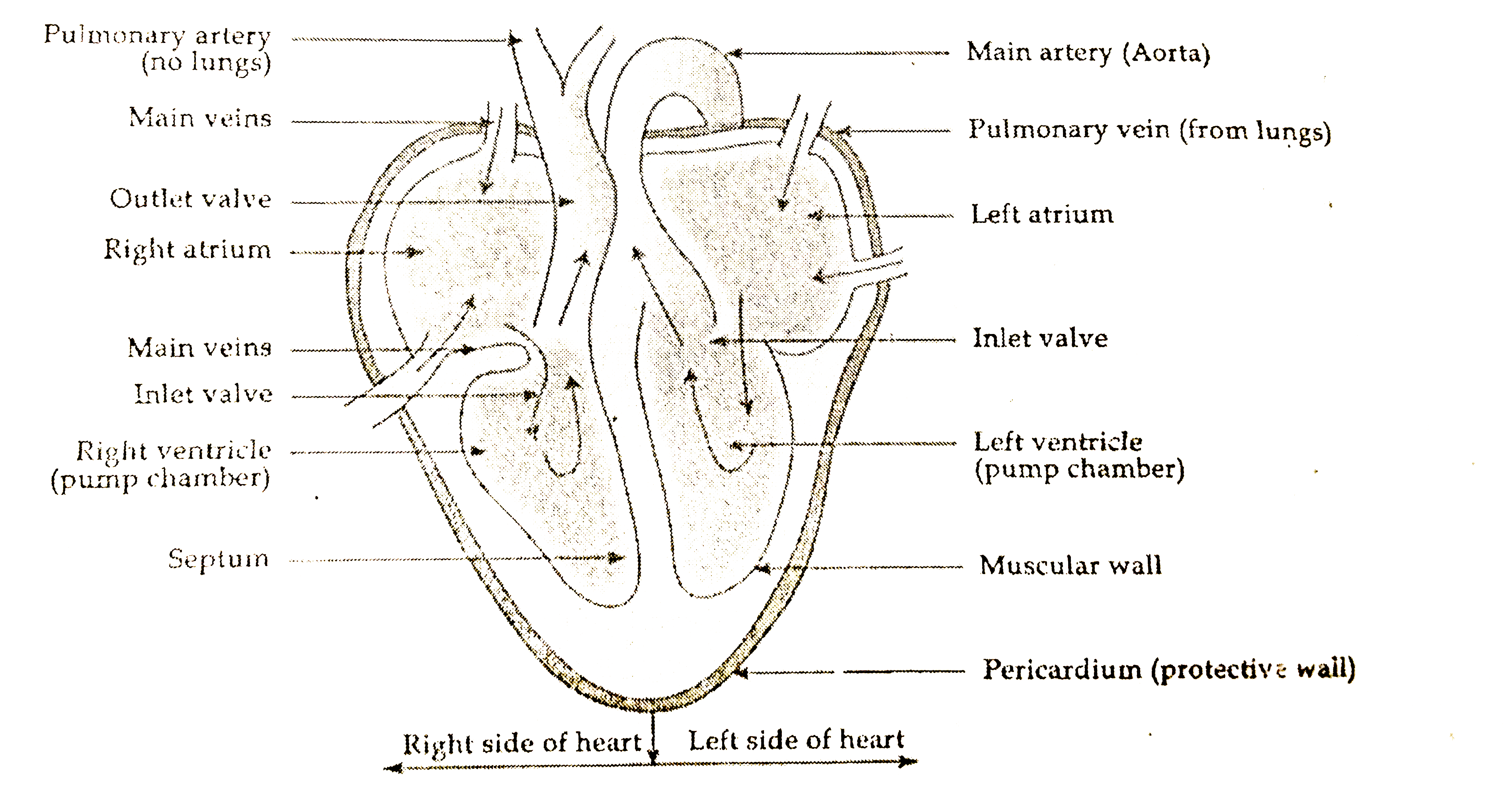
Draw A Labelled Diagram Of Human Heart
IG 263 describe the structure of the heart and how it functions DP 621 Draw and label a diagram of the heart showing the four chambers associated blood vessels valves and the route of blood through the heart.

Draw a labelled diagram of human heart and explain it. The heart though small in size performs highly significant functions that sustains human life. Science 29012020 1515 jack626259 Draw a diagram of human heart and label its parts. Draw a labelled diagram of Human Heart.
The heart lies in the thoracic cavity in the space between the lungs mediastinum anterior to the vertebral column and posterior to the sternum. In this video our mentor has showed how you can draw a basic anatomical diagram of human heartLike and comment your thoughts and feedback belowSubscribe. The heart is responsible for the circulation of blood in our body.
Find an image that displays the entire heart and click on it to enlarge itStep 2 Find a piece of paper and something to draw with. Heart rate varies depending on the phase of respiration. Draw Label Annotate Guidelines You are drawing labelling and annotating the digestive system.
It is not possible to draw the human heart dont u have in your text book or u can search in net. We will review the anatomy function and order of blood through the human heart using pictures of the atria ventricles tricuspid valve mitral valve pulmonary valve aortic valve superior and inferior vena cava pulmonary arteries and veins and aorta. The wall of the heart has three different layers such as the Myocardium the Epicardium and the Endocardium.
The heart consists of 4 chambers. Draw a labelled diagram of human ear and explain its working Get the answers you need now. Watch 1000 concepts tricky questions explained.
Correct answer to the question. FileDiagram of the human heart croppedsvg. There are two of them.
Drawings must be correctly proportioned and clearly drawn showing connections between structures. The drawing may show the heart without contraction or in any stage of contraction. This occurs even in normal human beings and hence it is quite physiological one.
This is known as sinus arrhythmia. This is a file from the Wikimedia Commons. A heart diagram labeled will provide plenty of information about the structure of your heart including the wall of your heart.
It is approximately the size of owners closed fist and weighs about 250-300 gm in female and 300-350gm in the male. Arrhythmia refers to irregularity in the beating of heart. Apne doubts clear karein ab Whatsapp par bhi.
Loading DoubtNut Solution for you. 611 600 pixels. Markscheme Remember up to TWO quality of construction marks per essay.
Every single part of our body is so well designed that it works continuously throughout our life. A well labeled human heart diagram given in this article will help you to understand its parts and functions. Step 1 To find a good diagram go to Google Images and type in The Internal Structure of the Human Heart.
Draw a labeled diagram of human heart. The heart one of the most significant organs in the human body is nothing but a muscular pump which pumps blood throughout the body. Draw a labelled diagram of human heart.
The human body is the best machine created by God. When the pacemaker is located in the SA node and heartbeats accordingly it is known as sinus rhythm. The anatomy of the heart is made easy in this post using labeled diagrams of the main cardiac structures and vascular system.
Human Heart Diagram Labeled Science Trends. Start with the pulmonary veins. AND 623 Explain the.
Draw a labelled diagram of the human heart showing the attached blood vessels. 26 K views 100 people like this. Draw the top vein slightly smaller than the bottom veinStep 3 Below the pulmonary veins and slightly to the right begin sketching the.
Heres more about these three layers. A Labeled Diagram of the Human Heart You Really Need to See. 10 Draw And Label A Human Heart.
Draw a table to show the functions of any two chambers of Human Heart. Exterior of the Human Heart. The human heart and its functions are truly fascinating.
244 240 pixels 489 480 pixels 782 768 pixels 1043 1024 pixels 2086 2048 pixels 663 651 pixels. Click here to get an answer to your question Draw a labelled diagram of human heart. Size of this PNG preview of this SVG file.
The lungs re-oxygenate. The atria receive blood while the ventricles pump blood.

How To Draw Internal Structure Of Human Heart Easy Version Human Heart Diagram Heart Diagram Heart Drawing
Apne doubts clear karein ab Whatsapp par bhi.

Explain the internal structure of heart with a neat labelled diagram. Earth s interior structure chapter 1 plate tectonics crust mantle core the structure of earth earthquakes volcanoes geo41 Internal Structure Of The EarthThe Structure Of Earth Earthquakes Discovering GeologyStructure Of The Earth Diagram ActivityDraw Neat Diagram Label Them And Explain The Interior OfEarthquakes Volcanoes Geo41Draw The Neat Diagram Showing Layers Of. Define blood pressure and discuss various factors regulating the blood pressure in human beings. Explain the external and internal structure of heart with neat labelled diagram - 18313931 lathapushpa9538 lathapushpa9538 13062020 Science Secondary School answered Explain the external and internal structure of heart with neat labelled diagram 2 See answers.
Draw a neat labelled diagram of the longitudinal section of the human heart and show the direction of the flow of blood through the different chambers. Diagram of the human heart created by Wapcaplet in Sodipodi. Blood then moves to the right ventricle where it is pumped to the lungs.
SHORT ESSAYS 5M Explain the normal ECG with a labeled diagram. Describe the internal anatomy of the heart with a neat labeled diagram. The outer layer of the heart wall is called epicardium.
The upper two chambers of the heart are called auricles. Explain coronary and portal blood. The inner layer of the heart wall is called endocardium.
The heart wall is made up of three layers. This is done following a detailed set of arithmetic instructions written in the main memory. The heart is divided into a right and left side by the septum.
Interauricular septum is relatively thin while interventricular septum is thick. Well-Labelled Diagram of Heart. Human heart is covered by a double layered structure which is known as pericardium.
Then fill in the base of the heart with the right and left ventricles and the right and left atriums. The lower two chambers of the heart are called ventricles. The middle layer of the heart wall is called myocardium.
The heart lies in the thoracic cavity in the space between the lungs mediastinum anterior to the vertebral column and posterior to the sternum. What is the cardiac cycle. The heart is divided into four chambers consisting of two atria and two ventricles.
The outer layer is associated with the major blood vessels whereas the inner layer is attached to the. Diagram the anatomical structure of the heart. It is approximately the size of owners closed fist and weighs about 250-300 gm in female and 300-350gm in the male.
There is no connectioncommunication at all between the right and left sides of the heart. It also uses the main memory for the memory. So as we discuss the various parts you keep checking out the parts simultaneously in the above given labeled diagram of the human heart.
Internal structure of human heart. Alan walker - Force NCS Releaselink. The heart consists of 4 chambers.
The right and left sides of the heart are separated by a partition called septum formed of myocardium covered by endocardium. Describe the internal stsructure of heart with a neat labelled diagram. Correct answer to the question.
Function and anatomy of the heart made easy using labeled diagrams of cardiac structures and blood flow through the atria ventricles valves aorta pulmonary arteries veins superior inferior vena cava and chambers. To draw the internal structure of the heart start by sketching the 2 pulmonary veins to the lower left of the aorta and the bottom of the inferior vena cava slightly to the right of that. Explain systemic and pulmonary circulation.
The heart has four chambers two relatively small upper chambers called atria and two larger lower chambers called ventricles. How to draw internal structure of heart illustrated Music. The right atrium receives blood from the superior and inferior vena cavas and the coronary sinus.
Describe various events of the cardiac cycle. The heart is roughly cone shaped hollow organ. This will help you to understand the functioning of its different parts.
The heart is made up of two chambers. Explain the internal Structure of heart with a neat labelled biagram. Cropped by Yaddah to remove white space this cropping is not the same as Wapcaplets original crop.
The Central Processing Unit CPU is the heart of the computer combined in the sys with the processing system of a computer. Labeled diagram Science Life Processes describe the internal structure of human heart with the help of well labeled diagram The human heart diagram shows a cross section of a healthy heart and its inside structures The blue arrow shows the direction in which Ask Questions for CBSE Class 10 Biology Life Processes April 9th 2019 - draw a neat. It has two atria towards the base.
Human heart is four chambered. Once you have the basic outline of the heart sketched out use an existing diagram. The CPU carries out actions with information help of Arithmetic-Logic Unit ALU.
The walls of the ventricles are relatively thicker than atrial walls.
Even mamono mana which has the power to erode other types of mana. Each woodcut was counted individually totaling 16494 Many woodcuts depict more than one object or image although all still remain part of an individual woodcut.

Water Cycle Wikipedia
9781420000948 1420000942 Percutaneous Absorption - Drugs Cosmetics Mechanisms Methods Robert L.

Explain the mechanism of water cycle with the help of well labelled diagram. The first attempt to present Capt. 37 Full PDFs related to this paper. Unlike gasoline- or diesel-powered generators natural gas generators must be able to burn a gaseous fuel rather than a liquid one.
To Which are Added Observations on the Dangerous Effects of Altering the Accustomed Diet of Patients and the Ill Effects of Salt Water Hemlock c. Добірки джерел і теми досліджень. For instance the two sites Wizernes and Evoisson which possess similar general indices poor water.
Indeed I think a good case might be made for thirty. The Memorial Art Gallery Rochester New York January nineteen hundred nineteen. Also a gun fitted with this type of lock.
The land use along the Gombak River has not changed very much over the years. The makers of our large dictionaries have been exceedingly crotchety in their choice of what. The computation of simple proportions revealed a great deal about the publishers priorities as well as those of the English religious authorities which were to encourage people to live well as Protestants in order to die well.
An Abstract of the Methods of Curing Diseases of the Eyes Legs and Breasts. As he explains it then if starless spheres as well as partial epicycles and eccentric spheres are not physically real they would be regarded as useful by astronomers for determining the positions and trajectories of the stars and planets given that the human mind was not endowed by God with the ability to apprehend the true quality of the celestial order though it was made able to access. The Anthropology of Globalization provides an ethnographic introduction to the world of flows and interconnections.
That would be to say that one-fifth of the miracles of the great cycles were artistic units in themselves and. A figure characterized by her deviant desires and sexual secrets depraved acts and dangerous agenda. Thus apoptosis and necrosis decrease could only be explained by total destruction of damaged cells without renewal.
The mana that all life forms discharge blends with the mana that fills the world and the mana that fills the world nurtures plants and animals while at the same time dwelling within them. Download Full PDF Package. With this storage time we showed that the selected immune biomarkers were able to discriminate between different sites which were similarly classified by general indexes such as water and ecological qualities and FBI.
The 1918 rotary exhibition of the American Water Color Society and a group of oils by Andrew T. It is concerned with tracking the paths taken by the various cultural flows that crisscross the globe as well as with exploring the. This could explain why the rate of ionic.
Список тез доповідей конференцій на тему Italian language Etymology Early works to 1800. Before that and since the days of their original printing only scattered bits had been republished for one or another reason -- on occasion even merely to disparage or glorify the man or what he wrote depending on the publishers bent. Schwartz University of Rochester Memorial Art Gallery and American Watercolor Society page images at HathiTrust 1960 joint economic report.
This chapter analyzes the mechanism that produces a witchthe output so to speak of the witch trial scene. The slow but resolute pace of English as the language of science. In this manner the mana that fills the world continuously cycles.
Maibach 9782287003899 2287003894 Le Labyrinthe Du Continu - Colloque De Cerisy Springer Publishing. Firearms now historical an early type of flintlock usually of English manufacture having an external safety in the form of a pivoted hook which engages a notch in the rear underside or breast of the cock. A short summary of this paper.
This requires a carburetor -- a device that blends a precisely metered amount of fuel and air and injects the mix into the engines cylinders --. The affectively supercharged cycle by which this occurs is projective and attributive. I have gone carefully through the four English cycles with Professor ten Brinks censures in mind and I conclude that at least twenty of the individual plays have central motive consistent action and well-rounded dramatic plot.
In short the production-through-demonization of the witch has a particularly queer shape. CD4 T cell functions include activating other immune cells releasing cytokines and helping B cells to produce antibodies. Наукові публікації для бібліографії з повним текстом pdf.
A major portion of the land along the Penchala River was considered as squatter areas which have been replaced by housing estates and office buildings in the last few years. Preying on plants and animals aids the predators in producing new mana. From knowledge to power.
Bronaugh Nina Dragicevic Howard I. If Mr Bond would share the grounds for explaining the diminution instead of injuring himself we could perhaps help him somewhat63. And on the Cure of Cancerous Scrophulous and other Chronic Diseases by Mild Internal Remedies.
John Smiths works objectively and with sympathetic understanding of their character was made by Edward Arber in 1884. The Gallery 1919 by Andrew T. The slow but resolute pace of English as the language of science.
This may explain why the levels of salt pollution are generally higher for the Penchala River. They help to shape activate and. He has to explain also that a few words will probably be noticed in the Slang and Cant Dictionary that are questionable as coming under either of those designations.
MHC Class II molecules interact with CD4 on the T helper cells which helps identify this cell type. Newbery 1774 by William Rowley page images at. These have been admitted because they were originally either vulgar terms or the compiler had something novel to say concerning them.
Experiments on lodestones such as immersing them in water or cutting pieces off them were reported62 and offers of help made. From knowledge to power.
A kidney is composed of an enormous number of uriniferous tubulesThey are also known as nephrons or renal tubules or kidney tubules. Learning a quick mnemonic VAD can help you remember these structures renal Vein renal Artery D.
Draw A Diagram Of An Excretory Unit Of A Human Kidney And Lable The Following Bowman S Capsule Glomerulus Collecting Duct Renal Artery Studyrankersonline
The kidneys remove wastes from the blood and are the effectors for controlling the water potential of the blood.

Explain the structure of kidney with the help of labelled diagram. Homeostasis in mammals requires complex systems to maintain internal conditions near constant. Gross Structure Of Kidney Kidneys with blood vessels the one on to right is a longitudinal section of the kidney. Each kideney is covered by semi-liquid fatty tissue called adipose capsule.
It codes for enzyme or protein for structural functions. A kidney is a bean-shaped organ that is reddish-brown in color. In other words you take your data and give it a visual.
They are protected in the retroperitoneal space by the renal fat pad and overlying ribs and muscle. Each kidney is formed of about 1 million nephrons. Blood from the glomerulus is carried away by an 5 artery.
Since they are located deep. With the help of a neat and a labelled diagram explain the detail structure of a flame 2 See answers rky2494021234 is waiting for your help. It is the outer region of kidney.
This will also help you to draw the structure and diagram of kidney. असल बत यह रह थ मन अपन दन पर तल उगल दबत ह ह सथ म ह त. Outer renal cortex and inner renal medulla.
Each kidney of an adult human measures 10-12 cm in length 5-7 cm in width 2-3 cm in thickness with an average weight of 120-170 g. It is the region where transcription ends. Ii The medulla of kidney is divided into a few conical masses called 2 projecting into the 3 iii Glomerulus is a tuft of capillaries formed by the 4 artery.
Add your answer and earn points. As to the use cases diagrams and other visuals perfectly fit for describing trends making a comparison or showing relationships between two or more items. If we wanted to examine someones kidneys with ultrasound we definitely must know where to find them.
Towards the centre of the inner concave surface of the kidney is a notch called hilum through. Rendered from our 3D model of the kidney with texture map this diagram is available in various sizes depending on the needs of the buyer. Labeled Diagram of the Human Kidney The human kidneys house millions of tiny filtration units called nephrons which enable our body to retain the vital nutrients and excrete the unwanted or excess molecules as well as metabolic wastes from the body.
The kidney is packed with around a million structures called nephrons. The long axes of the kidneys are aligned with that of the body but the upper end of each kidney pole is tilted slightly inward toward the backbone vertebral column. Syllabus 2016-2018 141 Homeostasis in mammals.
It has a dimension of 11 cm in length 6 cm in width and 3 cm in thickness. These nephrons start in the. When it comes to presenting and explaining data charts graphs and diagrams you should help people understand and memorize at least the main points from them.
Find labeled diagram of the human kidney stock images in HD and millions of other royalty-free stock photos illustrations and vectors in the Shutterstock collection. This diagram shows where the renal artery enters the kidney and where the renal vein leaves. Nephrons are held together by a connective tissue.
They are your bodys very own laboratories that test your. In LS the kidney shows two regions within the capsule. Nephrons are the structural and functional units of the kidney.
As noted previously the structure of the kidney is divided into two principle regionsthe peripheral rim of cortex and the central medulla. The two kidneys receive about 25 percent of cardiac output. It is the binding site for RNA polymerase for initiation of transcription.
Your kidneys are silent workhorses toiling 247 to clean your blood of impurities and toxins that build up from the bodys metabolism. Learn more about the anatomy of the kidneys and the urinary system with our urinary system quizzes and labeled diagrams. This capsule maintains the kidneys shape and protects the inner tissues.
This waste fluid which we know better as urine is then excreted. Thousands of new high-quality pictures added every day. However the kidneys role extends to well beyond just making urine.
Kidneys are reddish brown bean shaped structures situated between the levels of last thoracic and third lumbar vertebra close to the dorsal inner wall of the abdominal cavity. Outer covering of this capsule is made up of tough fibrous connective tissue called renal fascia. Schematic structure of a transcription unit.
Situated in the middle of the medial concave border is a deep vertical cleft the hilus which leads to a cavity within the. They lie on the posterior abdominal wall. In addition they also play an important role in maintaining the water balance of our body.
A discuss the importance of homeostasis in mammals and explain. A thin connective tissue called the renal capsule surrounds each kidney. I Towards the centre of the inner concave surface of the kidney is a notch called 1 through which ureters blood vessels and nerves enter.
The anatomy of the human kidney seen in cross section begins with hilum or opening of the medial. The diagrams below show the histology structure of tissues of the kidney. After reading this article you will learn about the structure of the kidney.
The bean-shaped kidneys have an outer convex side and an inner concave side called the renal hilus where the renal artery vein and ureter are found. The kidneys are two in number which are situated one on each side of the verteral column and in-front of the last ribs. The right kidney is placed slightly lower- than the left due to the presence of liver which occupies much space on the right side.
Kidney Labeled Diagram The Human Kidney Diagram is a highly detailed representation identifying the major structures of kidney as seen in cross section.