The heart pumps enriched blood cells. The pattern mentioned earlier consists of eight hearts and eight arrows that can be seen in a diamond of superb cut quality.

Normal Cardiac Anatomy The Blue Arrows Give The Flow Of Oxygen Poor Download Scientific Diagram
Oxygen-rich blood cells travel to the heart from the lungs.

Heart diagram with arrows. Now see How to Draw a Love Heart Ribbon. Then the blood enters the right atrium chamber of the heart. Diagram of the cardiac anatomy showing the top chambers atria and bottom chambers ventricles.
The blood enters the heart from the body through the superior vena cava and the inferior vena cava. A diagram shows a cross-section of a heart between two lungs. The atria are smaller than the ventricles and have thinner less muscular walls than the.
This is what youll see by putting the diamond into a hearts and arrows. The right side of the heart has less myocardium in its walls than the left side because the left side has to pump blood through the entire body while the right side only has to pump to the lungs. A Atria comes before V Ventricles so the atria will be located on topbefore the ventricles.
It is located in the middle of the chest and slightly towards the left. They are cut to ideal proportions with good optical symmetry polish and a specific faceting pattern. As shown on the left image below each pavilion main facet shown in face down view when inverted becomes visible as both the shaft part of one arrow A directly below and is also reflected 180 to produce the arrowhead part B of a second arrows figure.
HttpyoutubecVe2YUfOAPkSee a simple way to draw a love heart with a ba. Arrows show the path of blood flow in the human heart. The small cardiac vein parallels the right coronary artery and drains the blood from the posterior surfaces of the right atrium and ventricle.
Find premium high-resolution illustrative art at Getty Images. Consult your anatomy book to draw arrows that show where the blood enters the heart the valves it moves through and the direction it exits the heart. Hearts and Arrows diamonds are precision-cut variations of the traditional 57 faceted round brilliant cut.
Heart Diagram Use this labelled diagram of a human heart major blood vessels to answer these questions. 55101315 - Heart symbol diagram for graph infographic presentation with. For the ideograph of a heart pierced by an arrow see Heart symbol.
Add to Likebox 79213210 - heart rate decrease. The heart pumps around 57 litres of blood in a day throughout the body. If you clench your hand into a fist this is approximately the same size as your heart.
115142712 - Cupid with heart arrows and bow vector greeting card of Valentines. A heart diagram labeled will provide plenty of information about the structure of your heart including the wall of your heart. The wall of the heart has three different layers such as the Myocardium the Epicardium and the Endocardium.
On the left side of the picture above is a flawless diamond displaying eight symmetrical hearts. The middle cardiac vein parallels and drains the areas supplied by the posterior interventricular artery. The heart contains 4 chambers.
Heres more about these three layers. Exterior of the Human Heart. The right atrium left atrium right ventricle and left ventricle.
View top-quality illustrations of Heart Diagram With Arrow. The coronary sinus is a large thin-walled vein on the posterior surface of the heart lying within the atrioventricular sulcus and emptying directly into the right atrium. How to Draw a Basic Heart with a n Arrow.
Make small arrows that show the flow of blood for another study aid. Blue arrows show the path of oxygen-poor blood. The average male heart weighs around 280 to.
The structure of the heart. Blue arrows show the path of oxygen-poor blood. Use the alphabet A before V to remember the atria are on top and the ventricles are on the bottom.
On average the heart beats about 100000 times a day ie around 3 billion beats in a lifetime. Though not as complex as the heart pattern the creation of arrows is no small task. Red arrows show the path of oxygen-rich blood cells.
They travel through the arteries to the body. A diagram shows a cross-section of a heart between two lungs. Red arrows show the path of oxygen-rich blood cells.
If youre learning about how blood circulates through the body and the heart draw tiny arrows within the hearts segments. The blood then moves through the tricuspid valve shown as two white flaps into the right ventricle chamber of the heart. Chambers of the Heart.
Black arrows indicate direction of blood flow Blue structures contain deoxygenated blood Red structures contain oxygenated blood A B С С D N M E K I O T. The heart is situated at the centre of the chest and points slightly towards the left.
In what direction does blood flow through the heart. This prevents blood from flowing backward into the atria while the ventricles contract squeeze.

Circulatory Systems In Animals Transport Systems In Animals Siyavula
In systemic circulation oxygenated blood moves from heart to all parts of the body.

What is the order of blood flow in the heart. After the blood oxygenation process. First area of heart blood flows through. 55k views Reviewed 2 years ago.
When the ventricles are full the tricuspid valve shuts. Right atrium right ventricle left atrium left ventricle Humans have double circulation. Blood moves in two directions simultaneously.
It comes from the superior and inferior venae cavae. Blood Flow Sequence Activity The purpose of this activity is to understand the sequence of blood flow through the heart lungs and body. Click card to see definition.
Capillaries are small blood vessels that connect the arteries and veins. The order of blood vessels is great vessels arteries arterioles capillaries venules veins and back to the cavae. Blood flows from your right atrium into your right ventricle through the open tricuspid valve.
It flows into the lungs. Then it goes into the tricuspid vale and then to the right ventricle. To aorta to the body.
Blood enters the heart through two large veins the posterior inferior and the anterior superior vena cava carrying deoxygenated blood from the body into the right atrium. THIS SET IS OFTEN IN FOLDERS WITH. Through the aortic SL valve.
Leading into the right atrium is the superior ve. As the atrium contracts blood flows from your right atrium into your right ventricle through the open tricuspid valve. Superior and inferior vena cavae and the coronary sinus 2.
De-oxygenated blood enters the right side of the body and is pumped towards the lungs to pick up oxygen and start the process again. When the ventricle is full the tricuspid valve shuts. In pulmonary circulation deoxygenated blood is pumped by heart to get oxygenated in lungs.
Tap again to see term. Second area of heart blood flows through. The blood from the lower body is forced upward in the veins of the legs and then into the inferior vena cava where it eventually goes to the heart.
V veins Toward Blood is moving toward the heart. Blood flows from the right atrium into the right ventricle through the tricuspid valve. The blood flow in the heart begins from the right atrium that receives deoxygenated blood from the body to the right ventricle which pumps blood to the lungs.
In any case the heart is a 4 chamber organ which comprises of the right atrium and ventricle and the left atrium and ventricle. First the blood comes into the right atrium of the heart. The Aorta is the main highway that receives blood from the heart.
Figure 15 illustrates different parts of the heart involved in the blood flow. Oxygen-rich blood enters the heart on the left side and is pumped out to the cells of the body. 1 body 2 inferiorsuperior vena cava 3 right atrium 4 tricuspid valve 5 right ventricle 6 pulmonary arteries 7 lungs 8 pulmonary veins 9 left atrium 10 mitral or bicuspid valve 11 left ventricle 12 aortic valve 13 aorta 14 body.
Blood flows through the heart in the following order. Blood flows from the right atrium into the right ventricle through the tricuspid valve. Blood enters the heart through two large veins the inferior and superior vena cava emptying oxygen-poor blood from the body into the right atrium of the heart.
Pathway of the Heart - 9th - 2017. The process of the flow of blood through the heart is a complicated one. Click again to see term.
It requires a joint functioning of all the cardiac chambers to make proper circulation possible. Tap card to see definition. Blood enters the heart through two large veins the posterior inferior and the anterior superior vena cava carrying deoxygenated blood from the body into the right atrium.
It splits into Arteries these split into smaller arterioles then even smaller capillaries. Depends if you are talking about the heart or the circulatory system. That blood then goes out the left ventricle to the body and returns via the vena cavae.
Similarly one may ask what is the correct order of blood flow through the heart. Beginning with the superior and inferior vena cavae and the coronary sinus the flowchart below summarizes the flow of blood through the heart including all arteries veins and valves that are passed along the way. The left and right sides of the heart function jointly in pumping the de-oxygenated and oxygenated blood.
From the heart it is pumped back through the aorta into the body and the whole process repeats. One is systemic circulation and other is pulmonary circulation. The blood from upper body flows into the superior vena cava where it eventually goes to the heart.
Blood Flow Through the Heart. Then it flows through the pulmonary semilunar valves and into the pulmonary trunk.
This quiz has tags. What is Anatomy and Physiology.

Map Quiz 3 33 Anatomy Of The Heart
Human Heart Anatomy Multiple Choice Quiz.
(230).jpg)
The heart anatomy quiz. MJK7221995617AM on February 14 2015 660 plays. From the quiz author. The Human Anatomy and Physiology course is designed to introduce students pursuing careers in the allied health field to the anatomy and physiology.
Human Heart Anatomy 15. The cells that carry oxygen to body cells are. With more than 55-questions on such topics your.
Anatomy of the Kidneys Regulation of Urine Concentration Urethra Quiz. Test your knowledge about the anatomy of the Human Heart and blood. Science Quiz Human Heart Anatomy Random Science or Anatomy Quiz Can you find the different parts of the human heart.
Random Quiz Create A Quiz. Heart Anatomy Quizzes Trivia Elvis sang that he didnt have a wooden heart showing that not only was he the king of rock and roll he also had a passable knowledge of heart anatomy. Heart Anatomy Test Answers.
This is an online quiz called Heart Anatomy Quiz. Inner layer of the pericardium. Anatomy And Physiology Quiz.
Test your knowledge of the location shape and size of the heart as well as the anatomy of surrounding features in this quiz. 11th grade Anatomy Quiz on The Anatomy of the Heart created by Shannan Muskopf on 24022015. The number of valves in the heart is.
There is a printable worksheet available for download here so you can take the quiz with pen and paper. You are comparing audio recordings of the heartbeats of three different patients. Hence you can not start it again.
The major blood vessel that carries oxygenated blood out of the heart is the. You have already completed the quiz before. 0 of 10 Questions completed.
9th - University grade. Quiz Chapter 11 Cardiovascular System. Based on this information blood from the right ventricle is on its way to the _____.
Free interactive quiz for students biology anatomy and physiology. The human heart has taken on an almost mystical quality with its associations to love and bravery but it is also an essential and functional part of our physiognomy. Click on the tags below to find other quizzes on the same subject.
The distal end of the heart Preview this quiz on Quizizz. Play this game to review Human Anatomy. The Human Bunker 29.
By timmylemoine1 Plays Quiz Updated Jan 21 2016. The sac of tissue that protects the heart is the. An online interactive study guide to tutorials and quizzes on the anatomy and physiology of the heart using interactive animations and diagrams.
Respiratory System Map 18. Forced Order Support Sporcle. Test prep cardiovascular system.
This quiz is about the Cardiovascular System or says the lifeline of the body which consists of vessels called arteries veins and capillaries. This is a free printable worksheet in PDF format and holds a printable version of the quiz Heart Anatomy QuizBy printing out this quiz and taking it with pen and paper creates for a good variation to only playing it online. In Patient Three you notice that the second.
How many chambers are in a human heart. The Heart Previous The Heart. Anatomy of the Heart.
Blood Flow and Parts. Structure of the heart. This quiz is all about the Heart and the passage of Blood through the heart.
How Blood Type is Determined The blood type test is used to determine which antigenic molecules are present on RBCs. The layer of simple squamous epithelium that lines the inside of the heart is called myocardium. Atoms Molecules Ions and Bonds Quiz.
Top Quizzes with Similar Tags. The fluid that surrounds the heart and prevents friction when it beats is. 1 The right ventricle is the chamber of the heart that pumps blood for the pulmonary circulation.
Rate 5 stars Rate 4 stars Rate 3 stars Rate 2 stars Rate 1 star.
There are four heart valves in a healthy human heart. The valves help to maintain proper blood flow through the heart keeping blood moving efficiently and smoothly and in the right direction.

Heart Valve Wikipedia
After passing through the tricuspid valve blood.
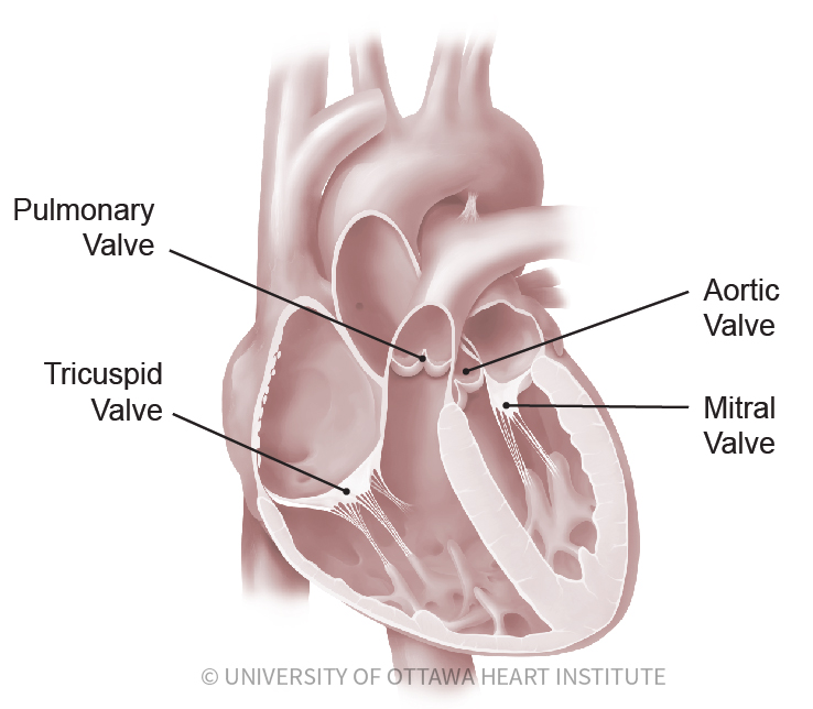
What are the four heart valves called. All four valves open and close to help move blood from one area to another. Good morning Dhruval Thank you for asking me. The leaflets open to let blood move forward through the heart during half of the heartbeat.
The valves are made of strong thin flaps of tissue called leaflets or cusps. Types of heart valves. The heart valves play a vital role in the function of the heart.
Normal valves have 3 flaps leaflets except the mitral valve. The tricuspid valve lies between the right atrium and the right ventricle and opens to allow blood to flow into the right ventricle from the right atrium. Keep reading to know more about these four valves and their functioning.
The left side of the heart can be thought of as the hearts powerful engine. Causes of recurrent boils. The 4 heart valves are.
The tricuspid valve is the first of the heart valves through which blood flows to the. The four valves in order of circulation are. The mitral valve and tricuspid valve are located between the atria upper heart chambers and the ventricles lower heart chambers.
Tricuspid valve located between the right atrium and right ventricle Pulmonary or pulmonic valve between the right ventricle and the pulmonary artery. The four heart valves are. Each side of the heart has a collecting chamber on top atrium and a pumping chamber on the bottom ventricle.
These valves are actual flaps that are located on each end of the two ventricles lower chambers of the heartThey act as one-way inlets of blood on one side of a ventricle and one-way outlets of blood on the other side of a ventricle. CHD a prior heart attack uncontrolled hypertension abnormal heart valves congenital heart disease heart defects present at birth and heart muscle disease. There is a valve between the upper and lower chamber and there is a valve between the lower pumping chamber and the body.
The valves prevent the backward flow of blood. The four valves of the heart include the tricuspid valve the mitral vale the pulmonary valve and the aortic valve. Conditions that can be associated with the development of heart failure include.
The tricuspid valve is named because it has three flaps called cusps or leaflets. I am a bit perplexed by the variety of topics in medicine and in the complexity or otherwise of your Qs. The four types of heart valves play an important role in ensuring that blood flows in only one direction and they are as follows.
Has three leaflets or cusps. Normally the mitral valve closes just before the tricuspid valve and when the two different sounds are detectable it is called a split S1 A split S1 may be indicative of certain conditions affecting the heart. In addition to the valves there are four heart chambers the upper chambers are called the left and right atria the lower chambers are the left and right ventricle.
The heart has four valves - one for each chamber of the heart. Separates the top right chamber right atrium from the bottom right chamber right ventricle. The four heart valves make sure that blood always flows freely in a forward direction and that theres no backward leakage.
The first sound S1 is generated by vibrations created by the closing of these two valves. There are 4 valves of the heart 2 on the left side and 2 on the right side. I thought initially that you might be a medical student but from the very empirical nature of.
Valves are actually flaps leaflets that act as one-way inlets for blood coming into a ventricle and one-way outlets for blood leaving a ventricle. Two of the valves the mitral and the tricuspid valves move blood from the upper chambers of the heart the atria to the lower chambers of the heart the ventricles. The heart has four heart valves the aortic mitral pulmonary and tricuspid valves.
Blood flows through this valve after leaving the right atrium. They close to keep blood from flowing backward during the other half of the heartbeat. The valves keep blood moving through the heart in the right direction.
The aortic valve and pulmonic valve are located between the ventricles and the major blood vessels leaving the heart. 4 heart valves decide the pathway for efficient blood flow through the heart. The heart valves keep blood flowing in one direction through the heart.
Opens to allow blood to flow from the right atrium to the right ventricle. It only has 2 flaps.
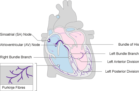
Anderson RH Taylor IM. Anatomy of the conduction system Sinus Node AV Node right ventricle left ventricle left atrium right atrium.

5 Conduction System Of The Heart 21 Download Scientific Diagram
Conducting System of Heart.
The conducting system of the heart pdf. The general topography of this system is closely similar in these two classes of warm-blooded vertebrates. LCA is larger and the most to get affected and responsible for the main pump of the heart RCA is the one that is engaged in blood supply of the conducting system. These special cells are able to generate an action potential on their own self-excitation and pass it on to other nearby cells conduction including cardiomyocytes.
Two set of veins empty directly into Rt Atrium Venae cordis minimi Ant cardiac vein st Rt marginal vein also CS - dilatation of Great Cardiac Vein located in post part of AV groove Opens into Rt atrium bw IVC and Tricuspid opening guarded. Each of these structures consists of cardiac muscle modified enough in structure to. CONDUCTION SYSTEM OF THE HEART The anatomy of four structures that compose the conduc-tion system of the heartsinoatrial SA node atrioven-tricular AV node AV bundle and Purkinje systemwas discussed briefly in Chapter 18.
Niranjan Murthy HL Asst Prof of Physiology SSMC Tumkur Two types of muscle fibers-contractile and conducting Contractile fibers in atria and ventricles- form two functional syncytia due to presence of gap junctions Conducting system includes SA Node internodal tracts AV Node Bundle of His Bundle branches and purkinje fibers Conducting system has- i less cross-striations ii less glycogen The Conduction System. As the electrical activity spreads along the hearts conduction system it initiates myocardial contraction in the surrounding myocardial tissue. Chambers of the heart.
The cardiac conduction system is a collection of nodes and specialised conduction cells that initiate and co-ordinate contraction of the heart muscle. The cardiac conduction system involves the spread of electrical activity from the sinoatrial node to the atrioventricular node down the bundle of His and along the Purkinje fibres. Apart from their interest in the cardiac conduction system the participants shared the opinion that their contribu tion should be relevant to the understanding and treatment of patients with cardiac arrhythmias.
Anderson RH Arnold R Wilkinson JL. Development of atrioventricular specialized tissue in human heart. The main differences can.
Components of the cardiac conduction system. The cardiac conduction system is the electrical pathway of the heart that leads to atrial and ventricular contraction. Per minute calculated by multiplying stroke volume by heart.
The heart is an essential pumping organ in the cardiovascular system where the right heart pumps deoxygenated blood returned from body tissues to the lungs for gas exchange while the left heart pumps oxygenated blood returned from the lungs to tissues cells for sustaining cellular respiration. THE CONDUCTING SYSTEM OF THE HEART Dr M Idris Siddiqui. The volume discharged from the ventricle.
6 Intrinsic Conduction System Function. II-1 are not only initiation and rate of the heartbeat but its coordinated transmission to the entire heart resulting in maximum mechanical efficiency. System of the heart Dr.
Nodal tissue and Purkinje fibres is limited to mammals and birds. The conducting system in congenitally corrected transposition. It comprises specialised cardiac cells which initiate and conduct impulses providing a stimulus for myocardial contraction.
And thats why LCA branches are more affected than RCA branches Overall important notes. This has resulted in a book in which the cardiac specialized tissues are discussed by different specialists. THE CONDUCTING SYSTEM The heart is able to contract on its own because it contains specialized cardiac muscle tissue that spontaneously forms impulses and transmits them to the myocardium to initiate contraction.
The conduction system consists of pacemaker cells that generate spontaneous action potentials and then deliver those impulses throughout the heart. Heart is drained by CS - empties into Rt Atrium. A cardiac conducting system consisting of specialized muscle ie.
The conducting system of heart. The conduction system in the heart is an intrinsic system wher eby the cardiac muscle is automatically stimulated to contract with-out the need for external stimulation Waugh Grant 2007. 4 Intrinsic Cardiac Conduction System Approximately 1 of cardiac muscle cells are autorhythmic rather than contractile 70-80min 40-60min 15-40min.
It consists of a complete series of systolic and diastolic events. Initiate distribute impulses so heart depolarizes contracts in orderly manner from atria to ventricles. The cardiac conduction system is a network of specialized cardiac muscle cells that initiate and transmit the electrical impulses responsible for the coordinated contractions of each cardiac cycle.
It supplies the pump of the heart. The Conduction System The heart is controlled by the ANS it increasesdecreases contraction but it does NOT initiate it. The electron-microscopist anatomist pathologist physiologist physicist and clinician.
Copyright 1978 Publisher Springer Netherlands Copyright Holder Martinus Nijhoff Medical Division The Hague eBook ISBN 978-94-009-9726-4 DOI 101007978-94-009-9726-4 Softcover ISBN 978-90-247-2080-4 Edition Number 1 Number of Pages XII 708. The Conduction System of the Heart Book Subtitle Structure Function and Clinical Implications Editors. CONDUCTION SYSTEM ANATOMY The functions of the electrical system of the heart Fig.
The heart has its own regulating system conduction system The conduction system is composed of specialized muscle tissue. The initiation and distribution of impulses through the myocardium that coordinates the cardiac cycle. It is controlled by the autonomic nervous system.