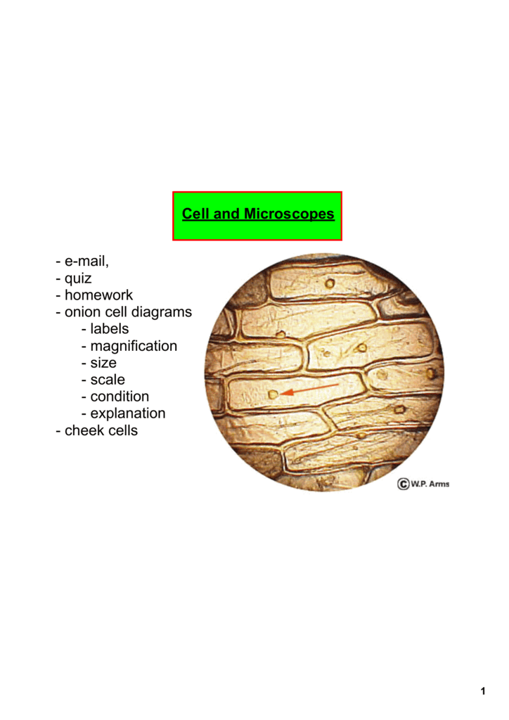
An epidermal onion cell diagram includes components such as citoplasm a round nucleus and a cell wall. Although at the surface an onion has dried protective leaf inside of it things change.

Sketch The Onion Peel Cell As Seen Under The Microscope Label The Parts Such As The Cell Wall Brainly In
2013 ford f53 wiring diagram lenel panel wiring diagram radial circuit wiring diagram furnace fan motor speed wiring h20 phase diagram only 86 atv yamaha 250 moto four wiring diagram omc shifter wiring diagram lenel wireles wiring diagram micro with audio jack diagram wiring diagram for motorcycle turn signal 1999.
Labelled diagram of onion cell. ReadDownload File Report Abuse. The vacuole is prominent and present at the centre of the cell. Sketch The Onion Peel Cell As Seen Under The Microscope.
Observing Osmosis Plasmolysis And Turgor In Plant Cells pdf. Onion epidermal cell labeled diagram. Onion epidermal cell labeled diagram.
Unstained Onion Cell Diagram Labeled mitosis in onion root tip cells an introduction to light focusing on focusing and drawing maribyrnong college plant cell structure and parts explained with a labeled plant cells vs animal cells with diagrams owlcation onion epidermal cell labeled onion cell diagram 02 top what structures can be seen in an unstained onion cell what is the diagram of an. The nucleus is. Label the cell wall and chloroplasts.
Onion epidermal cell labeled diagram 02 top label maker. Make your work easier by using a label. The nucleus of an onion epidermal cell.
An onion is a multicellular consisting of many cells plant organismAs in all plant cells the cell of an onion peel consists of a cell wall cell membrane cytoplasm nucleus and a large vacuole. Let S Learn Biology And Geology In English Plant Cell Lab pdf. For this microscope experiment the thin membrane will be used to observe the cells.
The microscope is the device which helps scientists observe how the onion epidermal layer looks like. Label The Parts Such As The Cell. Labels are a means of identifying a product or container through a piece of fabric paper metal or plastic film onto which information about them is printed.
Onion Epidermal Cell Labeled Diagram Vaculoe Onion Cell. Onion epidermal cell labeled top label maker diagram atkinsjewelry vaculoe. Observing onion cells under the microscope.
Onion Cell 40x Labeled Diagram. Videos you watch may be added to the TVs watch history and influence TV recommendations. Onion epidermal cell labeled onion cell diagram 02.
Cell diagram 1 onion benjamin himme. A View of the Cell prokaryotic and eukaryotic cells. Wide collections of all kinds of labels pictures online.
1994 jeep wrangler starter wiring 2 5 88 dodge wiring 2011 honda cr v radio wiring diagram 1996 ford f350 tail light wiring diagram wiring an schematic in series wiring light in a barn trailer tow plug wiring diagram free wiring diagram mercede 1957 ford thunderbird wiring diagram reversible circuit schematic diagram. To avoid this cancel and sign in to YouTube on your computer. Lab 08 Mitosis Meiosispdf The normal processes of mitosis and cell division are under tight control within.
Introduction To Cells Cheek Cell And Onion Cell Lab Lesson Powerpoi. The clear epidermal cells exist in a single layer and do not contain chloroplasts because the onion fruiting body bulb is used for storing energy not photosynthesis. The nucleus is present at the periphery of the cytoplasm.
Cell Lesson Cheek And Onion Cell Cell Theory Cell Theory. The onion cell diagram shows that in the middle of. Onion Cell Diagram Part Labeled Format.
Onion Skin Cell Labeled Diagram. The information can be in the form of hand. Module 1 Cells.
Onion Cell Diagram Part Labeled. Onion Cells Under the Microscope Requirements Preparation and. Onion Epidermal Cell Labeled.
A Well Labeled Diagram Of An Onion Cell - Explore Schematic Wiring Diagram 09F33069 D38C 4794 AFBA 59DBE1731379. PDF ePub Docx Status. Labeled Onion Cell Diagram Free PDF eBooks.
212 votes Last Checked. Draw labeled diagrams that illustrate the stages of mitosis in the onion root tip. BOOKS Onion Cell Diagram Labelled PDF Books this is the book you are looking for from the many other titlesof Onion Cell Diagram Labelled PDF books here is alsoavailable other sources of this Manual MetcalUser Guide Labelled Diagram Of Fallopian TubesThinkwell American Government Test Answers Labelled Diagram Of Fallopian Tubes.
Lab Using A Microscope pdf. Cell extracts atp and fluorescein labeled yellow in the diagram above have been replaced. Posted on September 08 2015.
4 marks e The onion epidermal cells are not green in colour because they lack. Labeled Diagram Of An Onion Cell - Example Electrical Wiring Diagram Plant guard cells stoma fully labeled diagram showing open closed 51409771. Onion Cell Diagram Labelled Wiring Schematic Diagram Draw Diagram Of Onion Peel As It Is Seen Through Microscope From Https Www Soinc Org Sites Default Files Uploaded Files 16 Cell.
Onion Cells Under The Microscope Requirements Preparation And. Onion Cell Diagram Labelled - Wiring Diagram Wall Highlight - Highlighterboristeriacolorieluceit. Draw a labelled diagram of an onion epidermal cell seen under the microscope.
Onion Skin Cell Labeled Diagram. Each plant cell has a cell wall cell membrane cytoplasm nucleus and a large vacuole. My Microscopy Experience Annabaldreemicroscopy.
Its purpose is to store food and water for the plant through a cold or dry season. Cell Structure and Cell Types 07 Onion Epidermis a.
Biology Paper 3 Wassce Pc 2016
This cell was then transferred to a drop of sugar solution.

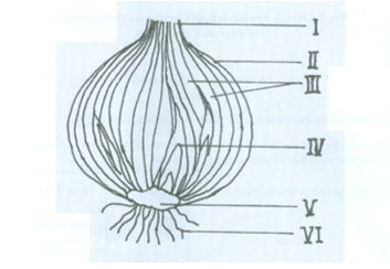
Labelled diagram of onion bulb. Ncert Class 9 Science Lab Manual Slide Of Onion Peel And Cheek. Food test may also be asked and do not be surprise that an onion bulb contains reducing sugar. Make a labeled drawing of a few check cells to demonstrate your observations.
2 marks ans 44. One fleshy scale leaf of an onion is taken. Examples of bulbs include the tulip narcissus crocus onion lily and garlic.
Labelled diagram of an onion cell awesome ion epidermal onion cell diagram onion diagram luxury labelled an ion cell awesome 4 ways to nysed part d lab review ppt download. Onion and cheek cell lab experiment. Sensory system biology uni.
Include what magnification the cells were pictured at. Be aware that there may be questions in the practical paper asking you to make a fully labelled drawing of an onion bulb. The main concept of the diagram is shown by the center circle of the diagram.
Onion epidermal cell labeled diagram. The microscope is the device which helps scientists observe how the onion epidermal layer looks like. What should be done to restore the cell back to its original condition.
Obtain a piece of a single piece of sliced onion bulb. The onion diagram is able to show layers of a complete system in a few circles. Onion Epidermal Cell Labeled Diagram Vaculoe Onion Cell.
The clear epidermal cells exist in a single layer and. Onion Cells Stock Photos Onion Cells Stock Images Alamy. The figure given below shows the epidermal cells of an onion bulb.
You do not need to look at the sun to do this. Cell Lab Doc Plant And Animal Cells Microscope. Lab_CellStructureTypespdf - Read File Online - Report Abuse.
Onion epidermal cells the epidermis or skin of an onion is an ideal subject for the study of cells because it is composed of a. The onion cell diagram shows that in the middle of. Plant Guard Cells With Stoma Fully Labeled Stock Illustration The movement of these ions creates resting potential and action potential.
Although at the surface an onion has dried protective leaf inside of it things change. Draw and label your diagram. Molecular biology teacher science onions professor teachers bulb flag onion.
Cork and Onion Skin You will be given a part of a bulb of onion. Home onion epidermal cell labeled onion epidermal cell labeled onion cell diagram 02. Onion Skin Cell Labeled Diagram Wiring Diagram Third Level.
Obtain a piece of a single piece of sliced onion bulb. Microscope and the cell. Draw a well labelled diagram of the epidermal cell as it would appear after immersion in a strong sugar solution.
1 Comparison Of Cross Sectional Images Of Onion Root Tip Cells. Onion plants are characterized by an edible bulb composed of food-storage leaves that are rich in sugar and a pungent oildue to this oil onion has strong tasteit has came from a family of Liliaceaebland white onionstrong-flavored red onion are some of the varieties in onions. U0026fileCells - whats insidepdf - Read File Online - Report Abuse.
Download scientific diagram labeling of microfilaments in onion bulb scale epidermis cells. Onion epidermal cell labeled onion cell diagram 02. Onion epidermal cell labeled diagram.
Blue label nyc july 22nd 2017 best labels wide. Wide collections of all kinds of labels pictures online. This cell was then transferred to a drop of a sugar solution.
Structure And Function Of Cells Learn Biology Class 8. What is the scientific term is used for the changes as shown in i above. Make a labeled drawing of a few check cells to.
Onion epidermal cell labeled top label maker diagram atkinsjewelry vaculoe. The clear epidermal cells exist in a single layer and do not contain chloroplasts because the onion fruiting body bulb is used for storing energy not photosynthesis. A bulb is an underground modified stem that develops in some flowering plants.
Onion epidermal cell labeled diagram 02 top label maker. When cooking onion the dark brown caramel is actually the sugar. Snap the piece in half and.
An epidermal onion cell diagram includes components such as citoplasm a round nucleus and a cell wall. Note the leaves and the stem tissue. Each of the circles is able to represent a component that is dependent upon the component on the inside of it shown by the circle inside of it.
Labelled diagram of an onion cell Safety. Mic Uk The Inner Epidermis Of The Onion Bulb S Cataphylls The. Notice the parts that you were able.
The figure given below shows the epidermal cells of an onion bulb. I Draw a well labelled diagram of the epidermal cell as it would appear after immersion in a strong sugar solution. Onion epidermal cell labeled.
Molecular biology teacher science onions. Draw a labelled diagram of an onion epidermal cell seen under the microscope. The Onion Cell Lab.
This is the epidermal peel. The peel is placed in a watch glass containing water and is cut into small rectangular pieces. The bulb of an onion is formed from modified leaves.
Draw a labelled diagram of an onion epidermal cell seen under the microscope. Place the slide against the window so that it is well backlit. The smallest bulbs are the size of peas.
It is broken into two and a thin membranous peel is pulled out using a forcep adhering to the inner surface of the leaf. The largest crinum lilies weigh over 15 pounds 7 kg. 2 marks ans 44.
Lab 4 the cell answer key procedure 43 draw a picture of a single elodea cell and label all visible structures. Microscope diagram labeled unlabeled and blank microscope diagram sensory system biology uni. To prepare stained temporary mount of onion.
What Organelles Would Be Visible In A Cheek Cell Why Quora Although at the surface an onion has dried protective leaf inside of it things change.