Prominent blood vessels Palpation 1. Position of mediastinum a.

A Draw A Labelled Diagram Of Human Respiratory System B Explain The Transportation Of Oxygen And Carbon Dioxide In Human Beings
It ranges from 20-25mm in diameter and 10-16cm in length.

Fully labelled diagram of the respiratory system. The hairs present in the nose filter out particles in the. Dimitrios Mytilinaios MD PhD Last reviewed. Movements with respiration a.
Human Respiratory System Diagram showing different parts of the Respiratory Tract. When air passes through the nose it is warmed moistened and filtered. The lungs are covered by a lining called the pleura which has two layers.
In the throat the trachea or windpipe filters the air. Labeled diagram of respiratory system. The human respiratory system is adapted to allow air to pass in and out of the body and for efficient gas exchange to happen.
Millions of small air sacs in the lungs where oxygen and carbo tubes which branch off from. View Original Image at Full Size. It is made up of several organs and structures that transport air into and out of the lungs exchanging oxygen with carbon dioxide.
But fear not - with a bit of practice and dedication you can. This is an online quiz called Respiratory System Labeling Interactive. Compare and measure both sides 3.
Click on the tags below to find other quizzes on the same subject. Interactive for 5th Graders This quiz has tags. May 31 2021 Reading time.
The lungs are divided into areas called lobes. They supply oxygen to the organs and tissues of the body. Respiratory System Examination Inspection 1- Shape of the chest 2.
The respiratory system allows people to breathe. Features of the Human Respiratory System. Respiratory system quizzes and labeled diagrams Author.
Your Skills Rank. The lungs are the parts of the body that we use to breathe. All your KS2 students have to do is drag and drop the various labels to identify the nasal cavity larynx alveoli diaphragm and more.
Position of mediastinum a. How we can help. There is a separate diagram explaining how breathing works breaking down the process of inhalation and exhalation.
Air enters the nose through the nostrils. The respiratory system which includes air passages pulmonary vessels the lungs and breathing muscles aids the body in the exchange of. Air a mix of oxygen and other gases is inhaled.
It includes the main parts of the respiratory system such as the trachea bronchi bronchioles alveoli and the diaphragm. This is a brilliant resource. The trachea is surrounded by.
In the throat the trachea or windpipe filters the air. The right lung has three lobes and the left lung has two lobes. Fill in the blanks.
Use this resource as a handout or transparency for science class. Name Description Function 1 2 3 4 5 6 7 8 9 10 11 Name Function 1 nose A. The gas exchange process is performed by the lungs and respiratory system.
You should make a label that represents your brand and creativity at the same time you shouldnt forget the main purpose of the label. The energy is generated by the breakdown of glucose molecules in all living cells of the human body. The entire respiratory tract passage consists of the nose pharynx larynx trachea bronchi and bronchioles.
The airway the lungs and the muscles of respiration. Image 37789 is a 1125 by 1408 pixel PNG Uploaded. Use this engaging interactive activity to label the respiratory system.
2 minutes Full to the brim with interesting but arguably complex anatomy the respiratory system may feel a little confusing to the anatomy apprentice. From the quiz author. Labeled diagram of the lungsrespiratory system.
The human respiratory system consists of a pair of lungs and a series of air passages leading to the lungs. The inner membrane of the trachea is covered in tiny hairs called cilia which catch particles of dust which we can then remove through coughing. Respiratory System Label Diagram Diagram Labels Label Gallery Get some ideas to make labels for bottles jars packages products boxes or classroom activities for free.
The respiratory system provides oxygen to the bodys cells while removing carbon dioxide a waste product that can be lethal if allowed to accumulate. Also known as the windpipe this is the tube that carries air from the throat into the lungs. An easy and convenient way to make label is to generate some ideas first.
There are 3 major parts of the respiratory system. Exercise and smoking both affect the lungs and circulatory system. Ideal for putting knowledge into practice respiratory system interactive activities can be a great addition to your Science lessonsChildren also have the chance to correct any mistakes.
Oxygen is inhaled and is transported to various parts and are used in the process of. The respiratory system in humans has the following important features. The respiratory system transports oxygen from the air we breathe through a system of tubes into our lungs and then diffuses it into the bloodstream whilst carbon dioxide makes the opposite journey.
The airway which includes the nose mouth pharynx larynx trachea bronchi and bronchioles carries air between the lungs and the. 15 rows Printable Blackline Diagram of The Respiratory System Test yourself. There is a printable worksheet available for download here so you can take the quiz with pen and paper.
Molly Smith DipCNM mBANT Reviewer. Lower respiratory tract organs.
Skin diagram coloring and labeling. Download 75 Plant Cell Labeled Stock Illustrations Vectors Clipart for FREE or amazingly low rates.

Plant Cell Structure And Parts Explained With A Labeled Diagram Biology Wise
As plants are eukaryotic organisms their cells have many similarities with other eukaryotic cells such as those found in animals.

Fully labelled diagram of plant cell. The structure of the plant cell is. Plant Cell Structure And Parts Explained With A Labeled Diagram. Plant Cell SEN Labelling.
Download Diagram Of A Typical Plant Cell And Label Fully Pics. Provides - a simple labelled diagram of a plant cell - a blank plant cell for colouring - instructions to colour parts of the cell specific colours and copy labels. What is a plant cell.
In addition plant cells differ from animal cells in a number of key ways. Plant Cell Diagram Vacuole. Some of these differences can be clearly understood when the cells are examined under an electron microscope.
Fully labelled diagram of the alveolus in lungs showing gaseous exchange vector by doethion generalized structure of animal cell plant under microscope parts shown in the plant cell diagram generalized structure of animal cell under light microscope. The typical characteristics that define the plant cell include cellulose hemicellulose and pectin plastids which play a major role in photosynthesis and storage of starch large vacuoles responsible for regulating the cell turgor pressure. Recently Viewed and Downloaded.
Draw a labelled diagram of a plant cell. Calcium carbonate deposits around the stalk and fully developed cystolith appears as grape-like clusters with a stalk that is attached to the cell wall from where it hangs into the cell lumen Fig. Here lets study the plant cell.
Definition Eukaryotic cell Diagram. Note going down the left the numbers are not sequential this is to match the numbering on others in the series. During development all epidermal cells have similar appearance.
How To Draw A Plant Cell Plants Botany Easily Quickly Well Labelled Diagram Youtube Plant cell. Plant cells contain many organelles such as ribosomes the nucleus the plasma membrane the cell wall mitochondria and chloroplasts. Plant guard cells with stoma fully labeled stock illustration.
Posted by unknown at 2209. Print and provide both sheets to student. Plant Cell Diagram Labeled Of A With.
As the central vacuole shrinks it leaves the cell wall unsupported. Draw a labelled diagram of a plant cell. We are aware that all life stems from a single cell and that the cell is the most basic unit of all living organisms.
Simple diagram of plant cell numberssvg. Examining a diagram of the plant cell will help make the differences clearer. A bacteria diagram clearly enables us to learn extra approximately this single cell organisms that have neither membrane-bounded nucleolus or organelles like mitochondria and.
The cell being the smallest unit of life is akin to a tiny room which houses several organs. Ideal for SEN students. New users enjoy 60 OFF.
167866268 stock photos online. Class 10 Solved Question paper 2020. A Labeled Diagram of the Plant Cell and Functions of its Organelles.
The plant cell is rectangular and comparatively larger than the animal cell. The Plant Cell is the basic structural and functional unit found in the members of the kingdom Plantae. We are aware that all life stems from a single cell and that the cell is the most basic unit of all living organisms.
A Labeled Diagram of the Plant Cell and Functions of its Organelles. Draw A Labelled Diagram Of A Plant Cell. Class 12 Solved Question paper 2020.
On this page we will learn about what is a plant cell definition structure model labeled plant cell diagram its cell organelles and the difference between plant cell and animal cell. As a plan to show the arrangement of any distinct regions of the tissue for example the tissues in a plant root to show the outline of individual cells that make up. A low power diagram is used.
Lets go over the individual components of plant cells. The cell being the smallest unit A Brief Comparison of Plant Cell Vs. Lab Manual Exercise 1a.
It shows the cytoplasm nucleus cell membrane cell wall mitochondria permanent vacuole and chloroplasts. Here is a comparative study of a plant cell and an animal cell so as to have a better understanding of the similarities. A simple diagram of a plant leaf cell labelled with numbers.
Even though plant and animal cells are eukaryotic and share a few cell organelles plant cells are quite distinct when compared to animal cells as they perform different functions. This will also help you to draw the structure and diagram of plant cell. A vacuole is a membrane bound structure found in the cytoplasmic matrix of a cell.
Later cells destined to become lithocyst can be.
Label Diagram Of Kidney Guide About Wiring Diagram Labeled diagram of the human kidney. The two kidneys act as filters for the blood removing harmful toxins.

Draw Well Labelled Diagram Of L S Of Human Kidney
The kidney is packed with around a million structures called nephrons.
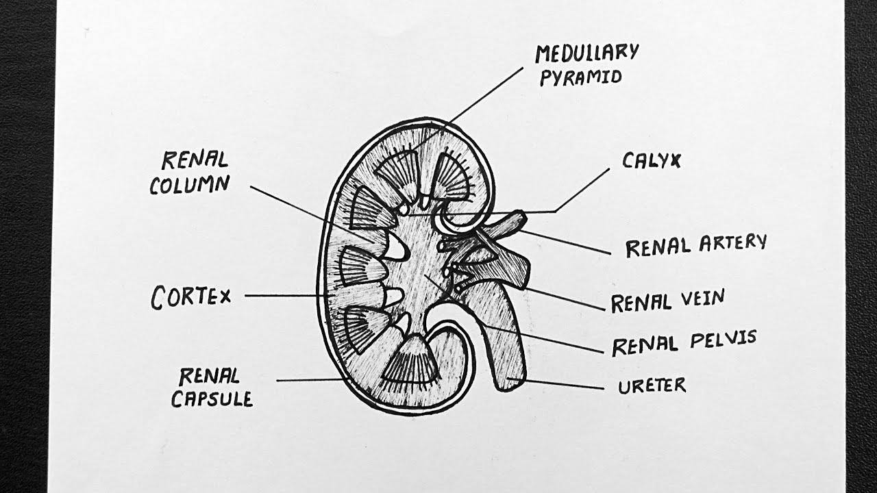
Fully labelled diagram of kidney. There are two kidneys one on each side of the abdomen and each contains between one and two million nephrons loosely embedded in connective tissue and amply supplied with blood. Because of all of the vital functions the kidneys perform and the toxins they encounter the kidneys. Kidneys are supplied with blood at arterial pressure by renal arteries which branch off the abdominal aorta.
They lie on the posterior abdominal wall. Learn more about the anatomy of the kidneys and the urinary system with our urinary system quizzes and labeled diagrams. The right kidney is placed slightly lower- than the left due to the presence of liver which occupies much space on the right side.
The nephron is one of the most important parts of our body and also one of the smallest functioning units. The nephrons are surrounded by veins but they do not touch. We have a particular passion for languages but also a very strong science background.
The kidneys are two in number which are situated one on each side of the verteral column and in-front of the last ribs. The kidneys are important for blood filtration excretion and osmoregulation. Blood flow through the kidneys is extremely high because of the large number of capillaries.
Start studying Kidney Anatomy Labeling. To understand the functions of the mammalian kidney and how it does this we must. Try these curated collections.
Search for labeled diagram of the human kidney in these categories. The medulla has a high osmolarity because it contains lots of salt. If we wanted to examine someones kidneys with ultrasound we definitely must know where to find them.
The medial-facing hila are tucked into the convex indentation of the kidney. Dehydration a blockage in the urinary tract or kidney. Blood leaves the kidneys through the renal vein into the inferior vena cava.
The nephron can be regarded as the functional unit of the kidney performing both functions of excretion and osmoregulation. Learn vocabulary terms and more with flashcards games and other study tools. 103 labeled diagram of the human kidney stock photos vectors and illustrations are available royalty-free.
These organs use up almost 25 percent. All the blood in the human body is filtered many times a day by the kidneys. A frontal section through the kidney reveals an outer region called the renal cortex and an inner region.
Kidneys filter blood and purify it. Use this interactive 3-D diagram to explore the kidney. Name the cavity in which the kidney sits.
The cortex is the dark outer. Labeled diagram of the human kidney bodytomy each kidney contains more than 800 000 nephrons each of which serves as the basic functional unit by performing three essential processes filtration reabsorption and secretion the nephron essentially prises renal corpuscle and renal tubule labeled diagram of a nephron and its location and functions the human kidney contains. 1133 Annotate a diagram of a glomerulus and.
Current diagram shows a little bit about human excretory system and a detailed diagram of Kidney which is the most important part of Excretory system. Creating high quality resources across a variety of subjects. See labeled diagram of the human kidney stock video clips.
Resources are being uploaded regularly but make a take. The kidney This diagram shows where the renal artery enters the kidney and where the renal vein leaves. The kidneys take time to do this.
Labeled diagram of the human kidney bodytomy each kidney contains more than 800 000 nephrons each of which serves as the basic functional unit by performing three essential processes filtration reabsorption and secretion the nephron essentially prises renal corpuscle and renal tubule labeled diagram of a nephron and its location and functions the human kidney contains more than 1 million. Each kidney looks like the kidney bean and the renal hilum is the entry and exit site for structures servicing the kidneys. WorksheetActivity no rating 0 reviews.
Since they are located deep retroperitoneally the easiest way to examine them is from the patients back. Kidneys are dark brown in colour. In addition they also play an important role in maintaining the water balance of our body.
Image of a kidney and nephron with the major structures labeled. This will also help you to draw the structure and diagram of kidney. The kidneys are located.
Vegetarian urine tends to be more basic or alkaline carnivore urine tends to be more acidic. The kidneys illustrated in Figure 1 are a pair of bean-shaped structures that are located just below and posterior to the liver in the peritoneal cavity. Labeled Diagram of the Human Kidney The human kidneys house millions of tiny filtration units called nephrons which enable our body to retain the vital nutrients and excrete the unwanted or excess molecules as well as metabolic wastes from the body.
I Need To Teach That. Kidney Structure The kidneys are made up of millions of nephrons which act as tiny filtering units. 1132 Draw and label a diagram of the kidney.
The body normally wants a blood pH between 735-745 and it will cause the kidneys to secrete H or bicarbonate to balance it. Vessels nerves lymphatics and ureters. Create a Labelled Diagram.
Figure 2512 Left Kidney. The adrenal glands sit on top of each kidney and are also called the suprarenal glands. In addition the food you eat does affect the urine pH.
They have six jointed legs two pairs of wings and two antennae. Labelled Diagram Of Grasshopper A Precise Explanation of the Life Cycle of a Grasshopper May 12th 2019 - Usually female grasshoppers are larger in size than the males Another feature of the females is the presence of two pairs of triangular structures valves at the end of the abdomen which they use for digging the sand during egg laying In case of male grasshoppers there is a single unpaired.
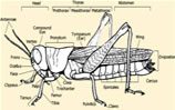
Grasshopper Anatomy Body Pictures Diagram
Labelled Diagram Of Grasshopper grasshopper anatomy like all insects the grasshoppers have three main body parts the head the thorax and the abdomen they have six jointed legs two pairs of wings and two antennae their body is covered with a hard exoskeleton grasshoppers breathe through a series of holes called spiracles which are located along the sides of the 1 answer to a very well labelled.

Fully labelled diagram of grasshopper. Antennae like all insects grasshoppers have 2 segmented antennae that sense touch and odours. Labelled Diagram Of Grasshopper grasshopper diagram labeled inspirational color diagrams of insect organs and internal structures so if you want to obtain all these awesome shots regarding grasshopper diagram labeled just click save button to store the shots for your laptop therere all set for download if you love and want to have it just click save symbol in the learn the parts that. Label Body Parts Of Grasshopper.
Grasshopper Diagram Labeled Learn Schematic Diagram May 15th 2019 - grasshopper diagram labeled Inspirational Color Diagrams of Insect Organs and Internal Structures So if you want to obtain all these awesome shots regarding Grasshopper Diagram Labeled just click save button to store the shots for your laptop Therere all set for download if you love and want to have it just click save. Home grasshopper diagram labeled learn schematic diagram reproductive system of grasshopper with diagram biology 172l general biology lab ii laboratory 09 grasshopper information report text images music video label grasshopper anatomy printout enchantedlearning com biology 2 jb004 k12 sd us grasshopper dissection diagram labeled michaelhannan co insect printouts allaboutnature. How to tell the difference between an adult male and.
In this article we will discuss about the structure of Locust Schistocerca with the help of a diagram. Labelled Diagram Of Grasshopper Free Books READ Labelled Diagram Of Grasshopper PDF Books this is the book you are looking for from the many other titlesof Labelled Diagram Of Grasshopper PDF books here is alsoavailable other sources of this Manual MetcalUser Guide Labelled Diagram Of Fallopian TubesThinkwell American Government Test Answers Labelled Diagram Of. Schematic diagram biology 2 jb004 k12 sd us parts of an insect grasshopper well labelled diagram of grasshopper pdfsdocuments2 com label the grasshopper anatomy diagram ednet ns ca free download here pdfsdocuments2 com labelled diagram of male grasshopper anatomy structure diagram of a male grasshopper concept design home definition swa cabel label an labelled diagram.
Abdomen - the segmented tail area of a grasshopper which contains the heart reproductive organs and most of the digestive system antennae - like all insects grasshoppers have 2 segmented antennae that sense touch and odors. The presence or absence of a throat peg is also often used to differentiate between species. They are found in arid regions in tropical parts of the world.
Displaying top 8 worksheets found for - Label Body Parts Of Grasshopper. Grasshopper InfoPrintout Read the definitions then label the grasshopper anatomy diagram below. Well labelled diagram of grasshopper plc ladder logic diagram for traffic light uml diagram on human resource management system 03 camry air intake diagram labeled diagram of plant cell bmw e90 fuses diagram entity relationship diagram for faculty management system john deere excavator fan belt diagram re4 r01a diagram venn diagram of rational vs irrational numbers.
The shape and markings of the thorax and the colours and patterns of the femur and tibia of the hind leg are often used as diagnostic characters in this guide. Join the popular membership section. Their body is covered with a hard exoskeleton.
Added by Sim Pern Chong 0 Comments 0 Likes Broken Spring Pavilion Rhino Grasshopper Tutorial. Compound eye grasshoppers have 2 faceted eyes made up of many hexagonal lenses. Most grasshoppers are green brown or.
Labelled Diagram Of Grasshopper on the cutting edge grasshopper dissection carolina com grasshopper information report text images music video grasshopper dissection diagram labeled michaelhannan co solved diagram of a cockroach grasshopper lizard grasshopper external purposegames arthropod morphology parts of a grasshopper amnh free download here pdfsdocuments2 com grasshopper. Great for new teachers student teachers homeschooling and teachers who like creative ways to teach. Diagram of a grasshopper with label.
Grasshoppers breathe through a series of holes called spiracles which are located along the sides of the body. Thorax legs first 2 pairs are for walking the last pair are for jumping wings forewings have a leathery. Abdomen the segmented tail area of a grasshopper which contains the heart reproductive organs and most of the digestive system.
Diagram of a grasshopper with label. Test your knowledge about grasshopper external anatomy with this online quiz. A Diagram Of A Grasshopper By Dazzel Almond On Deviantart There are three sections of the thorax.
Draw and label in figure 2. Body can be differentiated into head thorax. They are commonly known as short- horned grasshoppers and are gregarious insects spending their life in two phases A Solitary non migratory phase B migratory phase.
A quality educational site offering 5000 FREE printable theme units word puzzles writing forms book report formsmath ideas lessons and much more. Grasshopper 3D Tutorial. Grasshopper Anatomy Like all insects the grasshoppers have three main body parts the head the thorax and the abdomen.
Toggle Selecting From 2 Options. Some insects may have a single pair of wings while others may be wingless. The main body parts of a typical adult grasshopperlocust are named below.
Some of the worksheets for this concept are Draw and well label of grasshopper My insect report insect anatomy insect habitat insect life Labelled ant diagram for kids Diagram of ant to label Grasshopper anatomy and dissection answer key Labelling a bee diagram kindergarten.
Diagram Of Euglena. Each flagellum arises from a basal granule.

Structure Of Euglena With Diagram Zoology
What is taxonomy for Euglena.

Fully labelled diagram of euglena. Euglena viridis is elongated and spindle-shaped in appearance. The body is covered by thin and flexible pellicle. Diagram Of Euglena Labled.
While they can manufacture their own food a characteristic seen in plants they. The presence of pyrenoids is used as an identifying feature of the genus separating it from other euglenoids such as Lepocinclis and Phacus. Draw a well labelled diagram of Euglena.
How to draw diagram of euglena easy steps. Get a handful labeled euglena diagrams to assist your study about euglena anatomy. An Overview Of The Euglena Classification That U0026 39.
Euglena Under The Microscope. These diagrams include some organs and can give you some detailed information about the structures of an euglena. Euglena viridis is about 40-60 microns in length and 14-20.
Enrich Your Mind With These Mindblowing Euglena Facts. I Its pellicle is made of proteins and not of cellulose as in plants. Euglena is generally green in color due to the presence of chloroplast the organelle.
In particular they share some characteristics of both plants and animals. Euglena is a motile single-celled unicellular organism that is commonly found in aquatic habitats. The flagellum is located on the anterior front end and twirls in such a way as to pull the cell through the water.
These 101 Diagramss are provided to guide you in studying the structure of the organism. Euglena are single celled organisms that belong to the genus protist. The taxonomic classification of Euglena is Kingdom Protozoa.
Structure Morphology and Classification. They are found in freshwater saltwater marshes and also. Euglena is a unicellular eukaryote.
Euglena is single-celled organisms that belong to the genus protest and neither plants animal or fungi. Diagram of Euglena Euglena chloroplasts contain pyrenoids used in the synthesis of paramylon a form of starch energy storage enabling Euglena to survive periods of light deprivation. Posted by COMCOACHBAGSCOUK on.
Provided here is a collection of Euglena Diagrams labeled to help you. The pellicle has oblique but parallel stripes called myonemes. Thats complete full scientific classification of.
Diagram Of Euglena Labled. Provided here is a collection of Euglena Diagrams labeled to help you learn the structures and parts of a Euglena for your test quiz or biology class. Kumpulan Info Lowongan Kerja Terbaru Bulan Mei 2015 Untuk SMA SMK semua jurusan serta Lowongan S1 D3 Lowongan Kerja BUMN Lowongan Kerja Bank.
In the middle of anterior end lies a cytostome leading into a small cytopharynx. Flagellum- A long mobile filament that the Euglena uses to propel itself in its environment Reservoir- The part used for storage of nutrients Stigma- A light sensitive-spot that allows the Euglena to detect light so that it may move towards it in order to conduct photosynthesis Chloroplast- Organelle that allows. Euglena viridis is about 40-60 microns in length and 14-20 microns in breadth at the thickest part of the body.
A small free living and freshwater form. They were among the first organisms in the kingdom Protista to be seen under the microscope looking like a tiny particle making small movements in the water. The structure And diagram of Euglena Stock Vector - Illustration of stigma biological.
In this article we will discuss about the structure of euglena. There are around 1000 species of Euglena found. As such they are not plants animal or fungi.
2021-05-15 Diagram Of Euglena Labled COMCOACHBAGSCOUK. Euglena Viridis shows many characters of plants such as chloroplasts with chlorophyll and holophytic nutrition but it is regarded as an animal due to the following facts. Euglena has plastids and performs photosynthesis in light but moves around in search of food using its flagellum at night.
Ii Presence of blepharoplasts comparable to centrioles. It is the best known and most widely Diagram of Euglena. These diagrams include some organs and can give you some detailed information.
The diagrams are provided in the following images. Euglena is a genus of single cell flagellate eukaryotics. Weve gathered our favorite ideas for Euglena Labelled Diagram Explore our list of popular images of Euglena Labelled Diagram Photos Collection with high resolution.
Iii Presence of stigma and paraflagellar body the photosensitive structures. What is the taxonomy of Euglena. Euglena has two flagella usually one long and one short.
They have both plant and animal characteristics. Diagram Of Euglena Labled image gallery euglena labeled keywordsuggest org the structure and life cycle of amoeba with diagram euglena labeled parts by ducknatucta issuu protist cell diagram labeled 2003 ford expedition wiring spirogyra diagram microbiology projects to try draw a well labelled diagram of volvox. Euglena is a large genus of unicellular protists.
A Brief Understanding Of Euglena Movement. The anterior end is blunt the middle part is wider while the posterior end is pointed. On the right is a diagram of a Euglena displaying its Organelles which include.
What are Euglena taxonomy levels. 2007 ford f 250 radio wiring diagram diagram of busines peugeot cruise control diagram 4 prong dryer plug wiring diagram electric golf cart ezgo pd wiring diagram honda trx400ex wiring diagram 07 murano fuse diagram icm head pressure control wiring diagram 1994 toyotum pickup wiring harnes diagram 07 c230 fuse box 6 pin wire connector wiring diagram 2015. Euglena is a genus of single-celled organisms that are found in freshwatera pond a swimming pool or even a quiet puddle.
Body spindle shaped and usually green in colour and is surrounded with tough elastic membrane the pellicle. It is without a cellulose cell wall. Euglena has characteristics of both plants and animals.
Image will be Uploaded Soon. This will also help you to draw the structure and diagram of euglena. Labeled Euglena Line Drawing.
What is the classification of Euglena. The apical end bears an. Euglena is a free-living unicellular flagellate protist.
A taxonomic rank of Euglena is Genus.