Diagram Of Digestive System. Every single part of our body is so well designed that it works continuously throughout our life.
Explain Digestion Draw A Well Labelled Diagram Of A Human Digestive System And Its Associated Gland Also Mention The Role Of Every Organ Shown In The Diagram Snapsolve
Advertisement Remove all ads.

Well labelled diagram of a human being. When air passes through the nose it is warmed moistened and filtered. In addition they also play an important role in maintaining the water balance of our body. In human females a pair of ovaries is located in the abdominal cavity near the kidney.
Asked Jan 7 2019 in Class X Science by muskan15 -3980 points the excretory system. Asked Jul 18 2018 in Chemistry by Anukriti bharti 381k points life processes. Name the excretory organs in fish.
Air enters the nose through the nostrils. A Draw a well labelled diagram of human alimentary canal and label the following parts. The entire respiratory tract passage consists of the nose pharynx larynx trachea bronchi and bronchioles.
Diagram of the Human Lymphatic System Infographic By Ross Toro 05 August 2013. Asked Oct 12 2019 in. 2 is a long tube which collects urine from kidney.
Draw excretory system in human beings and label the following organs of excretory system which perform following functions. Iii What is the significance of the testes being. The human body is the best machine created by God.
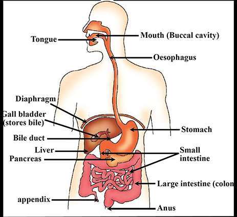
The important respiratory organs in living beings include- lungs gills trachea and skin. The diagram below shows the structure and functions of the human digestive system. The lymphatic system helps keep the body healthy by eliminating infections and diseases.
A well labeled human heart diagram given in this article will help you to understand its parts and functions. The human respiratory system consists of a pair of lungs and a series of air passages leading to the lungs. Digestion of food starts in the mouth.
With the help of a well labelled diagram explain how the accumulation of toxic substances increases as we move up the food chain. Name the process by which ammonia is excreted by fish. Draw a well labelled diagram of human alimentary canal and label the following parts.
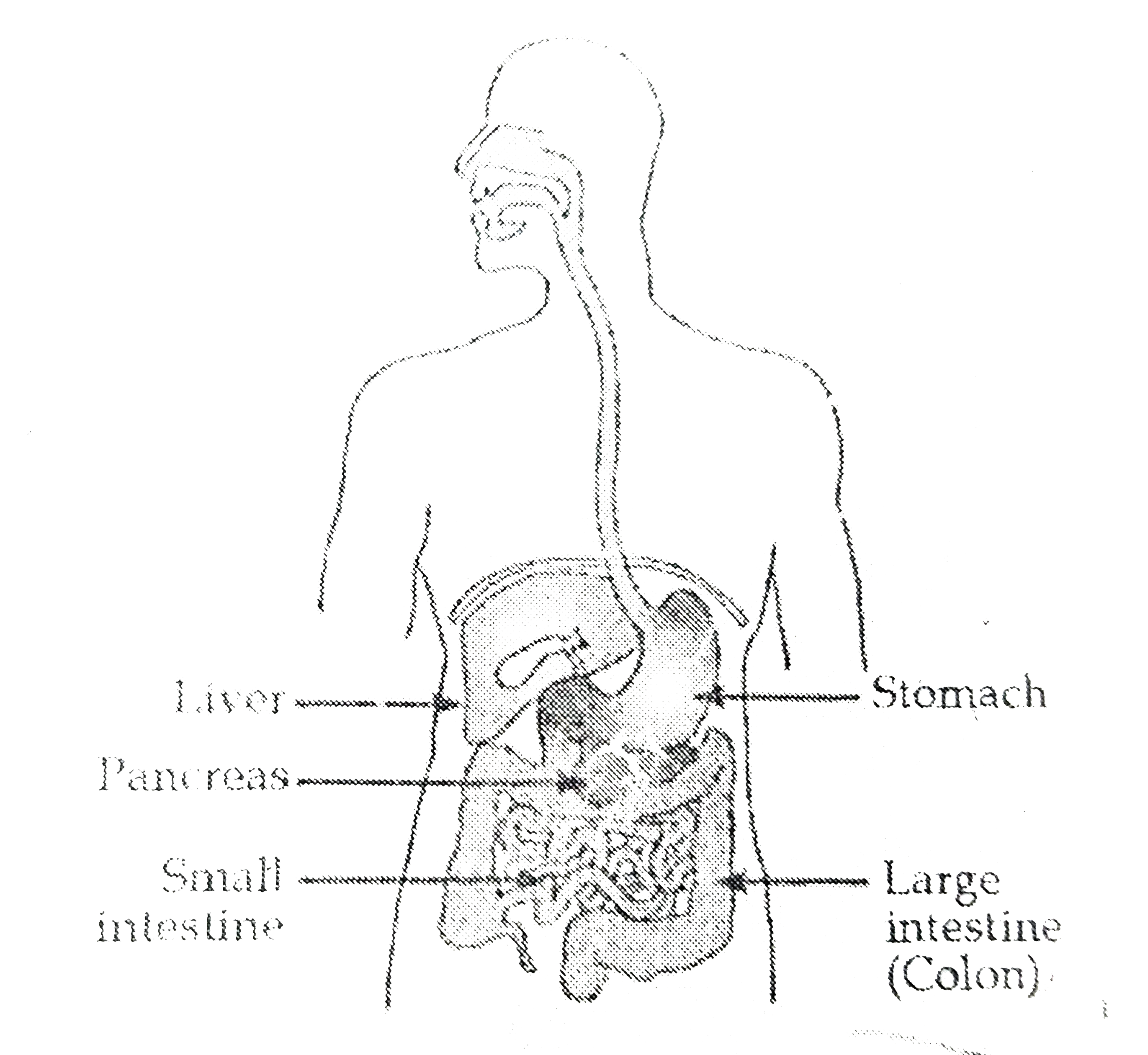
ILabel the parts numbered 1 to 4 in the diagram. Asked Jan 7 2019 in Class X Science by aditya23 -2137 points the excretory system. I Liver ii Pancreas iii Small intestine iv Large intestine.
View Answer Bookmark Now. Name the major forms of nitrogenous wastes which are excreted by animals. I production of female gamete ovum and ii secretion of female hormones estrogen and progesterone.
It is one among the few important topics which are repetitively asked in the board examinations. A Draw a labelled diagram of the respiratory system of human beings with diaphragm at the end of expiration. The diagram shows the Excretory System of a Human being.
The human respiratory system is a system of organs responsible for inhaling oxygen and exhaling carbon dioxide in humans. Labeled Diagram of the Human Kidney. I Digestion in mouth.
Here the food is. A Given below is a diagram of the lateral section of the testis of a man. Draw a diagram of.
B List four conditions required for efficient gas exchange in an organism. Draw a well-labeled diagram of the human excretory system. Ii State the functions of the parts labelled 1 and 3.
With the help of this diagram describe the process of digestion of food in man humans. Name any two classes of uricotelic animals of the phylum Chordata. The human kidneys house millions of tiny filtration units called nephrons which enable our body to retain the vital nutrients and excrete the unwanted or excess molecules as well as metabolic wastes from the body.
Following is the process of digestion of food in a human being. 1 answer a Draw a diagram depicting Human Alimentary Canal and label on it. Gall bladder Liver and Pancreas.
All major organs of the body like brain heart stomach kidney liver etc work in. The ovaries perform dual function of. Draw the well-labelled diagram of the human excretory system.
Draw a labelled diagram of the human digestive system. The diagram of the human digestive system is useful for both Class 10 and 12. State any two effects each of soil pollution on human beings and environment.
Study the same and answer the questions which follow. Name the nitrogenous waste product which requires a large amount of water for its elimination. 3 store urine until it is passed out.
Human eye is nearly a spherical structure. Structure Of Human Eye With Diagram Human Body Draw A Neat And Labelled Diagram Of Structure Of T Toppr Com Human Eye Anatomy Quiz.
With The Help Of A Neat And Labelled Diagram Describe The Anatomy Of Human Eye Explain The Mechanism Of Vision Sarthaks Econnect Largest Online Education Community
Working of Human eye human eye consists of various parts which help us in seeing the objects the function of various parts are.
Neat and labelled diagram of human eye. It is an opaque tissue that is usually known as white of the eye. Draw a neat labelled diagram of a normal human eye. It is the transparent membrane which refracts the light entering our eye.
The lacrimal gland is situated in the orbit on the superior lateral surface of the eyeball. Draw a Neat and Labelled Diagram of Structure of the Human Eye. Describe its parts briefly.
Human eye in humans specialized sense organ capable of receiving visual images which are then carried to the brain. Structure of Human Eye. The iris the colored part of the eye.
Human eye consists of various parts which helps us in seeing the objects the function of various parts are. For today we compile some pictures of human eye labelling parts and each of them giving you some fresh ideas. Draw a neat labelled diagram of a normal human eye.
The lacrimal apparatus of each eye consists of a lacrimal gland and its numerous ducts the superior and inferior canaliculi a lacrimal sac and a nasolacrimal duct. Eye In Cross Section Anatomy The Eyes Have It. Draw a neat labelled diagram to show the structure of the human eye.
Aryansharma99047 aryansharma99047 07112020 Chemistry Primary School answered Draw a neat and labelled diagram of human eye 2 See. Draw a neat labelled diagram to show the structure of the human eye. The cornea is a thin membrane which forms a transparent bulge on the surface of the eye ball.
Anatomically the eye comprises two components fused into. Eye Sketeh and label anatomical structure of human eye Draw the neat and labelled diagram of the anatomy of human eye. The front transparent part of the sclera is called cornea.
Advertisement Remove all ads. It is the transparent membrane which refracts the light entering our eye. Light enters the eye through the cornea.
Human eye labelling parts labelled diagram anatomy hd cross section of the eye diagram eyeball labeled human omtex classes draw a neat labelled diagram of normal human eye eye diagram labeled system eyeball. Iris controls the size of pupil. Cross section Question २७ असर २०७८ आइतवर 11 Jul 2021 Sun.
Describe its parts briefly. Cross section Question १२ सउन २०७८ मङगलवर 27 Jul 2021 Tue. Human Eye Diagram Labeled Labelled Diagram Of Human Eye Human Eye 1000 Human Eye Diagram Stock Images Photos Vectors Shutterstock.
Draw a neat labeled diagram of human eye and explain the working of each part of it. NCERT DC Pandey Sunil Batra HC Verma Pradeep Errorless. Explain the mechanism of vision.
Human eye in humans specialized sense organ capable of receiving visual images which are then carried to the brain. This simple introduction the subjects of the eye and visual optics includes a simple diagram of the eye together with definitions of the parts of the eye labelled in the illustration. It is composed of sclera and cornea.
It allows the light entering our eye. With The Help Of A Neat And Labelled Diagram Describe The Anatomy Of Human Eye. Draw a neat and labelled diagram of the structure of the human eye.
Maharashtra State Board SSC English Medium 10th Standard Board Exam. With the help of a neat and labelled diagram describe the anatomy of human eye. Human Eye Labeled Diagram Of The Eye Written By JupiterZ Saturday July 1 2017 Add Comment Edit Understanding The Different Parts Of Your Eye All About Eyes.
NCERT P Bahadur IIT. Working of Human eye. The human eye is a roughly spherical organ responsible for perceiving visual stimuli.
Maximum refraction of light rays entering the eye takes place. I Outer layer. Draw labelled diagrams of the following.
A human eye is roughly 23 cm in diameter and is almost a spherical ball filled with some fluid. Draw a neat and labelled diagram of human eye Get the answers you need now. It consists of the following parts.
Its wall is formed of three layers- sclera choroid and retina. It is the outer covering a protective tough white layer called the sclera white part of the eye. It is composed of a dense connective tissue.
Draw A Diagram Of The Human Eye As Seen In A Vertical Section And Label The Parts Which Suits The Following Descriptions Relating To The Studyrankersonline. It is enclosed within the eye sockets in the skull and is anchored down by muscles within the sockets. A problem with the curve of your cornea.
Structure And Functions Of Human Eye With Labelled Diagram. Anatomy of Human eye.
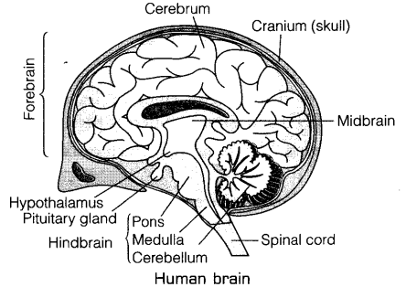
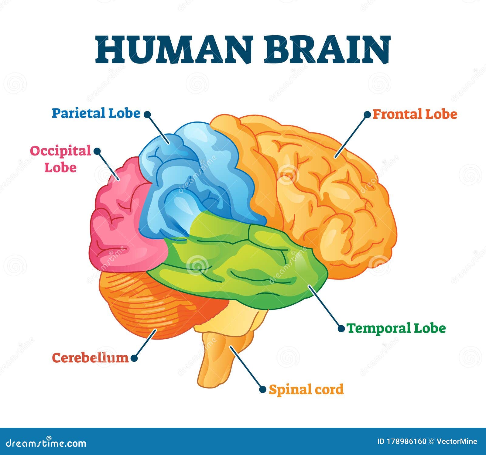
The frontal lobe is located in the forward part of the brain extending back to a fissure known as the central sulcus. It is the anterior part of the brain.
Draw A Labelled Diagram Of Human Brain And Mention The Functions Cbse Class 10 Science Learn Cbse Forum
Consisting of largely of three paired structures the thalamus hypothalamus and epithalamus the diencephalon plays a vital role in integrating conscious and unconscious sensory information and motor commands.
Labeled parts of human brain. The forebrain the midbrain and the hindbrain are the three main parts of the brain. The brainstem is made of three regions. The three regions of the hindbrain coordinates.
The parietal lobe takes care of sensation and perception managing how we react to sensory input. Midbrain Pons and Medulla oblongata. Nervous System - Label the Brain.
The forebrain is responsible for a number of functions related to thinking perceiving and evaluating sensory information. The forebrain is the anterior part of the brain which comprises the cerebral hemispheres the thalamus and the hypothalamus. See labeled brain anatomy stock video clips of 27 brain diagram with labels hypothalamus vector brain diagram pons cerebrum and cerebellum brain pons brain anatomy amygdala brain labelled amygdala brain human midbrain diagram pons.
It also consists of two subdivisions called the telencephalon and diencephalon. 2 frontal occipital parietal temporal. The medulla oblongata the pons and the midbrain.
Choose the correct names for the parts of the brain. The cerebrum the largest part handles vision movement hearing language and touch. The brain is a 3-pound organ that contains more than 100 billion neurons and many specialized areas.
The forebrain has two major parts called the diencephalon and the telencephalon. There are 3 main parts of the brain include the cerebrum cerebellum and brain stemThe Cerebrum can also be divided into 4 lobes. Lobes of the Brain The four lobes of the brain are the frontal parietal temporal and occipital lobes Figure 4.
Surrounded by the cerebral hemispheres the diencephalon forms the central core of the brain. Smallest and central part of the brain. It has the following parts.
The temporal lobes are involved with memory and hearing. Consisting of or divided into three major partsmedulla pons and midbrainit serves to develop the connection of spinal cord with the cerebellum and cerebrum. Frontal lobes parietal lobes temporal lobes and occipital lobesThe brain stem consists of three major parts.
The brain and its parts can be divided into three main categories. It helps the body survive by controlling the basic functions like breathing digesting food sleeping as well as heart rate. Which performs thinking reasoning speech intelligence and usage of information.
A net-like structure of mixed gray and white matter known as the reticular. The brain is surrounded by a layer of tissue called the meninges. The lower part of the brain.
The brain stem is located in front of the cerebellum and connects to the spinal cord. The part of the brain that makes up the spinal cord is called the brain stem. Where lobes are responsible for detecting the smell from different receptors.
Parts of Human Brain Forebrain Largest part of the brain. The forebrain midbrain and hindbrain. Many of the most basic survival functions of the brain are controlled by the brainstem.
The brain is so complex that even to simply discuss it certain distinctions have to be made regarding its structure and for that reason scientists divide the brain up into three major portions with each of these portions divided into many subregions and structures. It consists of three major parts. 1 cerebrum cerebellum brain stem spinal cord.
The occipital lobes contain the brains visual processing system. This science quiz game will help you learn the 7 parts of the brain. The human brain is the central organ of the human nervous system and with the spinal cord makes up the central nervous systemThe brain consists of the cerebrum the brainstem and the cerebellumIt controls most of the activities of the body processing integrating and coordinating the information it receives from the sense organs and making decisions as to the instructions sent to the.
The main portions of the brain are the cerebrum the cerebellum and the brainstem. 3 frontal occipital parietal temporal. The brainstem is the place from which ten of the twelve cranial nerves originate.
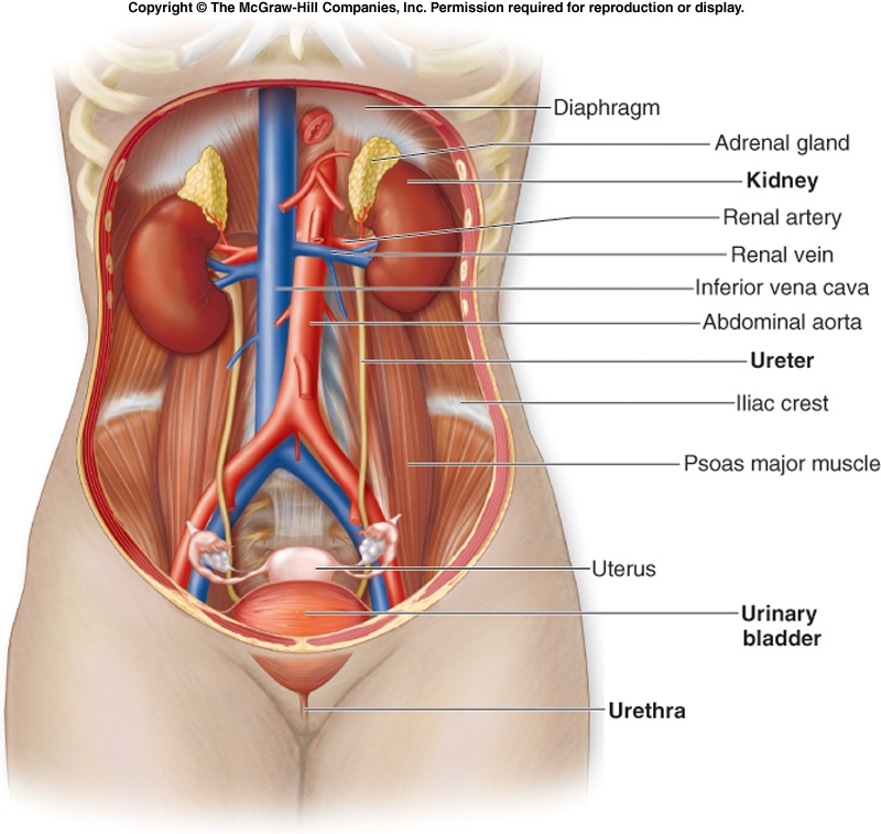
The right kidney is placed slightly lower- than the left due to the presence of liver which occupies much space on the right side. The kidneys come in a pair much like the lungs.
Associate Degree Nursing Physiology Review
The kidneys are located on either side of spine.

Where is the kidney located in the human body diagram. The kidneys are bean-shaped organs about 11 cm x 7 cm x 3 cm that are located against the back muscles in the upper abdominal area. They lie on the posterior abdominal wall. Your kidneys are bean-shaped organs that are located in the middle of your back against the back muscles with one on either side of your spine.
Left kidney is located a bit higher than your right kidney because the right kidney is below the liver. Both kidneys are located on opposite sides. They are beneath the rib cage with one kidney on either side of the spine.
Labeled Diagram of the Human Kidney. Kidney consist of two parts - Outer cortex and Inner medulla. The kidneys are two small organs that are located near the vertebral column at the posterior or the back portion of the body.
The kidneys are a pair of bean-shaped organs on either side of your spine below your ribs and behind your belly. Located toward the rear of the body the kidneys are a pair of organs that cleanse blood and regulate water levels in the body. Each kidney is approximately three vertebrae in length.
The kidneys are responsible for cleaning the body and removing waste products from it as well as regulating blood pressure. Our LATEST youtube film is ready to run. They typically extend from T12 to L3 although the right kidney is often situated slightly lower due to the presence of the liver.
The kidneys are some of the most important organs. They are usually reddish brown and are situated between the levels of last thoracic and third lumbar vertebra with a strong attachment to the dorsal inner wall of the lower abdominal cavity. The two kidn ey lie on the posterior wall of the abdomen outside the peritoneal cavity.
In this image you will find rib cage diaphragm esophagus adrenal gland kidney renal artery renal vein inferior vena cava aorta pelvis ureter bladder prostate gland urethra in kidney location bladder location in the human body. Pain in your upper abdomen or back and sides is also called flank pain or kidney pain. Most people are born with two kidneys which are bean-shaped organs on each side of the middle back.
Each kidney is about four or five inches long. Location of Kidneys and Anatomy. It contains the glomerulus and convoluted tubules.
Each kidney is about 4 or 5 inches long roughly the size of a large fist. An average kidney weighs around 120 170g in dependent on the ideal health of the human. The renal cortex is surrounded on its outer edges by the renal capsule a layer of fatty tissue.
The right kidney however sits a little lower. In the human body the kidneys are located vertically in the middle of the abdomen near the back wall. Other mammals also have kidneys in the rear abdominal area.
The primary function of the kidney is to extract water accompanied with other constituents from the blood. The kidney is a filter for the blood and works to remove waste materials. The kidneys lie retroperitoneally behind the peritoneum in the abdomen either side of the vertebral column.
Kidney Location Bladder Location In Human Body. They sit opposite each other on both the left and right side of the body. Waste matter is extricated from the system in the form of urine.
The human kidneys house millions of tiny filtration units called nephrons which enable our body to retain the vital nutrients and excrete the unwanted or excess molecules as well as metabolic wastes from the body. In the medical field kidney pain is usually called flank pain because it is noted most in the middle back or flank area. The renal cortex is the outer part of the kidney.
The kidneys are two in number which are situated one on each side of the verteral column and in-front of the last ribs. Kidneys filter waste out of the bloodstream and maintain the bodys level of water and minerals. The kidneys lie retroperitoneally behind the peritoneum in the abdomen either side of the vertebral column.
Your kidneys provide a vital function for your body to filter blood and produce urine. However the one of the left is placed a little higher compared to the one on the right. The kidneys are shaped like beans and are found right below the rib cage on both sides of the body.
In addition they also play an important role in maintaining the water balance of our body. Location of Kidneys. Every healthy human body has two kidneys the left and the right.
Human alimentary canal Biology Notes for IGCSE 2014. Find an answer to your question A Draw a neat and labelled diagram of a human alimentary canalB Label the following glands shown in the figureمسز.
A Draw A Diagram Of Human Alimentary Canal And Label The Following Parts I Largest Gland Ii Gland That Secretes Digestive Enzymes And Hormone Iii Part Where Hcl Is Produced Iv Part
Draw a well labelled diagram of human digestive system and explain the diges tive process.
Draw a neat labelled diagram of human alimentary canal and explain its associated glands. Draw Labelled Diagram Of Alimentary Canal - Fun for my own blog on this occasion I will explain to you in connection with Draw Labelled Diagram Of Alimentary CanalSo if you want to get great shots related to Draw Labelled Diagram Of Alimentary Canal just click on the. Ii gland that secretes digestive enzymes and hormone. Ii gland that secretes digestive enzymes and hormone.
Draw The Diagram Of Alimentary Canal Of Man And Label The. B What are villi. The saliva has salivary amylase or ptyalin enzyme which acts on the starch and converts it into maltose sugar.
Explain its mechanism. I Liver ii Pancreas iii Small intestine iv Large intestine. 6 1 4 Draw and label a diagram of the digestive system CHAPTER 23 THE DIGESTIVE SYSTEM HUMAN ANATOMY APRIL 28TH 2018 - THE DIGESTIVE SYSTEM IS YOU WILL BE PRESENTED WITH THE UNIQUE CONCEPT THAT FOOD INSIDE THE ALIMENTARY CANAL.
Iv part where digested food is absorbed. Explain their function in. Describe the alimentary canal of a man Freeshiksha.
Ii Part in which bile is stored. Iv Part in which water is absorbed. A Draw a diagram depicting Human Alimentary Canal and label on it.
Human digestive system comprises the alimentary canal and various digestive glands. Organs of the Alimentary Canal. The digestive system plays a significant role in the digestion process which is composed of the alimentary canal and other associated glands.
Food is crushed and cut in the mouth with the help of teeth and is mixed with saliva that is secreted by three salivary glands one below the tongue and two at the side of the jaw to make it wet and slippery this process is known as mastication. The Mouth and Oral cavity. C Name the organ which performs the following functions in humans.
Explain their function in the digestive system. Iii Part where HCl is produced. Iv Part where digested food is absorbed.
Ii State the role of liver and pancreas. A Draw a diagram of human alimentary canal and label the following parts. Iv part where digested food is absorbed.
Iii Part where HCl is produced. The partially digested food reaches the stomach where the food gets mixed with gastric juice secreted by gastric glands. Draw a labelled diagram of the alimentary canal of a.
A Draw a well labelled diagram of human alimentary canal and label the following parts. It is one among the few important topics which are repetitively asked in the. B State the roles of Liver and Pancreas.
Explain the process of digestion of food in mouth stomach and small intestine in human body. The digestive system graphic organiser Digestion Human. Ii Saliva contains amylase that breaks down complex carbohydrates and the tongue helps in pushing.
Draw a diagram of human alimentary canal and label the following parts1. B What are villi. B What are villi.
The various processes involved in the digestion of human. Iii Name the organs which perform the following functions in humans. These glands secrete intestinal juice.
Draw and label the alimentary canal in human beings. A Roach Gut Organizational Biology Other Thoughts. Mouth Oesophagus Stomach Intestine Or How do carbohydrates proteins and fats get digested in human beings.
Layers of the Alimentary Canal Tutorial Sophia Learning. The gastric juice contains large quantities of mucus HCl and pepsin. There are numerous small glands in the walls of the small intestine.
Iii Part in which nutrients are absorbed. Log in to add comment. Draw the diagram of alimentary canal of man and label the following parts.
B Mention the role of hydrochloric acid in the stomach. B What is peristaltic movement. The alimentary canal is a muscular tube which extends from the mouth to the anus.
The complete digestion of the remaining food material takes place in the ileum. I Draw a diagram depicting human alimentary canal and label the components gall bladder liver and pancreas in it. The alimentary canal in human beings measures about 8 to 10 meters in length.
The structure and functions of these organs are discussed below. Largest gland2gland that secret digestive enzymes and hormones3part where HCL produced4part where digestive food is absorbed. I Absorption of digested food ii Absorption of water.
Ask questions doubts problems and we will help you. A Draw diagram of human alimentary canal and label the following. The digestive enzymes in the intestinal juice break small peptides into amino acids disaccharides into monosaccharides lipids into fatty acids and glycerol and nucleic acids into nucleotides.
Draw the diagram of alimentary canal of man and label the following part. C What function is served by the following. Step by step video text image solution for a Draw a diagram of human alimentary canal and label the following parts.
The diagram of the human digestive system is useful for both Class 10 and 12. The main organs of the alimentary canal are. The alimentary canal is divided into five main parts- mouth esophagus stomach small intestine small intestine and lastly large intestine.
BECE JUNIOR HIGH SCIENCE SYLLABUS GHANA. A Draw a well labelled diagram of human alimentary canal and label the following parts. OR Draw a neat labelled diagram of digestive.
Labelled Diagram Of Human Alimentary Canal PDF Download. I Part in which starch digestion starts. Draw a neat and labelled diagram of human alimentary canal.
Gall bladder Liver and Pancreas. Iii Part where HCl is produced. File Digestive system diagram en svg Wikipedia.
B The digestion of carbohydrates starch starts in the mouth. How to Draw Human Digestive System Drawing for kids YouTube. Draw a neat labelled diagram of the human alimentary canal.