Simple Microscope Parts Functions Diagram And Labelling. Photo Compound Microscope With Labels Image 3850568.

How To Draw Compound Of Microscope Easily Step By Step Youtube
Compound Microscope Parts Made Easy.

Easy labelled diagram of microscope. Microscope Diagram Easy To Draw Microscope Labeled Diagram Binocular Microscope Sketch At Paintingvalley Com Explore Labeling Microscope Worksheet Teaching Resources Comparing And Contrasting The Different Parts Of The Microscope Free Microscope Drawing Download Free Clip Art Free Clip Art On The Compound Microscope Microscope Structure Uses Functioning Processes Of Simple Microscope. It is useful in various fields of sciences such as physical and biological science nanotechnology metallurgy and forensic analysis. An easy and convenient way to make label is to generate some ideas first.
All you will need is a pencil an eraser and a sheet of paper. Basic Microscope Diagram Schematic Diagrams. Tsetse Biology Systematics And Distribution Techniques.
Compound Microscope With Label Clipart. The optical parts of a simple microscope are centered on the specimen lighting and magnification. Microscope Clipart Part Microscope Part Transparent Free For.
The real size of the cell shown above is 005 mm 50 μm. It looks like a magnifying glass because it has the ability to magnify between 200 and 300 times. You should use the Course adjustment only to focus the microscope.
Easy Simple Compound Microscope Diagram. They are as follows. You should make a label that represents your brand and creativity at the same time you shouldnt forget the main purpose of the label.
You should always start on the lowest power objective lens and should always leave the microscope on the low power lens when you finish using it. In a book a photomicrograph of the cell measured 100 mm. Microscope With Labels Clip Art At Clker Com Vector Clip Art.
Labeled Easy Compound Microscope Diagram Wiring Schematic Simple Microscope Drawing At Getdrawings Com Free For Blank Microscope Diagram Optics Binoculars Drawing Diagram Illustration Of Compound Microscope With Labels Microscope Diagram Labeled Unlabeled And Blank Parts Of A Free Microscope Drawing Download Free Clip Art Free Clip Free Microscope Drawing. Despite the fact that they are rudimentary imaging devices simple microscope. Easy Labelled Diagram Of Compound Microscope Written By MacPride Thursday July 13 2017 Add Comment Edit.
Easy Well Labelled Diagram Of Compound Microscope Written By MacPride Sunday November 4 2018 Add Comment Edit. Diagram of the simple scanning electron microscope incorporating a carbon- fibre field-emitterelectron source. If you wish to examine the subject being studied at a different.
Online Label The Microscope Quiz Microscope Parts Biology. Compound Microscope Drawing At Paintingvalley Com. It is a type of optical microscope that uses visible light and lens to magnify objects.
Binocular Microscope Sketch At Paintingvalley Com Explore. It allows the use of different types of objective lenses by simply rotating the top part of the turret. Pin By Microscope World On Science Activities With Microscopes.
Microscope Structure Uses Functioning Processes Of Simple. Easy Labeled Diagram Of Compound Microscope Written By JupiterZ Thursday March 14 2019 Add Comment Edit. You should always carry a microscope with two hands one on the arm and the other under the base.
Microscope With Labels Clip Art At Clker Com Vector Clip Art. The image above is a confocal microscope. Text magnification frac text 100.
Unlock AD FREE and PRINTABLE drawing and coloring tutorials. Mirror You can distinguish a simple microscope from other microscopes because of its mirror. The Microscope Image courtesy of.
Parts Of Compound Microscope Botany. Compound microscope important questions for class 12 physics cbse optical light microscopy clipart simple labeled microscope diagram fluorescence microscope bacillus subtilis oral cavity 18. The image above is a stereo microscope.
A simple microscope works on the principle that when a tiny object is placed within its focus a virtual erect and magnified image of the object is formed at the least distance of distinct vision from the eye held close to. Diagram microscope labeled simple photos compound studiootb. Simple Microscope Diagram Parts labelled Principle Formula and Uses.
Would you like to draw a cartoon microscope. It is the revolving part of the microscope. To calculate the magnification.
This easy step-by-step cartoon object drawing guide is here to show you how. It has a plano-convex mirror that focuses the surrounding light on the subject being studied. Diagram Of Microscope With Full Label Diagram Labels Label Gallery Get some ideas to make labels for bottles jars packages products boxes or classroom activities for free.
Microscope Diagram Labeled Unlabeled And Blank Parts Of A. Microscope labeled diagram 1. Simple Microscope Parts Functions Diagram And Labelling.
If you liked this tutorial see also the following drawing guides. 1 2 3 and 4 Picture 1. I will show you How to draw compound of microscope easily - step by step Please watch carefully and try this okayThanks for watchingmicroscopedrawi.
By Editorial Team Last updated on July 1 2021 Dating back to the 14th century simple microscope is the most basic of the various microscopes available. Cartoon Scientist Glasses and Magnifying glass. Kostenlose Labeled Diagram of Microscope ClipArt in AI SVG EPS und CDR Finden Sie auch verzierte buchstaben des alphabets oder diagramm der menschlichen anatomie Clipart kostenlose Bilder unter 73061 Vektoren.
Parts of dissecting microscope botany. The body tube part of a microscope is where the ray of light is bent to allow the object being viewed to enlarge by the scope.
Once you find your worksheet s you can either click on the pop-out icon or download button to print. Light and electron microscopes.
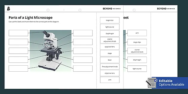
Parts Of A Light Microscope Cut And Stick Worksheet Twinkl
Write the letter on the line that represents each part of the microscope.

Label microscope diagram worksheet. Head This is also known as the body it carries the optical parts in the upper part of the microscope. Free microscope parts Oct some of the worksheets displayed are microscope lab microscope mania label parts of the microscope answers parts of the light microscope name the microscope. Can be rotated to change MAGNIFICATION.
Photo that shows the parts of the microscope from a photo can be used to grade microscope labeling worksheetMicroscope Labeling. Power 10 x 4 40 Power 10 x 10 100 Power 10 x 40 400 What happens as the power of magnification increases. Some of the worksheets for this concept are Labeling scientific tools microscope name Parts of the light microscope Labeling scientific tools microscope name Label parts of the microscope The microscope parts and use Label parts of the microscope answers Powerpoint work the microscope diagram.
Report this resource to let us know if it violates our terms and conditions. Each microscope layout both empty and the version with answers is available as a PDF download. When using the high power objective only the _____ knob should be used.
There are three structural parts of the microscope ie. Posted in Worksheet August 13 2020 by Kimberly R. Some of the worksheets displayed are The microscope parts and use Parts of the light microscope Powerpoint work the microscope diagram Label parts of the microscope answers Parts of the microscope quiz Parts of the microscope study An introduction to the compound microscope Plant and animal cells.
Arm - this attaches the eyepiece and body tube to the base. Microscope labeling worksheet answer key. NOSEPIECE microscope when carried Holds the HIGH- and LOW- power objective LENSES.
Body tube - the tube that supports the eyepiece. A straightforward worksheet in which students are required to identify the parts of a basic microscope. Microscope Diagram - Displaying top 8 worksheets found for this concept.
This activity has been designed for use in homes and schools. You should carry. Head base and arm.
Some of the worksheets for this concept are The microscope parts and use Parts of the light microscope Powerpoint work the microscope diagram Label parts of the microscope answers Parts of the microscope quiz Parts of the microscope study An introduction to the compound microscope. Coarse focus adjustment - a knob that makes large adjustments to the focus. It also carriers the microscopic illuminators.
You can use the word bank below to fill in the blanks or cut and paste the words at the bottom. This Pin was discovered by. 12 arm base body tube coarse adjustment knob diaphragm fine adjustment knob light source nosepiece objective lenses ocular lens stage stage clips.
Diagram of parts of a microscope. The type of microscope used in most science classes is the _____ microscope. Microscope Diagram Grade 8 - Free Printable Tests and Worksheets.
Using the terms listed below label the microscope diagram. When focusing a specimen you should always start with the _____ objective. Labeling the Parts of the Microscope.
Label microscope diagram worksheet This activity is designed for use in households and schools. Answer key net start class manualzz com. Microscope diagram worksheet cells and microscope worksheet and label microscope parts worksheet are some main.
Each microscope layout both blank and the version with answers are available as PDF downloads. Tes classic free licence. Label the parts of the microscope.
When we talk concerning Microscope Labeling Worksheet weve collected several similar images to complete your references. Here you can take a closer look at each part of the microscope. Label Microscope Diagram.
Base It acts as microscopes support. Can be used for practice or as a quizMicroscope Labeling KEY. Microsoft word microscope basics quiz basic doc.
Students label the parts of the microscope in this photo of a basic laboratory light microscope. Microscope G Stage D. Label the parts of the microscope.
Showing top 8 worksheets in the category - Microscope Diagram. Grade level course 7 life science lesson unit plan name labeling microscope worksheet by beci w teaching resources tes the microscope create a labelled diagram by ineedtoteachthat the microscope create a labelled diagram by ineedtoteachthat. How can a microscope help us study living things.
Some of the worksheets below are Parts and Function of a Microscope Worksheets with colorful charts and diagrams to help students familiarize with the parts of the microscope along with several important questions and activities with answers. On Labeling The Microscope - Displaying top 8 worksheets found for this concept. Base - this supports the microscope.
Label parts of the Microscope.
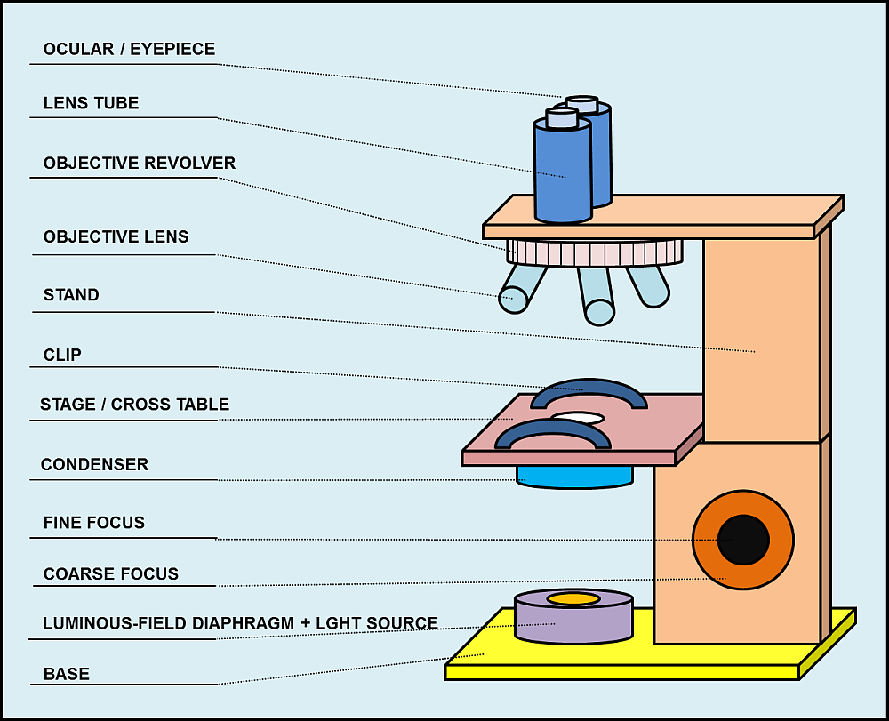
First the purpose of a microscope is to magnify a small object or to magnify the fine details of a. The parts of a compound microscope are of two categories as given below the eyepiece is a drum which fits loosely into the draw tube.

Parts Of A Microscope With Functions And Labeled Diagram
It is the topmost part of the microscope.
Draw and label the parts of a light microscope. 872018 2 Simple. 1 2 3 and 4 Let us take a look at the different parts of microscopes and their respective functions. Draw and label a light microscope.
Draw and label the different parts of a light microscopeIN THE PARTSpe post navel hirte in the wine The RealistaAmState Cupan Am KasTaphragmEyepieceFine Adjustment nobHigh Power CrveStat201aFigure T. Similar to a magnifying glass and has only one lens. Microscopy from www2nauedu.
It is a u shaped structure and supports the entire weight of the compound microscope. This forms the illuminator or light source of the microscope. Before exploring microscope parts and functions you should probably understand that the compound light microscope is more complicated than just a microscope with more than one lens.
Easy and simple step by step tutorial for beginnersThanks for Watching and Subscribing Minutes DrawM. Observe the skin cells under both low and high power of your microscope. Base As the name suggests the base is the lowest portion on which the whole structure of the microscope rests.
How to use a microscope. How to draw microscope slides. Animal cells under a light microscope.
How to draw a microscope diagram. BASE Supports the MICROSCOPE D. Light Microscope With Labeled Parts Written By MacPride Thursday October 22 2020 Add Comment Edit.
STAGE CLIPS HOLD the slide in place C. Microscope drawing and label at. Use the diagram to show how the microscope works ie.
Viewing Cells Using The Compound Light Microscope Stains Ppt. Draw and label the parts of the microscope. Light Microscope Drawing At Getdrawings Free Download.
Optical parts a mechanical parts of a compound microscope. COARSE ADJUSTMENT KNOB Moves the stage up and down for FOCUSING I. Remember to subscribe to my channel and turn on notification.
1 14 13 2 12 3 11 4 10 5 an 8 6 7. Draw it in a paper and label each part numbered 1-14. A light is needed to shine on the object and then reflected by the mirror into the lenses hence causing greater magnification.
Arm The arm connects the body tube to the base. Stereoscopic Microscope Gives a three dimensional. Draw your own light microscope and label the parts.
Parts of the Light Microscope T. FINE ADJUSTMENT KNOB Moves the stage slightly to SHARPEN the image G. Microscope Parts and Functions With Labeled Diagram and Functions How does a Compound Microscope Work.
Draw a cylinder at the base of the arm. Draw and label the parts of microscope. Body Tube It is the part of the microscope that holds the eyepiece.
Through the eyepiece you can visualize the object being studied. It also carriers the microscopic illuminators. Use straight lines to enclose a rectangular shape around the bottom of the microscope.
Microscope drawing and label at getdrawings com free for. Add notes to the diagram. Use straight parallel vertical lines for its sides and curved lines for its top and bottom.
Parts Of A Microscope With Functions And Labeled Diagram. Https Ntrs Nasa Gov Archive Nasa Casi Ntrs Nasa Gov 20170000349 Pdf. Microscope Parts and Functions Microscope One or more lenses that makes an enlarged image of an object.
872018 3 Compound Microscope Lets light pass through an object and then through two or more lenses. Microscope drawing - step 9. Label the parts of the compound light microscope in the figure below and then draw the path of light from the illuminator to your eye.
All the best Microscope Drawing And Label 33 collected on this page. EYEPIECE Contains the OCULAR lens J. The user must hold this part in order to move the microscope from one place to another.
Microscope diagram with labels and functions. Through the eyepiece you can visualize the object being studied.

Microscope Diagram Labeled Unlabeled And Blank Parts Of A Microscope
This part rotates to change which objective lens is active.

Labelled diagram of a microscope with functions. 872018 3 Compound Microscope. Compound Microscope Types Parts Diagram Functions And Uses. You should always carry a microscope with.
By Uncategorized 0 Comments. Its magnification capacity ranges between 10 and 15 times. It is the topmost part of the microscope.
Objects that are not visible to the human eyes. Bottom base of the microscope that houses the illumination supports the compound microscope. Labelled Diagram Of A Microscope And Their Functions - Fun for my own blog on this occasion I will explain to you in connection with Labelled Diagram Of A Microscope And Their Functions.
Electron microscope parts and functions. In a book a photomicrograph of the cell measured 100 mm. Give A Well Labelled Diagram Of Compound Microscope Using Of.
Microscopy is the science of investigating small objects and structures using. Label the microscope worksheet activities par. To calculate the magnification.
So if you want to get great shots related to Labelled Diagram Of A Microscope And Their Functions just click on the save icon to save the photo to your computer. Microscope Parts and Functions Microscope One or more lenses that makes an enlarged image of an object. The parts of a microscope labeled printable this diagram labels and explains the function of each part of a microscope.
Its actually not a toy microscope its. Microscope diagram with labels and functions. The Microscope Image courtesy of.
Parts of the optical parts are as follows. You should always carry a microscope with two hands one on the arm and the other under the base. Supports the microscope head and attaches it to the base.
Ncert Class 9 Science Lab Manual Slide Of Onion Peel And Cheek. This particular image Parts Of A Compound Microscope With Diagram And Functions with Diagram Of The Microscope Parts earlier mentioned is actually labelled along with. Its actually not a toy microscope its.
Text magnification frac text 100. 872018 2 Simple Compound Stereoscopic Electron Simple Microscope Similar to a magnifying glass and has only one lens. Published by simply CARPNY TEAM at May 7 2016.
Mirror A simple microscope has a plano-convex mirror and its primary function is to focus the surrounding light on the object being examined. You should always start on the lowest power objective lens and should always leave the microscope on the low power lens. The real size of the cell shown above is 005 mm 50 μm.
Diagram of microscope parts and function diagram of the microscope parts labeled diagram of microscope parts. Microbiology 3510l Dustman Flashcards Lecture 1. Microscopy is the science of investigating small objects and structures using.
Parts And Components Of Light Microscopes Light Microscope. Labelled diagram of compound microscope. Weve gathered our favorite ideas for Microscope Slide Diagram Explore our list of popular images of Microscope Slide Diagram Photos Collection with high resolution.
A Study Of The Microscope And Its Functions With A Labeled Diagram. Parts Of The Microscope And Their Functions. Microscope Labelled Diagram And Functions Written By JupiterZ Tuesday December 18 2018 Add Comment Edit.
What Is The Function Of The Pointer On A Microscope. Electron microscope parts and functions. Holds the objective lenses attaches them to the microscope head.
Labelled Diagram Of A Microscope Optics Amp Binoculars Exe Microscope Side Vector Drawing With Parts Labelled Free Svg. Parts Of A Compound Microscope With Diagram And Functions Microscope Labeling News Information Microscope Labeling Biology. Compound Microscope Diagram Labelled.
Let us take a look at the different parts of microscopes and their respective functions. Lens The biconvex lens is placed above the stage and its function is. Shop compound microscopes compound microscope definitions for.
Microscope labeled diagram 1.
All the best Microscope Drawing And Label 33 collected on this page. Can be rotated to change MAGNIFICATION.
Free Microscope Drawing Download Free Microscope Drawing Png Images Free Cliparts On Clipart Library

Human eye functions on the same principal as that of a bright-field or light microscope ie one can see objects because of the light reflected by them.

Draw a labelled diagram of a light microscope. NOSEPIECE microscope when carried Holds the HIGH- and LOW- power objective LENSES. The following labeled drawings must be completed. ProductsServices for Labelled Diagram Of A Light Microscope.
Make your work easier by using a label. Used to examine material mounted on microscope slides usually thinly sectioned stained Provides total magnification of 40x-1000x. Draw A Large Diagram.
Observe the cheek cells under both low and high power of your microscope. I will show you How to draw compound of microscope easily - step by step Please watch carefully and try this okayThanks for watchingmicroscopedrawi. LIGHT SOURCE Projects light UPWARDS through the diaphragm the SPECIMEN and the LENSES H.
Represent by means of a labelled accurate diagram or graph using a pencil. DIAPHRAGM Regulates the amount of LIGHT on the specimen E. Anything closer than this distance will appear like a.
Labels are a means of identifying a product or container through a piece of fabric paper metal or plastic film onto which information about them is. Microscopes - 704 companies. Compound Light Microscope Drawing.
No space for dissection. STAGE Supports the SLIDE being viewed K. Write any one expression for its magnifying power.
Draw cell structures as seen with the light microscope. Always start at 4x Coarse focus Fine focus. Onion Cell drawing.
Record the microscope images using labelled diagrams or produce digital images. Draw a diagram of one cheek cell and label the parts. ARM Used to SUPPORT the B.
It was first invented in the early 1600s. Draw and label a diagram showing a transverse section of the ileum as seen under a light microscope. Thank you watching more videosplease subscribe my channel.
Get 10 free Shutterstock images - PICK10FREE. The type of microscope in our drawing guide is known as a compound microscope an optical microscope or a light microscope. A ruler straight edge should be used for straight lines.
Ray diagram of a compound microscopeWhen the final image is formed at the least distance of distinct visionFor the image formed at infinity ue feand By making focal length of the objective small the magnifying power can be increased. Draw a labelled diagram of an image formed by a compound microscope with the image at least distance of distinct vision. When first examining cells or tissues with low power draw an image at this stage even if going on to examine the.
Diagrams should be drawn to scale. Wide collections of all kinds of labels pictures online. The human eye has a resolving power about 025 mm in the sense two dots situated 025 mm or more apart can only be seen as two dots.
You should observe the cell membrane nucleus and cytoplasm Observation. Isolation of Halobacterium salinarum retrieved directly from halite brine inclusions. The word microscope comes from ancient Greek words meaning small and to see Microscopes are used to investigate small objects that are too little to be seen with the naked eye.
A Well Labelled Diagram Of A Light Microscope. Graphs should have points correctly plotted if appropriate and joined in a straight line or smooth curve. Drawings MUST be completed neatly using a pencilcolored pencil.