Well-Labelled Diagram of Animal Cell The cell membrane is a double-layered membrane made up of phospholipids that surrounds the entire cell. Labelled cell surface membrane with carrier proteins phospholipid bilayer etc.

Structure Of A Plant Cell Labelled Diagram Of The Structure Of A Plant Cell Canstock
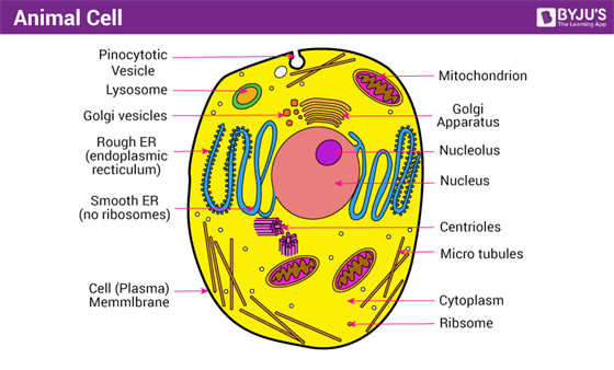
Diagram of the human cell illustrating the different parts of the cell.

Labelled diagram of a cell. About the Well Labelled Diagram of Animal Cell A Well Labelled Diagram of Animal Cell. Cytoskeleton is the. To draw a well labelled diagram of an animal cell the cell membrane has to be drawn.
In a plant cell the cell wall is made up of cellulose hemicellulose and proteins while in a fungal cell it is composed of chitin. Printable plant and animal cell labelled diagram of a generalised. Plant cells are eukaryotic cells but unlike animal cells which have a cell membrane plant cells have cell walls.
Labelled diagram - Drag and drop the pins to their correct place on the image. The cell membrane is semipermeable and flexible. Labelled diagram of a plant cell under microscope posted on march 18 2011 by admin onion cells stained with methylene blue look at the images of onion cells as they would be seen under a microscope draw each magnification label appear high picture plant and animal cell diagrams.
The Rate Of Red Blood Cell Labeling Using 55 Co Dtpa At 37 C N 5. A Labeled Diagram of the Plant Cell and Functions of its Organelles. It is evident from the introduction that an animal cell is a representation of.
Labelled Diagram Definitions and Structure Published by Admin on July 28 2021 July 28 2021. Labelled xylem cell diagram. Dr alban March 17 2021 Diagram No Comments.
Eukaryotes are sophisticated cells with a well defined nucleus and cell organelles. White Blood Cell Diagram Labeled Beautiful White Blood Cell Diagram. Answer verified by Toppr Upvote 0.
Cytosol is the fluid present within a cell that is made up of water and ions such as potassium proteins and small. During cellular division plant cells grow a new cell wall down the middle separating one cell into two. Unicellular to multicellular in nature and evolved 1 billion years ago.
Cell Membrane The cell membrane is the outer coating of the cell and contains the cytoplasm substances within it and the organelle. A cell wall is multilayered with a middle lamina a primary cell wall and a secondary cell wall. The cell is the basic unit of structure function and organization in all organisms.
These cells reproduce both asexually and sexually. Middle lamina contains polysaccharides that provide adhesion and allows binding of the cells to one another. Structure of Plant Cells.
The red blood cells those that you can see in the cell diagram are the most common type of blood cells. Labelled diagram of a cell wall. A level standard labelled diagram of an animal cell with structure and function of all organelles.
The significant differences between plant and animal cells are also shown and the diagrams are. It is a rigid covering made up of cellulose which a complex substance is providing structural support to the plants. Unlike xylem phloem vessels contain cytoplasm and this goes through the holes in the sieve plates from one cell to the next.
The synthesis of cell wall in controlled by golgi bodies. The cell being the smallest unit of life is akin to a tiny room which houses several organs. Animal Cell Diagram Labeled And Functions Inspirational Animal Cell.
Here lets study the plant cell. Plant Cell Diagram 9th Class Labeled. A nucleosome is a basic unit of DNA packaging in the nucleus of a eukaryote cell which consisting of a segment of DNA wound in sequence around histone protein cores.
Plant Cell Diagram 9th Class. Structure in a plant cell the cell wall is made up of cellulose hemicellulose and proteins while in a fungal cell it is composed of chitin. The most important structures of plant and animal cells are shown in the diagrams below which provide a clear illustration of how much these cells have in common.
It is a double-layered membrane composed of proteins and lipids. How to draw diagram of Animal Cell easily - step by step - YouTube. Gross structure of cell wall.
The cells are comparatively larger in size 10-100 μm. We are aware that all life stems from a single cell and that the cell is the most basic unit of all living organisms.
With the help of a clean spatula or a toothpick the inner side of the cheek is gently scrapped. They are found in the brain spinal cord and the peripheral nerves.

Solved Draw A Labelled Diagram Of Human Cheek Cells 3 Marks Self Study 365
Name the parts labelled 1 2 and 3.

Draw a labelled diagram of human cell. Name the stage prior to this stage and draw a diagram to represent the same. Label any three parts and write their functions. Labelled diagram of plant and animal cell are as follow---Both plant and animal cells belong to eukaryotic cells.
The cell membrane is semipermeable and flexible. Eukaryotes are sophisticated cells with a well defined nucleus and cell organelles. Unicellular to multicellular in nature and evolved 1 billion years ago.
This structure contains enzymes used for penetrating the female egg. It is a double-layered membrane composed of proteins and lipids. Identify the above stage and give a reason to support your answer.
The middle piece has a central filamentous core with many mitochondria. The cell organelles are membrane bound present within the cells. The sperm cells are the haploid gametes which are produced in the male.
462 378 pixels. Draw a diagram of the microscopic structure of human sperm. Diagram of the human cell illustrating the different parts of the cell.
Size of this PNG preview of this SVG file. FileDiagram human cell nucleussvg. The above diagram is of the sperm cell.
The structure of a neuron varies with their shape and size and it mainly depends upon their functions and their location. V Egg is the largest human cell. Mention the type of cells in our body where this type of cell division occurs.
A neuron is also known as the nerve cell. These cells reproduce both asexually and sexually. Contains the genetic material that the sperm has to pass on a haploid genome because it contains only one copy of each chromosome.
Vi Ovaries are located lower abdomen. There is the genetic material which is a haploid genome because it contains only one copy of each chromosome. The scrapped material is transferred into a drop of water and taken on a clean slide.
Draw A Labelled Diagram Of A Animal Cell And Plant Cell Ncert. State the functions of the two parts labelled. Draw a Neat and Labelled Diagram of the Human Ear.
With the Help of this Diagram Explain the Construction and Working of the Human Ear. Label the following parts in it and write their functions. This is a file from the Wikimedia Commons.
Fallopian tubes i Two thin tubes attached to the upper sides of uterus ii Tubes terminate near the ovaries but are not attached iii Fimbriae are finger-like structures on the end of each tube. Posted by Unknown at 2209. A neuron is a specialized cell primarily involved in transmitting information through electrical and chemical signals.
Solution For Draw a well labelled diagram of eye. Draw a diagram of a mature human sperm. The cells are comparatively larger in size 10-100 μm.
There are different parts of the sperm cell. 293 240 pixels 587 480 pixels 733 600 pixels 939 768 pixels 1252 1024 pixels 2503 2048 pixels. Information from its description page there is shown below.
Iii In what two ways is mitotic division in an animal cell different from the mitotic division in a plant cell. Lakhmir Singh Science Class 8 Solutions Chapter 8 Cell Structure And. Cell Membrane The cell membrane is the outer coating of the cell and contains the cytoplasm substances within it and the organelle.
It contains enzymes used for penetrating the female egg. 1 left and 1 on the right. With the help of a needle the material is uniformly spread.
Eukaryotic cells are one which have organised nucleus with a nuclear membrane and genetic material is organised into chromosomes. It is the inner layer and consists of three sub layers- ganglional cells bipolar cells and photoreceptor cellsThe innermost ganglionic cells give rise to optic nerve fibre that forms optic nerve in each eye and is connected with the brain. Draw a labelled diagram of the embryonic stage that gets implanted in the human uterus.
Human Cell Diagram Parts Pictures Structure and Functions parts of a human cell Diagram of the human cell illustrating the different parts of the cell.
Structure Of Bacterial Cell With Diagram. Plant Cells Vs Animal Cells With Diagrams Owlcation.
Bacterial Cell Structure And Function Youtube
Personalized AI Tutor and Adaptive Time TableSelf Study MaterialUnlimited Mock Tests and Personalized Analysis Reports24x7 Doubt Chat Support.
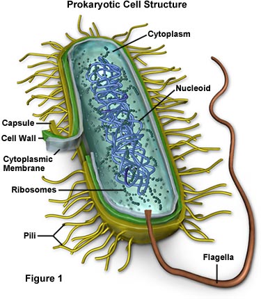
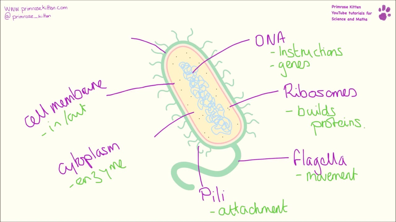
Simple labelled diagram of a bacterial cell. Cell Structure of Bacteria With Diagram. Just under the rigid cell wall is the more fluid cell membrane. This means they do not have a nucleus or any other structures which are surrounded by membranes.
It ranges in thickness around 002µ. A bacterial cell fig. Cheek Cell Diagram Labeled Simple Wiring Diagram Images Gallery.
Bacterial Cell Structure And Function Youtube. It shows the cytoplasm nucleoid cell membrane cell wall mitochondria which bacteria lack plasmids flagella and cell capsule. File Simple Diagram Of Bacterium En Svg Wikimedia Commons It shows the cytoplasm nucleoid cell membrane cell wall mitochondria which bacteria lack.
There are different types of bacteria with various sizes shapes and structures. Bacteria cell diagram simple. Bacteria Diagram Photograph By Monica Schroeder.
The single circular double-stranded chromosome is the bacterial genome. LPS teichoic acid etc surrounding the bacterium like a shell and lies external to the cytoplasmic membrane. Posting Lebih Baru Posting Lama Beranda.
Bacteria Cell Labelling Chromosomal Dna Plasmid Dna Flagellum. Every cell consists of an intricate system of different structures which all work together to allow the cell to function. Structure Of Bacteria With Diagram Microbiology.
The cell wall Fig. It lacks all membrane bound cell organelles such as mitochondria lysosome golgi endoplasmic reticulum chloroplast peroxisome glyoxysome and true vacuole. The structure of bacteria is known for its simple body design.
Draw It Neat How To Draw Bacteria. Other structures include cytoplasmic membrane mesosomes. The bacterial cell with a capcule normally does not bear flagella.
A simple diagram of a bacterium labelled in english. A bacterial cell fig. A simple diagram of a bacterium labelled in english.
Labels are a means of identifying a product or container through a piece of fabric paper metal or plastic film onto which information about them is printed. Plant and bacterial cell walls provide structure and protection. Review For Cell Test.
Gram Positive Vs Gram Negative Bacteria. File Simple Diagram Of Bacterium En Svg Wikimedia Commons. Membrane Structure Cell Structure Structure And Function Plasma Membrane Cell Membrane Extracellular Fluid College Physics Cell Biology Health.
Gonorrhea Bacteria Diagram Prokaryotic Cell Cell Lacking Wiring. File Simple Diagram Of Bacterium En Svg Wikimedia Commons Bacteria cell wall prevents osmotic lysis. It gives shape to the cell.
By Asegraf 25 Dec 2019 Post a Comment. The cell wall is tough though flexible. Interactive Bacteria Cell Model.
NEET Foundation Knockout NEET 2025 Easy Installment Personalized AI Tutor and Adaptive Time TableSelf Study MaterialUnlimited Mock Tests and Personalized Analysis. Structure Plant Cell Animal Bacteria Cells Prokaryotes Eukaryotes. The cytoplasm is enclosed by three layers the outermost slime or capsule the middle cell wall.
Even though viruses are smaller than bacteria viruses are not living cells. Labeled Simple Bacterial Cell Diagram Written By JupiterZ Sunday January 19 2020 Add Comment Edit. Structure simple bacterial cell diagram.
It is 10-25 nm in thickness. Bacteria are all single-celled. Start studying Labeling a cell membrane.
It shows granular structure and lacks microfibrils. Hence bacteria are the smallest of living cells. Belum ada Komentar untuk Simple Bacteria Diagram Labeled Posting Komentar.
The information can be in the form of hand-written or printed text or symbols and gives details about manufacturers name source of product shelf-life uses and the manner of disposal. Cheek Cell Diagram Labeled Simple Wiring Diagram Images Gallery. Labeled Simple Bacterial Cell Diagram File Simple Diagram Of Bacterium En Svg Wikimedia Commons Structure Of Bacterial Cell With Diagram 1000 Bacteria Cell Diagram Stock Images Photos Vectors Bacteria Lessons Tes Teach Bacterial Cell Structure And Function Youtube Draw It Neat How To Draw Bacteria Bacteria Cell Structure Youtube Collection Prokaryotic Cell Diagram Labeled.
The sizes of bacteria cells that can infect humans beings. It is a tough and rigid structure of peptidoglycan with accessory specific materials eg. Cell Membrane Drawing Labeled.
In the electron micrograph the cell wall is seen as a thin sharply defined envelope around the protoplast. A simple diagram of a bacterium labelled in English. The inert and somewhat rigid bacterial cell.
It shows the cytoplasm nucleoid cell membrane cell wall mitochondria which bacteria lack plasmids flagella and cell capsule. A simple diagram of a bacterium labelled in english. File Simple Diagram Of Virus En Svg Wikimedia Commons.
File Virus Stucture Simple. Unique Characteristics Of Prokaryotic Cells Microbiology. Labeled Diagram Simple Virus Cell.
Simple Bacterial Cell Diagram Labeled Ditulis JupiterZ Jumat 01 Januari 2021 Tulis Komentar Edit. The cytoplasm is enclosed by three layers the outermost slime or capsule the middle cell. Cheek Cell Diagram Labeled Simple Wiring Diagram Images Gallery.
Bacteria cells are the smallest living cells that are known. The cells are all prokaryotic. Supports and strengthens cell.
Herpes Simplex Virus Structure And Function Study Com. The types of bacteria and their labeled diagrams are shown below. File Simple Diagram Of Yeast Cell En Svg Wikimedia Commons.
Prokaryotic Cell Parts Functions Diagram. Hello Friends in this video I tell you about how to draw labelled diagram of bacterial cell step by step for beginners in easy wayso friends if you have pro. Larger bacterial cells may be.
Simple Labelled Bacterial Cell Diagram Written By JupiterZ Friday April 19 2019 Add Comment Edit. Clip Art Bacteria Labeled Diagram Simple Bacteria Cell. A bacteria diagram in actual fact helps us to learn extra about this single cell organisms that have neither membrane-bounded nucleolus or organelles like mitochondria.
Bacteria are single-celled microorganisms with prokaryotic cells which are single cells that do not have organelles or a true nucleus and are This diagram depicts the numerous shapes of bacteria.
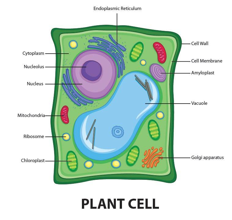
Draw a neat diagram of plant cell and label any three parts which differentiate it from animal cell. Get Draw A Neat Labelled Diagram Of Plant Cell And Label Its Parts BackgroundThe nitrates absorbed by the plant roots are converted into various types of amino acids about 20 types which sequences respectively to form polypeptide chains of various protein molecules in the ribosomes.
Draw A Welllabelled Diagram Of A Plant Cell Class 11 Biology Cbse
Answered Feb 6 2020 by KumariJuly 536k points selected Feb 6 2020 by.
Neat labelled diagram of plant cell class 11. How to draw animal cell labelled diagram Animal cell diagram for class 910 and 11 - YouTube. Compare The Location Of Nucleus In Animal Cell And Plant Cell Draw A. Draw a labelled diagram of a plant cell.
Labelled diagram of a plant cell. Find an answer to your question 1 Draw a neat labelled diagram of plant cell and list the function of each organalle. Energy is produced in the form of ATP in the process.
Compare Plant cell and Animal cell 4 Why Animal cell do not have cell wall for class 8 plz answer fast. The diagram given below represents a plant cell after being placed in a strong sugar solution. Link of our facebook page is given in sidebar.
If playback doesnt begin. Photosynthesis occurs in the chloroplasts of the plant cell. Draw a neat labelled diagram of animal cell.
Define plant cell and animal cell. Draw A Neat Diagram Of Plant Cell And Label Any Three Parts Which. ExplanationDiagram of plant cell and thier three parts Diagram of plant cell and thier three parts.
Study the diagram and answer the questions that follow. Answer verified by Toppr. Photosynthesis is the major function performed by plant cells.
Get Instant Solutions 24x7. 1 Answer 1 vote. Aaryansharmasml aaryansharmasml 20112020 English Secondary School answered Draw a neat labelled diagram of a plant cell.
The membrane is selectively permeable and allows only certain molecules to pass through. It is the process of preparing food by the plants by utilizing sunlight carbon dioxide and water. Ncert Solutions For Class 8 Science Chapter 8 Cell Structure And.
Cells under the microscope. Share It On Facebook Twitter Email. Rajubasak49041 rajubasak49041 28042020 Biology.
Class 11 Oscillations Redox. Cell Structure And Functions Class 11 Notes Biology Mycbseguide. Draw a labelled diagram of a plant cell.
Draw a neat diagram of the structure of chromosome and label the parts. A Centromere b p-arm. Plant and animal cell diagram class 8 NCERTHi friends In this video we will learn how to draw diagram of plant and animal cell this diagram is.
Draw a neat labelled diagram of the following cell organelles and explain their structure. Draw a neat and labelled diagram of a Plant cell and animal cell. It is very thin delicate elastic and selectively permeable membrane.
Class-11 Welcome to Sarthaks eConnect. Further plant cells are green in color due to the presence of special pigments that aid in photosynthesis. 13 Plant Cell Diagram Class 9Th PicturesThe class students can check below the diagram of plant cell and animal cell which can help them in understanding how to draw a cell diagram concept.
1st lab exam material biology 440 with dr. The plant cell is rectangular and comparatively larger than the animal cell. The cell membrane is a double-layered membrane made up of phospholipids that surrounds the entire cell.
Plant cells kept in hypertonic solution will get. Draw a neat diagram of plant cell and label any three parts which differentiate it form animal cell. The unit of life.
A Mitochondri b Centrioles. Students upto class 102 preparing for All Government Exams CBSE Board Exam ICSE Board Exam State Board Exam JEE MainsAdvance and NEET can ask questions from any subject and get quick answers by subject teachers. Draw a neat labelled diagram of an animal cell.
Class 8th 9th 10th 11th and 12th can use this diagram. Upvote 0 Was this answer helpful. 1 Draw the concept Map for the Cell 2.
How to draw a animal cell easy and step by step. Cytosol is the fluid present within a cell that is made up of water and. A unique platform where students can interact with teachersexpertsstudents to get solutions to their queries.
The cell wall is made up of cellulose which provides support to the plant. It is responsible for coordinating many of the important cellular activities such as protein synthesis cell division growth and a host of other important functions.
A Well Labelled Diagram Of Animal Cell With Explanation
Class 9 ScienceHome Page.

Labelled diagram of eukaryotic cell class 9. Draw well-labeled diagrams of any one. They are present in eukaryotic cell but absent in prokaryotic cells. A Draw a neat and labelled diagram of a prokaryotic cell.
Question Bank Solutions 7801. 9th Grade Prokaryotic Cell Diagram Class 9. Genetic material enclosed in nuclear envelope Genetic material not enclosed in nuclear envelope 3 Nucleolus and one or more chromosomes present.
Topic 1 2 Ultra Structure Of Cells Amazing World Of Science With. Viruses are smaller than prokaryotic cells. Name the kingdom in which they are placed.
Example plant cellanimal cell. Explain the structure and function of the nucleus. V Draw a labelled diagram of a prokaryotic cell.
These neat and well labelled diagram will ma. Draw a neat diagram of plant cell and label any three parts which differentiate it from animal cell. B Differentiate between a prokaryotic and eukaryotic cell any 4 points of difference.
A diagram of an eukaryotic nucleus is as given. Prokaryotic Cells Types Of Prokaryotic Cells Cell. B Differentiate between plant cell and animal cell.
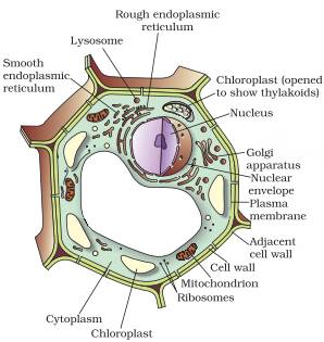
It is responsible for storing the cells hereditary material or the DNA. Compare and contrast distinguishing characteristics of all these cell types only on the basis of diagrams. A well labeled diagram of mitochondria Return to CBSE Class 9 Chapter Wise Notes Structure and Fundamental Unit of Life - Cell Posted on February 14 2018 by Manoj Saxena Posted in No Comments A well-labelled diagram of mitochondria is given below for your better understanding of the structure.
A Well Labelled Diagram Of Animal Cell With Explanation. Significantly bigger than the prokaryotic cells eukaryotic cells have diameter ranging from 10µm -100µm. Start studying 9th grade.
A labeled diagram of the plant cell and functions of its organelles we are aware that all life stems from a single cell and that the cell is the most basic unit of all living organisms. Chioroplast and a large centrally placed vacuole. You can draw flowcharts wherever necessary.
Genetic material in the form of single chromosome. Example plant cellanimal cell. Eukaryotic cells are exclusively found in plants animals fungi protozoa and other complex organisms.
Draw a labelled diagram of mitochondria. Long Answer Type 1. Advertisement Remove all ads.
Found in eukaryotic cell. Neither of the two. Explain the structure and functions of the chloroplast.
The examples of eukaryotic cells are mentioned below. Draw a well labelled diagram of mitochondria class 9. Cell wall is the non-living protective layer outside the plasma membrane in the plant cells bacteria fungi and algae.
ICSE Class 9 Biology Sample Question Paper 6 with Answers. They are the building block or smallest unit of life of organisms as simple as amoeba and protozoa to the most complicated plants and animals. Test your Knowledge on Nucleus -.
2018 in Class IX Science by saurav24 Expert 14k points the fundamental unit of life 1 vote. Found in prokaryotic cell. Draw a labelled diagram of a animal cell and Plant cell.
Genetic material in the form of single chromosome. Genetic material enclosed in nuclear envelope Genetic material not enclosed in nuclear envelope 3 Nucleolus and one or more chromosomes present. Prokaryotic and Eukaryotic Cell.
CBSE CBSE English Medium Class 9. Download free cbse sample paper for class 9 biology. How can you differentiate between prokaryotic cell and eukaryotic cell.
Draw a well labelled diagram of typical prokaryotic cell. Inside it are various cell organelles which performs individual functions. Write the functions of mitochondria.
8th Grade Science Class. Discuss the main components of a typical cell. Class 9 Cell Basic Unit of Life Long Questions.
Large collection of high quality biology pictures photos images illustrations diagrams and posters on marine biology cell biology microbiology. Animal Cell Diagram For Class 10In pairs discuss the different organs in. 4 Cell divides by mitosis and meiosis.
A bacteria diagram basically facilitates us to profit more approximately this single cell organisms that have neither membrane. The nucleus has 2 primary functions. Important Science Diagrams From All Chapters For CBSE Class 8.
Draw a neat diagram of plant cell and label any three parts which differentiate it from animal cell. Eukaryotic cell are the developed advanced and complex forms of cells. Ncert Solutions For Class 9 Science Chapter 5 The Fundamental Unit.
Comparison of chart of all class mates can be done. Found in eukaryotic cell. Draw a neat well-labelled diagram of an animal cell.
Well Labelled Diagram Of An Animal Cell. Sketch Plant Cell Diagram For Class 9. Draw a labelled diagram of mitochondria.
A list any two structural differences and two similarities between animal cell and plant cell. A Draw a well labelled diagram of a plant cell and label any 4 parts. 4 Cell divides by mitosis and meiosis.
Prokaryotic and Eukaryotic Cell video tutorial 000715. It initiates and regulates cell division. Draw a neat labelled diagram of an animal cell.
I It is plant cell as it contains cell wall. Asked Feb 5. Prokaryotic And Eukaryotic Cells Read Biology Ck 12 Foundation.
Found in prokaryotic cell. Also Read Different between Plant Cell and Animal Cell Though this animal cell diagram is not representative of any one particular type of cell it provides insight into the primary organelles and the intricate internal structure of most animal cells.