The Cell The Biology Primer. Find Human Cell Diagram stock images in HD and millions of other royalty-free stock photos illustrations and vectors in the Shutterstock collection.

Draw A Neat Diagram Of A Typical Animal Cell And Label The Following Organelles 1 The Organelle Called Science Cell Structure And Functions 10429465 Meritnation Com
Diagram of the human cell illustrating the different parts of the cell.

Labelled diagram of typical cell. Comparison of Prokaryotic Cells and Eukaryotic Cells and 2. In prokaryotic cell there is naked DNA without histone protein but in Eukaryotic cell there is DNA with histone protein. However it typically exists in three forms ie.
LPS teichoic acid etc surrounding the bacterium like a shell and lies external to the cytoplasmic membrane. It is a tough and rigid structure of peptidoglycan with accessory specific materials eg. You can also put your logo at the top or bottom corner of the label.
Structure and Functions With Diagram Let us make an in-depth study of the structure and functions of cell. Stock vector labelled diagrams of typical animal and plant cells with editable layers 222613513 in cell diagram simple the structure and contents of a typical animal cell every has membrane cytoplasm nucleus but not all cells have rat liver cell a scheme of the typical cell membrane structure with lipid bilayer integral proteins. Cell is a compartment where all the activities of life takes place.
Targeting Strategies for Multifunctional Nanoparticles in. A typical bacterial cell. Structure of a typical animal cell click to enlarge.
Prokaryotic cells article Khan Academy. Label Gallery Get some ideas to make labels for bottles jars packages products boxes or classroom activities for free. The Golgi Apparatus is the cell organelle mostly present in eukaryotic cells which is responsible for the packaging of macromolecules into vesicles so that they can be sent out to their site of action.
A typical bacterial cell - Labelled diagram free-floating DNA plasmid circular ring of DNA slime capsule stops the cell from drying out flagellum helps the bacterium move through liquids cell membrane and cell wall the label was too big to tell these apart. The lipid molecules on the outer and inner part lipid bilayer. Which biological activities will be hindered and why.
Most of the times we put the labels to show some specific information. Illustration of Labelled diagrams of typical animal and plant cells with editable layers. On the contrary plant cells lack centrioles and intermediate filaments which are present in animal cells.
Examining a diagram of the plant cell will help make the differences clearer. Labelled diagram of a plant cell under microscope posted on march 18 2011 by admin onion cells stained with methylene blue look at the images of onion cells as they would be seen under a microscope draw each magnification label appear high picture plant and animal cell diagrams. It is a double-layered membrane composed of proteins and lipids.
Labelled Diagram Of A Typical Animal Cell. In prokaryotic cell DNA is circular but in Eukaryotic cell DNA is double helix. Vector art clipart and stock vectors.
A Draw a fully labelled diagram of a typical receptor tyrosine cell surface recepto b Name the other classes of enzyme coupled receptors and describe their mechanism of action. Thousands of new high-quality pictures added every day. Labeled diagram of a typical plant cell.
Cisternae vesicles and tubules. Typical Animal Cell Diagram Labeled Best Of Typical Animal And Plant Animal Cell Coloring Page Lovely 25 Best Typical Plant Cell Ideas On Click The Animal Cell Diagram To Enlarge It Wiring Diagram Blog Eukaryotic Cells Biology I Typical Animal Cell Plant And Animal Cells S Cool The Revision Website Animal Cell Anatomy Diagram Structure With All Parts Nucleus Cells Alive Plant Cell. Typical Plant Cell Diagram Labeled Battery electricity Wikipedia.
1in prokaryotic cell there is primitive nucleus but in Eukaryotic cell there is well developed nucleus. After reading this article you will learn about. Room Full of Crazy TV Tropes.
Western Wood Products Association. A Draw an annotated diagram of a typical student microscope b Explain the. Unit 2 Cells Biology Junction.
The single circular double-stranded chromosome is the bacterial genome. An easy and convenient way to make label is to generate some ideas first. Cell Membrane The cell membrane is the outer coating of the cell and contains the cytoplasm substances within it and the organelle.
Labels are usually small in size so you should carefully choose the font of the texts to make sure it is readable. C Taxol from the Yew tree stabilises microtubules. Hello Guys In this Video Im going to teach you guys How to Draw Structure of a cell Labeled DiagramSubscribe to the Channel and also share this video.
Other structures include cytoplasmic membrane mesosomes. The structure of the Golgi Complex is pleomorphic. Labelled diagram of a typical animal cell.
INLINE PICTURES VERSION W1TP. Intro to eukaryotic cells article Khan Academy. It gives shape to the cell.
It is 10-25 nm in thickness. Structure and Components of a Human Cell. Human Cell Diagram Stock Vector Royalty Free 400272997.
It is brown to pale-grey in colour and measures 215-3 cm x 12-15 cm. This is the well labelled diagram of liver fluke.

How To Draw Liverfluke Fasciola Hepatica Easy Way Step By Step Youtube
In these organs they produce pathological lesions leading to parasitic diseases.
Labelled diagram of a liver fluke. Acoelomate a labeled diagram of a fluke labelled diagram of liver draw it a generalized life cycle of the liver fluke albendazole oral liver flukes troccap fluke flatworm britannica com liver flukes xanadufarms free download here pdfsdocuments2 com a well label diagram of a liver inner. The anterior body part is broader than the posterior part which is blunt in outline. Infections in humans usually occur after eating contaminated raw or undercooked freshwater fish or watercress.
Diagram Of Liver Fluke. Liver flukes are a group of about fifteen different species of parasites that share a number of characteristics during their life cycle and result in a similar etiology when they infect humans. Liver fluke disease is a chronic parasitic disease of the bile ducts.
The liver is a very hardworking organ that filters more than a liter of blood per minute. Lungworm and liver fluke to threaten livestock this autumn. Liver flukes are an important cause of acute and chronic disease in grazing sheep and cattle.
Add your answer and earn points. Liver fluke disease fasciolosis is caused by the trematode parasite Fasciola hepatica. Disease can result from the migration of large numbers of immature flukes through the liver or from the presence of adult flukes in the bile ducts or both.
Your risk of infection increases if you travel to parts of the world where the. Labeled Diagram Of Liver Fluke. Suggested in case of lung flukes.
The mouth of liver fluke is anterior and terminal surrounded by. Some selected sexual organs concerning with the passages of the spermatozoa and the eggs were obseved in detail. How to draw a liver fluke in exam is.
Draw A Neat Labelled Diagram Of Liver Fluke State The Phylum To Which It Belongs And Brainly In Fluke eggs which are passed in the faeces control of liver fluke disease should be an important part of a farm health plan drawn up with the witholding period information is carried on the product label and datasheet and advice should be. The conclusion of this study is that the Laurers canal may be the copulatory organ of the female reproductive system as Miyazaki et al. Fasciola hepatica also known as the common liver fluke or sheep liver fluke is a parasitic trematode fluke or flatworm a type of helminth of the class Trematoda phylum Platyhelminthes.
It may reach a size of 3 cm in length and 15 cm in breadth. They are principally parasites of the liver of various mammals including humans. Liver fluke can infect all grazing animals and man but mainly affects sheep and cattle.
640x480 well labeled diagram of liver fluke labelled diagram of liverLiver fluke in sheep also known as. A well-labeled pencil sketch diagram of liver fluke - 21306762 olubimioye olubimioye 22082020 Biology Secondary School answered A well-labeled pencil sketch diagram of liver fluke 1 See answer olubimioye is waiting for your help. Liver cell cardiac cell nerve cell skin cell.
640x480 well labeled diagram of liver fluke labelled diagram of liver. This is the well labelled diagram of liver fluke. Liver Fluke Labelled Diagram çšå¾çæ œççæžœ Chart Map Map Screenshot.
Body of liver fluke is soft flattened leaf-like with a triangular head lobe Fig. Draw A Neat Labelled Diagram Of Liver Fluke. Liver fluke is a collective name of a polyphyletic group of parasitic trematodes under the phylum Platyhelminthes.
In this article we will discuss about the external morphology of liver flukes. It is dorsoventrally flattened oval in shape like a leaf and faint brownish in colour. Free ncert and other textbook solutions.
From wikipedia the free encyclopedia. A related parasite fasciola. They are principally parasites of the liver of various mammals including humans.
For example the liver makes and processes many body fats. Draw a neat labelled diagram of Liver fluke State the phylum to which it belongs and its - 34158509. Fluke infection is estimated to cost the uk agriculture industry about 300 million a year.
Combination with levamisole have a specific. It infects the livers of various mammals including humans and is transmitted by sheep and cattle to humans the world over. A labeled diagram of a fluke labelled diagram of.
The body is covered with a cuticle the greater portion of which bears minute spines. While most infected persons do not show any symptoms infections that last a long. Capable of moving along the blood circulation they can occur also in bile ducts gallbladder and liver parenchyma.
Diagram of Liver Fluke How To Draw Liver Fluke Labelled Diagram Biology Diagram - YouTube. A labeled diagram of a fluke liver fluke anatomy gallery human anatomy learning. Diagram Of Liver Fluke With Label.
Diagram Of Liver Fluke With Label. 231 draw and label a diagram of the ultrastructure of a liver cell as an example of an animal cell. Beranda Diagram Of Liver With Labelling.
Liver fluke is a collective name of a polyphyletic group of parasitic trematodes under the phylum platyhelminthes. Anatomy of the human pancreas explained with labeled diagrams. Structure of Liver Fluke.
In this article we will discuss about the external morphology of liver flukes. Vector illustration in flat style isolated over white background.
Diagnostic Characters of Rosaceae Floral Formula and Floral Diagra. Learners will 1 identify the different parts of a flower and understand their function.
Dissect A Flower Scientific American
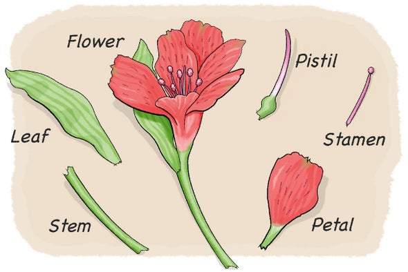
Parts of a Flower Diagram All the Flower Parts are Labeled Plant Parts and Their Function Including Diagrams.
Labelled diagram of rose flower. You will sometimes see the term quartered especially in reference to old garden roses. This fain lb has a great economic importance for mankind. Filament - the filament is the part of the flower that holds the anther and part of the stamen the male reproductive organs of the plant.
Looking at a diagram of a flower you can easily see the individual parts of the flower. In green rose the petals are leaf-like in structure and green in colour. Parts of a hibiscus flower complete flower fiower part sepal plant ovary flower anatomy diagram of flower parts of a flower flower diagram parts of the flower.
Refer children to the flower diagram chart. Floral formula and floral diagram Rose plant Economic Importance. Apr 26 2016 - Most plants in the same family have similar characteristics.
How to Identify Six Plant Families Using Their Flowers Parts of a flower Flower structure Diagram of a flower. Each member of corolla is called a petal. Rose family General characters floral formula floral diagram economic importance and common species January 31 2019 Angiosperm families Botany Rosaceae Rose family Rosaceae Rose family In this article we will discuss about.
The androecium is the male reproductive whorl of the flower. In cultivated roses many stamens gradually change into petals. Explain that each flower is unique with its own special beauty.
Answer verified by Toppr. It has great importance in temperate cold region. In Canna the stamens and the style become petaloid.
While flowers are composed of the same Location. See flower parts diagram stock video clips. What is the overhead for each department.
A rose with more than 25 petals in three or more rows is called double. Fresh flowers 1000 square feet. The five hairy red spots shown below is a close-up view of the tip or stigma female part of the flower.
Download a powerpoint showing labelled and unlabelled versions of these diagrams both parts of a plant and parts of a flower from the link on the right. Classroom with tables for children to work in small groups Objectives. The function of anther is to produce pollen grains that contain the male gametes.
This family is ranked third in the flowering families for commercial importance in thetemperate zone. 87 KB Rosa floral diagramsvg 670 768. It is the first or outermost protective whorl.
The pinkish-white hibiscus flower seen below is from one of my hibiscus plants. Fine china 700 square feet. The pistil and stamen in the middle of the flower are surrounded by brightly colored petals.
11 -14 KS3 14 -16 KS4 Post 16 Plant growth health and reproduction. Rosa floral diagramjpg 719 747. It is the second or attractive whorl present inner to calyx.
Draw a diagram showing longitudinal section of a flower and label on it. The stigma is located at the tip of the pistil. Individual member of calyx is called a sepal which is generally green.
Individual members of the whorl are referred to as stamens. This is the part of the flower thats sticky and collects pollen. Many fruits are cbtained from the plants of this family.
Pottery 300 square feet. Oxford Floral allocated overhead of 7815 by floor space. The water lily shows a gradual transition from sepals to petals and from petals to stamens.
Gifts 800 square feet. Youll recognize the pistil in a plant diagram because it looks like a small knob that protrudes from the flower. Both male and female organs are found on the same flower.
Each stamen consists of a stalk called the filament and sac like structure called the anther. 1201 flower parts diagram stock photos vectors and illustrations are available royalty-free. A rose with 13 to about 25 petals in two or three rows is said to be semidouble.
Try these curated collections. Diagram Label the Flower anther - the anther is the tip of a flowers stamen the male reproductive organs of the plant - it contains the pollen. A very full flower having more than 45 to 50 petals in numerous rows is known as very double.
20 KB Rose flower bud longitudinal cut section spanish labels ovario estilo estigma estambre pétalo sépalo receptáculo pedicelosvg 640 560. I have labelled it to illustrate the different part of a typical flower. Stigma ovary anther filament.
It contains the heart Malpighian tubules reproductive organs and most of the digestive system foregut hindgut and rectum. Egg larva pupa and adult.

Ant Printout Enchanted Learning Software
The eggs are very small soft and oval-shaped.

Labelled diagram of ant. And congrats on being most clicked this week. As you color the diagram showing the basic anatomy of the ant discuss and label the major parts. Ant To Label - Displaying top 8 worksheets found for this concept.
What a great printable and lesson. April 18 2014 at 509 pm. What are the mandibles used for.
Unfertilized eggs produce male ants. Where do you find claws. And each structure has its own special function.
To what part are the legs connected. A well labelled diagram of a spider ant labeled Label Gallery Get some ideas to make labels for bottles jars packages products boxes or classroom activities for free. Read below to quickly identify what type of ant may be on your property decide if you have ants or termites and learn the many different species of ants.
It is protected by an exoskeleton. Ants begin as eggs. Like all insects an ants body is divided into three main parts.
Apr 8 2021 - Use this ant diagram during a unit on insects. Color label and discuss. They can lift 10 times their own weight.
Imagine being the size of an ant. They are exceptionally strong for their size. Ant Body Parts Diagram Activity.
Some of the worksheets for this concept are My insect report insect anatomy insect habitat insect life Labelled ant diagram for kids Diagram of ant to label 2 ant bodies Insect diagram for children Draw and well label of grasshopper Ask a biologist Match and snap. Abdomen - The abdomen is the segmented tail area of an ant. Head thorax abdomen antennae and legs.
Students will label the five major parts of an ant. It can take a few weeks or as long as a year for the life cycle to be completed. Ants have many body parts that are normally hard to see without a magnifying glass or microscope.
Students will label the five major parts of an ant. Thanks for sharing it on Science Sunday. Ants life cycle includes four stages.
Be careful - a face-to-face encounter with an ant would be scary and potentially life-threatening. What a great ant anatomy diagram and with the cut and paste aspect even preearly writers can do this. This product is now both a printable AND a TpT EASEL activity for y.
One including a word bank and one without a word bankUPDATE. The life cycle of the ant consists of four stages. Ants range in size from about 008 inch 2 mm to up to about 1 inch 25 mm long.
Students learn to insert size and move textboxes. Egg larva pupa and adult. One including a word bank and one without a word bankUPDATE.
Some of the worksheets for this concept are Insects It s a bug s life lesson plans grades 12 written and My insect report insect anatomy insect habitat insect life The miracle of ant therapy master class work Ant anatomy activity Labelled ant diagram for kids epub Label the insect Insect study. At the bottom of this guide is a breakdown of the various. Read the definitions below then label the ant external anatomy diagram.
Use this resource to allow students to label the key parts of a red bull ant and identify how they differ from other animals in regards to their external featuresTags in this resource. The queen ant can lay many eggs at a time. Fertilized eggs produce female ants queens workers or soldiers.
An easy and convenient way to make label is to generate some ideas first. Head thorax abdomen antennae and legs. The head the thorax and the abdomen.
Student learn how to insert size and move clipart. Ant eggs are oval shaped and tiny they are on the order of 1. But if you avoided being eaten you could learn a lot about ant anatomy from a close-up view.
Thank you for sharing at Sharing Saturday. Ant Body Parts - Displaying top 8 worksheets found for this concept. The queen ant finds a good place to lay her eggs.
Labelled diagram - Drag and drop the pins to their correct place on the image. Use this ant diagram during a unit on insects. What might be found inside the head.
Ants have a hard waterproof exoskeleton which is made of a material called chitin. A fun activity for students to learn how to label and annotate diagrams using a word processor or slide presentation app. This product is now both a.
Some methods work better for certain ant species than other methods and not every ant killer product is labeled for all ant species.
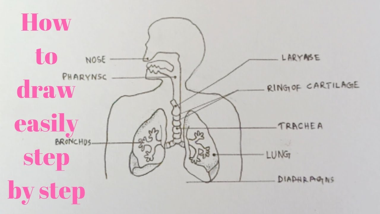
Click Share to make it public. Draw neat and well labelled diagram of Human respiratory system.
A Draw A Labelled Diagram Of The Respiratory System Of Human Beings With Diaphragm At The End Of Expiration Sarthaks Econnect Largest Online Education Community
This leaderboard has been disabled by the resource owner.
Human respiratory system with labelled diagram. View Original Image at Full Size. The respiratory system blank printable. The human respiratory system consists of a pair of lungs and a series of air passages leading to the lungs.
B List four conditions required for efficient gas exchange in an organism. Changes in the volume and pressure in the lungs aid in pulmonary ventilation. Transport of oxygen and carbon dioxide occurs with the help of respiratory pigment called hemoglobin.
Haemoglobin though is purple coloured but oxyhemoglobin is of bright red colour. Write postulates the cell theory. The air is exhaled back through the same pathway.
Features of the Human Respiratory System. Diagram of human respiratory system with labels. Many more lungs and other anatomical models available online via this link.
In this video we will study human respiratory system and its different parts which help in the process of brea. The respiratory system helps in breathing known as pulmonary ventilation. This leaderboard is currently private.
Take a look at the labeled diagram of the respiratory system above. Brainly User Brainly User New questions in Biology. Labeled Diagram Of The Respiratory System Of A Human was posted in June 7 2017 at 529 am.
This HD Wallpaper Labeled Diagram Of The Respiratory System Of A Human. Lungs are the respiratory organs in humans and other animals. For educational purposes only.
Follow me to draw. In this video Im going to draw the labelled diagram of Human Respiratory System. Put this list together in the correct order by writing their numbers below.
How to draw diagram of human Respiratory system easily - step by step - YouTube. Haemoglobin the iron-containing respiratory pigment is a red coloured pigment of blood which has a very high affinity for oxygen. The air inhaled through the nose moves through the pharynx larynx and trachea into the lungs.
Given below is a labeled diagram of the human lungs followed by a brief account of the different parts of the lungs and their functions. The route of air - on its proper way from the outside into the lungs and out again. As you can see there are several structures to learn.
All the main parts of the respiratory system labeled. Who proposed it hey friends help me with this only G_i_r_l. What is the benefits of neem tree.
Air enters the nose through the nostrils. Description from Labeled Diagram Of The Respiratory System Of A Human pictures wallpaper. Oxygen is transported from lungs to the body cells in the form of oxyhemoglobin.
The human respiratory system functions are mentioned below. Labeled diagram of the lungsrespiratory system. Learn how to draw diagram of human respiratory system using a very easy to understand method.
Labeled Diagram Of The Respiratory System Of A Human download this wallpaper for free in HD resolution. Choose from Respiratory System Labeled Diagram stock illustrations from iStock. Asked Oct 12 2019 in Biology by Suchita 664k points a Draw a labelled diagram of the respiratory system of human beings with diaphragm at the end of expiration.
Show more Show less. The Respiratory System diagram easily and step by s. This video shows the structure of lungs.
Find high-quality royalty-free vector images that you wont find anywhere else. Image 37789 is a 1125 by 1408 pixel PNG Uploaded. Use the mouse or tap the screen to label this diagram of the complete human respiratory system combining the head and thorax chest regions.
Share Share by Sumanbalasingh2. How to draw diagram of human Respiratory system easily - step by step. The structure of the lungs is created in such a way that it helps the exchange of gases.
Human Respiratory System Diagram If you carefully observe the respiratory system diagram you will be able to see the various organs involved in its functioning. The entire respiratory tract passage consists of the nose pharynx larynx trachea bronchi and bronchioles. 2 See answers TheRuhanikaDhawan TheRuhanikaDhawan Neat and well labelled diagram of Human respiratory system.
The gas exchange process is performed by the lungs and respiratory system. When air passes through the nose it is warmed moistened and filtered. Spend a few minutes reviewing the name and location of each one then try testing your knowledge by filling in your own diagram of the respiratory system unlabeled using the PDF download below.
This leaderboard is disabled as your options are different to the resource owner. Human respiratory system - Labelled diagram Nasal cavity Pharynx Trachea Lungs Bronchi Bronchioles Alveoli Diaphragm.