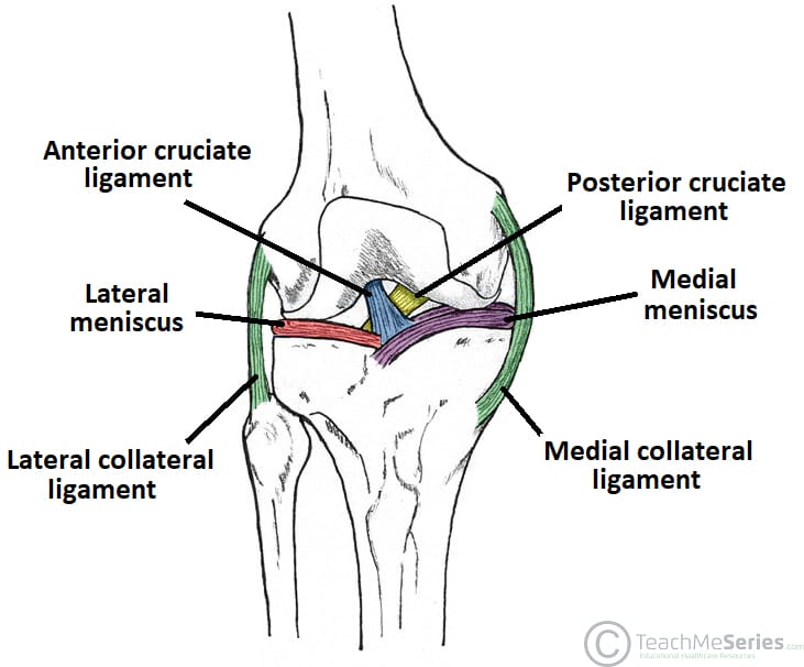
There are two sets of knee ligaments. These muscles work in groups to flex extend and stabilize the knee joint.
The Knee Joint Articulations Movements Injuries Teachmeanatomy
Following are the function and names of knee parts which work together and make the locomotion possible for a human being.

What are the parts of the knee joint. Tibiofemoral medial and lateral condyles of the femur articulate with the tibial condyles. A simple overview of knee anatomy and the knee joint Knee Bones and Joints. The ligaments of the.
The femur tibia and patella. The knee joint keeps these bones in place. Its role is to lubricate the joint.
These motions of the knee allow the body to perform such important movements as walking running kicking and jumping. Within the joint the bottom end of your thigh bone the femur encounters the top end of your. It is present in all joints in small quantities 1 to 2 ml in the knee for example.
There are usually four in each knee at the embryonic stage and help to bend the joints easily. It is the weight-bearing component of the knee joint. Anterior and posterior ACL PCL which sit inside the middle of the joint controlling forwards.
Two rounded convex processes known as condyles on the distal end of the femur meet two rounded concave condyles at the proximal end of the tibia. Medial and lateral MCL LCL that are found either side of the joint and control sideways. The knee joins the thigh bone femur to the shin bone tibia.
It ensures perfect sliding between the bone ends. Aka kneecap is the small bone at the front of the knee. Its an easy lyric to remember from the Dem Bones song many of us sang as kids.
The two largest knee bones the femur and the tibia join together to form what is known as the tibiofemoral joint and at the front of the knee the kneecap rests in a groove on the front of the femur known as the patellofemoral joint. The knee is a complex joint that flexes extends and twists slightly from side to side. Extending along the anterior surface of the thigh are the four muscles of the quadriceps femoris group vastus lateralis vastus medialis vastus intermedius and rectus femoris.
A plica is a fold in the thin synovial membrane that lines the knee joint. The femur thigh bone tibia shin bone and patella kneecap make up the bones of the knee. Knee joint Articular surfaces.
The joint capsule of the knee joint is one of a composite nature mainly formed by muscle tendons and. The joint surfaces are lined with hyaline cartilage and are enclosed within a single joint cavity. Joint fluid also called synovial fluid is secreted by the synovial membrane.
The ends of the bones or spots where the bones in. The knee is one of the largest and most complex joints in the body. About 50 of the population loose their plicae by absorption at the foetal stage.
The knee joint consists of two articulations tibiofemoral and patellofemoral. The smaller bone that runs alongside the tibia fibula and the. In its normal state.
Knee Bones Knee actually consists of three bones femur tibia and patella. The tibiofemoral joint is an articulation between the lateral and medial condyles of the distal end. The patella is a.
The knee is the meeting point of the femur thigh bone in the upper leg and the tibia shinbone in the. The knee also known as the tibiofemoral joint is a synovial hinge joint formed between three bones. Two large bones and a small bone meet in your knee.