Click Share to make it public. Draw neat and well labelled diagram of Human respiratory system.
A Draw A Labelled Diagram Of The Respiratory System Of Human Beings With Diaphragm At The End Of Expiration Sarthaks Econnect Largest Online Education Community
This leaderboard has been disabled by the resource owner.
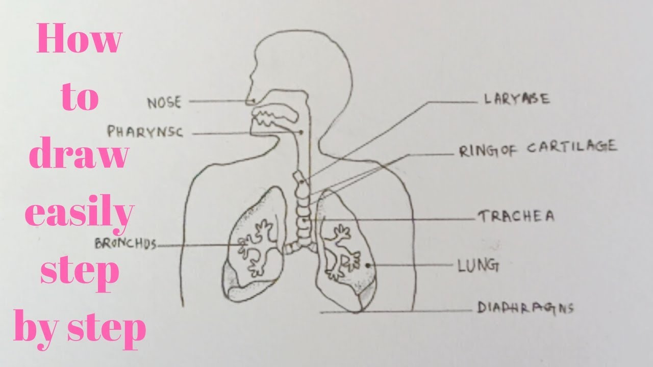
Human respiratory system with labelled diagram. View Original Image at Full Size. The respiratory system blank printable. The human respiratory system consists of a pair of lungs and a series of air passages leading to the lungs.
B List four conditions required for efficient gas exchange in an organism. Changes in the volume and pressure in the lungs aid in pulmonary ventilation. Transport of oxygen and carbon dioxide occurs with the help of respiratory pigment called hemoglobin.
Haemoglobin though is purple coloured but oxyhemoglobin is of bright red colour. Write postulates the cell theory. The air is exhaled back through the same pathway.
Features of the Human Respiratory System. Diagram of human respiratory system with labels. Many more lungs and other anatomical models available online via this link.
In this video we will study human respiratory system and its different parts which help in the process of brea. The respiratory system helps in breathing known as pulmonary ventilation. This leaderboard is currently private.
Take a look at the labeled diagram of the respiratory system above. Brainly User Brainly User New questions in Biology. Labeled Diagram Of The Respiratory System Of A Human was posted in June 7 2017 at 529 am.
This HD Wallpaper Labeled Diagram Of The Respiratory System Of A Human. Lungs are the respiratory organs in humans and other animals. For educational purposes only.
Follow me to draw. In this video Im going to draw the labelled diagram of Human Respiratory System. Put this list together in the correct order by writing their numbers below.
How to draw diagram of human Respiratory system easily - step by step - YouTube. Haemoglobin the iron-containing respiratory pigment is a red coloured pigment of blood which has a very high affinity for oxygen. The air inhaled through the nose moves through the pharynx larynx and trachea into the lungs.
Given below is a labeled diagram of the human lungs followed by a brief account of the different parts of the lungs and their functions. The route of air - on its proper way from the outside into the lungs and out again. As you can see there are several structures to learn.
All the main parts of the respiratory system labeled. Who proposed it hey friends help me with this only G_i_r_l. What is the benefits of neem tree.
Air enters the nose through the nostrils. Description from Labeled Diagram Of The Respiratory System Of A Human pictures wallpaper. Oxygen is transported from lungs to the body cells in the form of oxyhemoglobin.
The human respiratory system functions are mentioned below. Labeled diagram of the lungsrespiratory system. Learn how to draw diagram of human respiratory system using a very easy to understand method.
Labeled Diagram Of The Respiratory System Of A Human download this wallpaper for free in HD resolution. Choose from Respiratory System Labeled Diagram stock illustrations from iStock. Asked Oct 12 2019 in Biology by Suchita 664k points a Draw a labelled diagram of the respiratory system of human beings with diaphragm at the end of expiration.
Show more Show less. The Respiratory System diagram easily and step by s. This video shows the structure of lungs.
Find high-quality royalty-free vector images that you wont find anywhere else. Image 37789 is a 1125 by 1408 pixel PNG Uploaded. Use the mouse or tap the screen to label this diagram of the complete human respiratory system combining the head and thorax chest regions.
Share Share by Sumanbalasingh2. How to draw diagram of human Respiratory system easily - step by step. The structure of the lungs is created in such a way that it helps the exchange of gases.
Human Respiratory System Diagram If you carefully observe the respiratory system diagram you will be able to see the various organs involved in its functioning. The entire respiratory tract passage consists of the nose pharynx larynx trachea bronchi and bronchioles. 2 See answers TheRuhanikaDhawan TheRuhanikaDhawan Neat and well labelled diagram of Human respiratory system.
The gas exchange process is performed by the lungs and respiratory system. When air passes through the nose it is warmed moistened and filtered. Spend a few minutes reviewing the name and location of each one then try testing your knowledge by filling in your own diagram of the respiratory system unlabeled using the PDF download below.
This leaderboard is disabled as your options are different to the resource owner. Human respiratory system - Labelled diagram Nasal cavity Pharynx Trachea Lungs Bronchi Bronchioles Alveoli Diaphragm.
This activity has been designed for use in homes and schools. Microscopes R Us Pack Microscopic Biology Lesson Plans Biology Lessons High power objective 6.
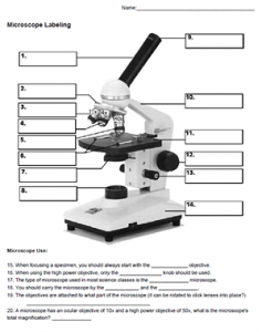
Microscope Labeling
Click to Download.

Label microscope parts worksheet with answers. Microscope parts and use worksheet answer key along with labeling the parts of the microscope blank diagram available for. Activity Students Label Parts. Nosepiece microscope when carried holds the high and low power objective lenses.
Using the microscopelabel the microscope determining the magnifying power working distance diameter of field of view brightness needed ocular objective magnification magnification total working magnification distance more diameter field of view wide. Some of the worksheets below are Parts and Function of a Microscope Worksheets with colorful charts and diagrams to help students familiarize with the parts of the microscope along with several important questions and activities with answers. Once you find your worksheet s you can either click on the pop-out icon or download button to print.
The labeling worksheet could be used as a quiz or as part of direct instruction where students label the microscope as you go over what each part is used for. Light microscope worksheet drag and drop worksheet on the parts of the microscope. View homework help - microscope worksheet pp with answers from bio.
Label parts of the Microscope. The study of living things. We tried to locate some good of Microscope Parts and Use Worksheet Answer Key Along with Labeling the Parts Of the Microscope Blank Diagram Available for image to suit your needs.
It was from reliable on line source and that we love it. Free worksheets for labeling parts of the microscope including a worksheet that is blank and one with answers. Microscope parts worksheet answers The rough adjustment knob b is the bigger one on your microscope.
1 86 Answer Key Science 1-7 71 57 43 29 14 0 1. NOSEPIECE microscope when carried Holds the HIGH- and LOW- power objective LENSES. Id language school subject science middle school age main content lab use of the microscope.
Label the parts of the microscope indicated and state the. Handphone Tablet Desktop Original Size There are many key components to understand when utilizing a microscope. The compound microscope uses lenses and light to enlarge the image and is also called an optical.
Text preview biology a laboratory scopes and cells worksheet name a. Parts of a eyepiece arm stageclips nosepiece focusing knobs illuminator stage objective lenses head base Label the parts of the microscope. Activity Students Label Parts.
Labeled microscope worksheet answers. Microscopes are often used in areas as diverse as plant and animal studies and anatomy. If you want to download the image of microscope parts and use worksheet answer key as well as life science teacher s edition simply right click the image and choose save as.
List of Microscope Labeling Worksheet Answers. Worksheet identifying the. This worksheet asks pupils to label the different parts of a microscope and then match up the with their function.
Each microscope layout both blank and the version with answers are available as PDF downloads. You can use the word bank below to fill in the blanks or cut. Microscope Parts and Use Worksheet Answer Key Along with Labeling the Parts Of the Microscope Blank Diagram Available for.
You can use the word bank below to fill in the blanks or cut and paste the words at the bottom. A basic microscope has a single convex lens such as those found in a magnifying glass. We tried to locate some good of microscope parts and use worksheet answer key along with labeling the parts of.
Can be rotated to change MAGNIFICATION. Microscope parts worksheet answer key. Microscope parts and use worksheet answer key along with labeling the parts of the microscope blank diagram available for.
View homework help - microscope worksheet pp with answers from bio biol at community college. Labeling the Parts of the Microscope. Live worksheets science lab equipment light microscope worksheet.
Label the Parts of the Microscope with answers A4 PDF print version. List of Microscope Labeling Worksheet Answers Pm company. Power 10 x 4 40 Power 10 x 10 100 Power 10 x 40 400 What happens as the power of magnification increases.
In this activity students identify and label the main parts of a microscope and describe their function. Microscope parts and use worksheet answer key along with labeling the parts of the microscope blank diagram available for worksheet january 13 2018 we tried to locate some good of microscope parts and use worksheet answer key along with labeling the parts of the microscope blank diagram available for image to suit your needs. Coarse adjustment knob 13.
We hope this graphic will likely be one of excellent reference. For a thorough review of each microscope part continue reading. Some of the worksheets shown are part microscope and use microscope mania microscope laboratory work part name of the student microscope from the light microscope review work filling the blank Wanganui High School.
By the end of this. Answers to my TEENrens crossword puzzles. Microscopes are often used in areas as diverse as plant and animal studies and anatomy.
G Labeling Scientific Tools Microscope G F E D C B A 1 Illuminator 2 Stage 3 Eyepiece 4 Focus Fine 5 Lense 6 Focus Course 7 Base Determine which letter best matches each microscope piece.
Read the following passage and answer the following questions. Start studying Kidney Anatomy Labeling.
Draw A Well Labelled Diagram Of Ls Of The Human Ki Class 12 Biology Cbse
Here you have to show the full process of digestion.

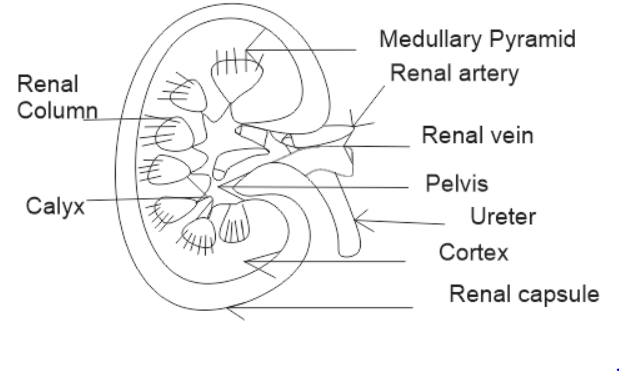
Diagram of kidney with labelling class 10. Veins of the abdomen and hepatic portal. Draw a well-labeled diagram of the human excretory system. Draw a labeled diagram of the human kidney as seen in a longitudinal section.
Each kidney is about 10 to 13 cm 4- 5 inches in long 6 cm. The diagram given below shows a section of human kidney. 2 ½ inches wide and 3 cm.
On the upper end of each kidney suprarenal glands are situated like a cap. Kidney diagram to label. Human Excretory System.
Label Diagram Of Kidney Wiring Diagram Pipe Activity Pipe Activity Fiocouture It. Draw the diagram of kidney recognize and label the following parts. Kidney diagram class 10 0 comments.
Dehydration a blockage in the urinary tract or kidney damage can cause acute renal failure which may be. A human brain is composed of three main parts-. Current diagram shows a little bit about human excretory system and a detailed diagram of Kidney which is the most important part of Excretory system.
Urinary bladder CBSE 2013 Answer. CBSE Class 10 Physics Important Diagrams. Kidney Diagram Class 10.
Label the parts numbered 1 to 4. Class-10 Welcome to Sarthaks eConnect. Sketch and label the structure of malphigian body.
In males and 135 gms in females. Why does part 2 have a striped appearance. Arteries Of Kidney Great For Anatomy Class Diagnostic Medical.
Q6 Draw A Diagram Of The Human Excretory System And Label Th Lido. Sketch and Label the Diagram. Draw A Well Labelled Diagram Of Human Excretory System And Write Name Of Filtering Unit Of Kidney Brainly In.
The human kidneys house millions of tiny filtration units called nephrons which enable our body to retain the vital nutrients and excrete the unwanted or excess molecules as well as metabolic wastes from the body. Kidney DiagramThe collecting duct labelled in the diagram above is part of the kidney nephron shown much enlarged. It allows small molecules such aswater to.
I Urinary bladder ii Left kidney iii Left ureter. Kidneys are dark brown in colour and are embedded in a mass of fat. What is the fluid that passes down 4.
The average weight of adult kidney is about 150 gms. Draw and label a diagram of the kidney. 101 labeled diagram of the human kidney stock photos vectors and illustrations are available royalty-free.
At September 11 2018 Labels. CBSE Class 10 Chemistry Important Reactions. Draw and label a diagram of the kidney ib biology syllabus.
Click hereto get an answer to your question Draw well labelled diagram of LS. Label the schematic drawing. Learn vocabulary terms and more with flashcards games and other study tools.
1 ½ inch in thickness. Diagram of human excretory system. In this diagram you have to show all the parts of the system which includes two kidneys urethra ureters etc.
Dialysistubing is a semipermeable membrane. The alignment of kidney are. Human Body Organs Diagram From The Back Koibana Info Human Body Organs Human Body Anatomy Body Organs Diagram Students drag labels to the structures on the slide.
You have to mention the mouth liver gallbladder stomach pancreas small and large intestine liver etc. Draw A Well Labelled Diagram Of The L S Of Kidney Label Any. The nephron is one of the most important parts of our body and also one of the smallest functioning units.
Study the diagram carefully and answer the questions that follow. Biology section 3 flashcards the hilus is present on the concave side of the kidney through which the uriters are attached to the kidney the part of the kidney where the renal artery and renal vain enter label the diagram the kidney and nephron below anatomy kidney structure and function the excretory system chegg label the diagram. Students upto class 102 preparing for All Government Exams CBSE Board Exam ICSE Board Exam State Board Exam JEE MainsAdvance and NEET can ask questions from any subject and get quick answers by subject.
Image of a kidney and nephron with the major structures labeled. A unique platform where students can interact with teachersexpertsstudents to get solutions to their queries. Thousands of new high-quality pictures added every day.
Featuring a professional light background with a large amount of vector format various symbols and some other important science elements you wont miss this illustrated kidney diagram template from. Use this interactive 3-D diagram to explore the kidney. Find labeled diagram of the human kidney stock images in HD and millions of other royalty-free stock photos illustrations and vectors in the Shutterstock collection.
1 ½ inch in thickness. Click to find video solution Book a free class. Name the main nitrogenous waste.
Ear Hair Cell Diagram Labeled Today Wiring Schematic Diagram. Pinna outer portion of ear used to collect sound waves 2.

Labeled Diagram Of The Ear Luxury Labelled Diagram Of The Ear Nursing Pinterest Ear Diagram Human Ear Diagram Ear Anatomy
The human body is like a big machine and various processes take place inside it.
Labelled ear diagram with functions. The ear catches sound waves and converts it into impulses that the brain interprets making it understandable and helps the human body differentiate between different sounds. Outer ear ear canal eardrum tiny bones in the middle ear cochlea auditory nerves. This ear-throat connection makes the ear susceptible to infection otitis media.
What Are The Main Parts Of The Ear Socratic. The latter fits into the fenestra ovalis. They pass through the external auditory meatus to the tympanic membrane which is caused to vibrate.
The external auditory canal links the exterior ear to the inner or the middle ear. The middle ear is a chamber located within the petrous portion of the temporal bone. Structure of the Ear Storyboard by oliversmith.
Image will be uploaded soon The Outer Ear. Parts of the Human Ear. What Are The Main Parts Of The Ear Socratic.
It vibrates striking the anvil to carry on the sound 5. With the help of the various organs and tissues it carries out some of the most marvelous tasks that are no less than a miracle. How Do We Hear.
Tinnitus Vector Illustration Labeled. Purchase Publications Vestibular Disorders Association. Equalizing air pressure ensures that the eardrum vibrates maximally when struck by.
Human Anatomy Ear Diagram With Functions. Anvil gets hit by the hammer striking the Stirrup 6. Ear Diagram Labeled And Functions.
Label Diagram Of Human Ear Labeled Diagram Of Human Ear Anatomy Human Human Ear Diagram Ear Anatomy Human Ear Anatomy Learn about the ears function in the body and test and. Ear Drum membrane that sends sound waves to hammer 4. Swallowing and chewing actions open the tube to allow air in or out as needed for equalization.
The tympanic membrane also known as the eardrum separates the outer ear from the inner ear. The external ear the middle ear and the internal ear Figure 2. Ear structure diagram parts sound waves label functions function human anatomy list different keywords storyboardthat each below oliversmith students windows.
Write Clearly and Concisely Grammarly. It is mainly concerned with detecting transmitting and transducing sound. If playback doesnt begin.
Anatomy And Physiology Of The Ear. The human ear consists of three parts. Structures within the middle ear amplify sound waves and transmit them to an appropriate portion of the internal ear.
Anatomy of the ear diagram labeled. Hammer first of 3 bones. The auricle or pinna is a flap of elastic cartilage shaped like the flared end of a trumpet and.
Auditory Canal tube that sound travels down 3. Pinnaauricle is the outermost section of the ear. Parts of the ear external outer ear.
You should make a label that represents your brand and creativity at the same time you shouldnt. The human ear consists of three different parts. Ear Anatomy Labeled Diagram Stock Photo Picture And Royalty Free.
The ear diagram is one of the important topics for Class 10 and 12 students of the CBSE board and in this article we will briefly explain the structure of the ear its different parts and their functions. The ear is divided into three anatomical regions. Ear Infection - Acute Otitis Media - ENT Clinic.
Weve gathered our favorite ideas for Ear Diagram Labeled Explore our list of popular images of Ear Diagram Labeled Photos Collection with high resolution. One such organ is the ear. Brain Areas And Their Functions Health24.
Structure And Functions Of The Ear Explicated With Diagrams Bodytomy. Let us have an overview of the structure and functions of the human ear. Human Ear Structure Function.
Special Senses Eyes And Ears Medical Terminology In A Flash A. An easy and convenient way to make label is to generate some ideas first. Structure and Functions of the Ear Explicated With Diagrams.
Label Gallery Get some ideas to make labels for bottles jars packages products boxes or classroom activities for free. How to Draw Human Ear Diagram With Labelling HumanEar - YouTube. Well-Labelled Diagram of Ear.
Mar 31 2016 - Image of the ear is colored according to the directions where structures such as the tympanum. Look no further this Bodytomy article gives you a labeled human ear diagram and also explains the Let us take a look at the human ear structure with the help of a diagram and understand its functions. The sound waves are collected by the external ear up to some extent.
Ear Diagram Book Wiring Diagram Images Gallery. Outer ear ear canal eardrum tiny bones in the middle ear cochlea auditory nerves. Draw A Neat Labelled Diagram Of The Human Ear Explain It S Working.
The perilymph of the internal ear. Stirrup vibrates against OVAL WINDOW carrying on sound. The ear is another extraordinary organ of the house of wonders that is the human body.
The Eustachian tube functions to equalize air pressure on both sides of the eardrum. Look no further this Bodytomy article gives you a labeled human ear diagram and also explains the functions of its different components. Ear diagram label quizlet labels ear functions diagram structure explicated diagrams function grade sponsored links quizlet ear diagram anatomy human ear throat nose ears hearing anatomy diagram ent porter county center ear diagram labeled tape label labels printable human notebook markcritz cavities ear labeled anatomy models ap classroom physiology eye diagram senses ear diagram worksheet.
Definition Of Structure And Function Of The Ear Chegg Com. Click on the score button to see how you did. The External ear or the outer ear consists of.
The ear performs the functions of hearing and balancing equilibrium. The vibrations are transmitted across the middle ear by the malleus incus and to the stapes bones. The ear is a sensitive organ of the human body.
Maintaining a sense of balance is another important function performed by the human ear. Pinnaauricle is the outermost section of the ear. Here we will discuss all parts of the ear.
Ear Diagram Labeled With Functions Written By JupiterZ Friday September 4 2020 Add Comment Edit. Wondering what is the structure of the human ear and how it performs the function of hearing. Normally the walls of the tube are collapsed.
The external ear is the visible portion of the ear and it collects and directs sound waves to the eardrum. The external auditory canal links the exterior ear to the inner or the middle ear. The Middle ear comprises.
Download clkers mature flower diagram clip art and related images now. 1 Stalk- The part by which a flower is attached to the branch is called stalk.

Q6 What Is A Flower Draw A Nea Lido
Labeled Diagram Of Flower Structure Simple Parts Of A Flower.
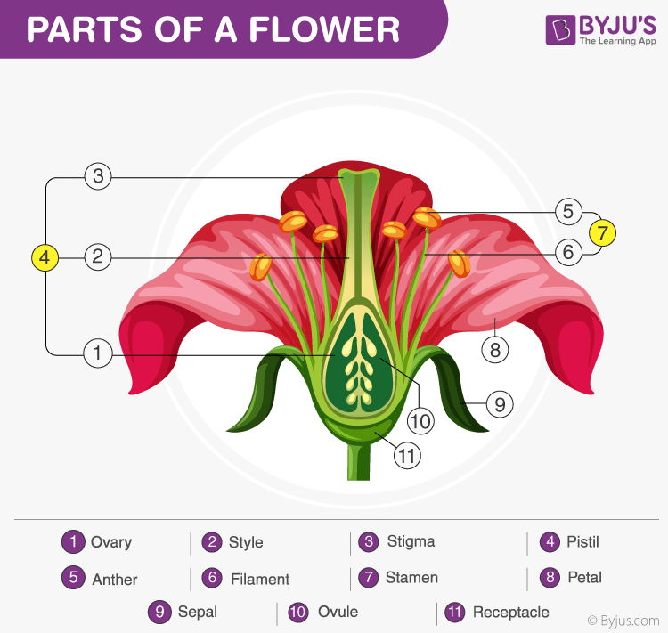
Explain the structure of flower with labelled diagram. A typical flower has four main partsor whorlsknown as the calyx corolla androecium and gynoecium Figure 1. Calyx and corolla are not directly involved in reproduction. When a flower has all the four floral parts it is called a complete flower.
Plant Cell Labeling Diagram Quizlet. Sugarcane is a tropical grass and one of our main sources of sugar. The male parts of the flower each consists of an anther held up on a filament Anthers.
Explain the structure of ts of mature anther with a neat labeled diagram. Flowers contain the plants reproductive structures. The gynoecium or pistil consists of carpels and is the female reproductive part.
The parts of a flower. Draw a mind map to show the different parts of the Human Skeletal System. After reading this article you will learn about.
List the different types of joints found in the human body. They are sterile and reproductive. Q4 What is meant by pollination.
Introduction to structure of a flower. Structure of China rose flower. A flower usually consists of four main parts.
Add your answer and earn points. A typical flower consists of four sets of floral parts or whorls. They are typically used with the floral formula of that flower.
However sepals are found to be fused in flowers such as periwinkle Hibiscus etc. Mention where are they located 5. Draw a labelled diagram to explain the different parts of a complete flower.
Youll recognize the pistil in a plant diagram because it looks like a small knob that protrudes from the flower. Floral diagram is a graphic representation of flower structure. Explain the structure of a flower with neat labeled diagram florencerobinaaron02 florencerobinaaron02 19092020 Science Secondary School answered 1.
Produce male sex cells pollen grains Stigma. 2 Sepals- The small green colored leaf shaped structures found on the outermost part of the flower are called sepals. The flower consists of an axis also known as receptacle and lateral appendages.
Parts Of A Plant Labelling Worksheets Sb12380 Sparklebox. They sometimes have fragrance also. The pistil contains the stigma style and ovary.
Petals are the often colorful parts of a flower that advertise it to insects and other pollinators. In the united states sugarcane is grown in hawaii and florida. They are mostly present in the leaf epidermis stem pith root and fruit pulp.
Explain the structure of germinating pollen grain with the help of a labelled diagram. Explain the structure of a flower with neat labeled diagram 1 See answer florencerobinaaron02 is waiting for your help. Labeled scientific diagram of a flower.
Changes In Atom Of 13 C In Leaves A Stems B And Flower Buds. 1 sepals 2 petals 3 stamen and 4 carpel each of them performing distinct functions. They can be clearly distinguished as separate structures in flowers such as rose marigold etc.
Different parts of the flower are represented by their respective symbols. Pictures Flower Labeled Diagram Vector Flower Parts Diagram. 1201 flower parts diagram stock photos vectors and illustrations are available royalty-free.
The whorls are arranged on the thalamus of a flower. 1 A typical flower consists of an outer whorl of green sepals calyx which protects the parts within. Parenchyma collenchyma and sclerenchyma.
Introduction to structure of a flower 2. They protect the flower when they are in the stage of bud. Describe the structure of flower with a neatly labelled diagram.
See flower parts diagram stock video clips. The lifecycle of angiosperms follows the alternation of generations explained previously. The stamens compose the androecium whereas the free or united carpels compose the gynoecium.
The top of the female part of the flower which. Write differences between pectoral and pelvic girdle. 2 Tha second whorl has petals corolla which are usually brightly colored.
Floral diagrams are useful for flower identification or can help in understanding angiosperm evolution. 4 The third whorl of the flower consists of stamens androecium which are the the male. Androecium and gynoecium are directly concerned with sexual reproduction.
Explain the movement and locomotion of. They are considered to be modified leaves. Parts of a hibiscus flower complete flower fiower part sepal plant ovary flower anatomy diagram of flower parts of a flower flower diagram parts of the flower.
Some flowers have only male or female organs and require a separate flower of the opposite gender to reproduce. Parts Of A Flower Azcars Info. They were introduced in the late 19th century and are generally attributed to A.
What are the Different Parts of a Flower A typical diagram of a flower is divided into four main parts. Pollination is the process in which the pollen grains from the anthers are transferred to the stigma of a flower of the same species. The haploid gametophyte alternates with the diploid sporophyte during the sexual reproduction process of angiosperms.
Free Unlabeled Flower Diagram Download Free Clip Art Free Clip. Download a powerpoint showing labelled and unlabelled versions of these diagrams both parts of a plant. The androecium is the male part of the flower and consists of stamens.
It shows the number of floral organs their arrangement and fusion. The appendages are known as floral parts or floral organs. Explain the structure of the rib cage.
A flower missing any one of them is called an incomplete flower. 3 Petals are soft and are useful to fasilitate cross pollination. The female part of the flower the pistil is located at the center of the bloom.
Try these curated collections. The outer parts of the flower often green and leaf like that enclose a developing bud. Labeled structure of flower diagram.
Mature flower diagram labeled. A typical flower contains the following parts. This process is called pollination.
They are the small leaf-like green structures of a flower. The sepals and petals which constitute the calyx and corolla respectively are the sterile parts. Calyx sepals corolla petals androecium stamens and gynoecium carpels.
Model Animal cell cake Cake Pinterest Models Animal.