The Cell The Biology Primer. Find Human Cell Diagram stock images in HD and millions of other royalty-free stock photos illustrations and vectors in the Shutterstock collection.

Draw A Neat Diagram Of A Typical Animal Cell And Label The Following Organelles 1 The Organelle Called Science Cell Structure And Functions 10429465 Meritnation Com
Diagram of the human cell illustrating the different parts of the cell.

Labelled diagram of typical cell. Comparison of Prokaryotic Cells and Eukaryotic Cells and 2. In prokaryotic cell there is naked DNA without histone protein but in Eukaryotic cell there is DNA with histone protein. However it typically exists in three forms ie.
LPS teichoic acid etc surrounding the bacterium like a shell and lies external to the cytoplasmic membrane. It is a tough and rigid structure of peptidoglycan with accessory specific materials eg. You can also put your logo at the top or bottom corner of the label.
Structure and Functions With Diagram Let us make an in-depth study of the structure and functions of cell. Stock vector labelled diagrams of typical animal and plant cells with editable layers 222613513 in cell diagram simple the structure and contents of a typical animal cell every has membrane cytoplasm nucleus but not all cells have rat liver cell a scheme of the typical cell membrane structure with lipid bilayer integral proteins. Cell is a compartment where all the activities of life takes place.
Targeting Strategies for Multifunctional Nanoparticles in. A typical bacterial cell. Structure of a typical animal cell click to enlarge.
Prokaryotic cells article Khan Academy. Label Gallery Get some ideas to make labels for bottles jars packages products boxes or classroom activities for free. The Golgi Apparatus is the cell organelle mostly present in eukaryotic cells which is responsible for the packaging of macromolecules into vesicles so that they can be sent out to their site of action.
A typical bacterial cell - Labelled diagram free-floating DNA plasmid circular ring of DNA slime capsule stops the cell from drying out flagellum helps the bacterium move through liquids cell membrane and cell wall the label was too big to tell these apart. The lipid molecules on the outer and inner part lipid bilayer. Which biological activities will be hindered and why.
Most of the times we put the labels to show some specific information. Illustration of Labelled diagrams of typical animal and plant cells with editable layers. On the contrary plant cells lack centrioles and intermediate filaments which are present in animal cells.
Examining a diagram of the plant cell will help make the differences clearer. Labelled diagram of a plant cell under microscope posted on march 18 2011 by admin onion cells stained with methylene blue look at the images of onion cells as they would be seen under a microscope draw each magnification label appear high picture plant and animal cell diagrams. It is a double-layered membrane composed of proteins and lipids.
Labelled Diagram Of A Typical Animal Cell. In prokaryotic cell DNA is circular but in Eukaryotic cell DNA is double helix. Vector art clipart and stock vectors.
A Draw a fully labelled diagram of a typical receptor tyrosine cell surface recepto b Name the other classes of enzyme coupled receptors and describe their mechanism of action. Thousands of new high-quality pictures added every day. Labeled diagram of a typical plant cell.
Cisternae vesicles and tubules. Typical Animal Cell Diagram Labeled Best Of Typical Animal And Plant Animal Cell Coloring Page Lovely 25 Best Typical Plant Cell Ideas On Click The Animal Cell Diagram To Enlarge It Wiring Diagram Blog Eukaryotic Cells Biology I Typical Animal Cell Plant And Animal Cells S Cool The Revision Website Animal Cell Anatomy Diagram Structure With All Parts Nucleus Cells Alive Plant Cell. Typical Plant Cell Diagram Labeled Battery electricity Wikipedia.
1in prokaryotic cell there is primitive nucleus but in Eukaryotic cell there is well developed nucleus. After reading this article you will learn about. Room Full of Crazy TV Tropes.
Western Wood Products Association. A Draw an annotated diagram of a typical student microscope b Explain the. Unit 2 Cells Biology Junction.
The single circular double-stranded chromosome is the bacterial genome. An easy and convenient way to make label is to generate some ideas first. Cell Membrane The cell membrane is the outer coating of the cell and contains the cytoplasm substances within it and the organelle.
Labels are usually small in size so you should carefully choose the font of the texts to make sure it is readable. C Taxol from the Yew tree stabilises microtubules. Hello Guys In this Video Im going to teach you guys How to Draw Structure of a cell Labeled DiagramSubscribe to the Channel and also share this video.
Other structures include cytoplasmic membrane mesosomes. The structure of the Golgi Complex is pleomorphic. Labelled diagram of a typical animal cell.
INLINE PICTURES VERSION W1TP. Intro to eukaryotic cells article Khan Academy. It gives shape to the cell.
It is 10-25 nm in thickness. Structure and Components of a Human Cell. Human Cell Diagram Stock Vector Royalty Free 400272997.
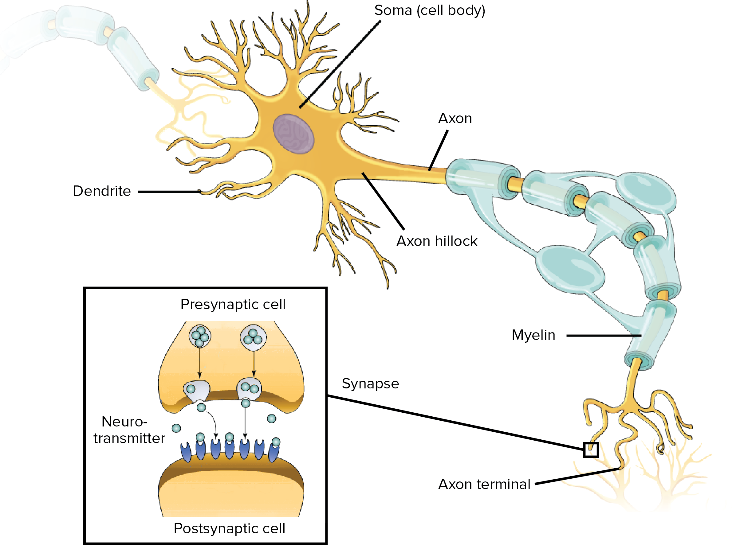
Supports and maintains the functioning of the neuron Think of the cell body as a small factory that fuels the neuron. The figure at the right shows a typical neuron.
Overview Of Neuron Structure And Function Article Khan Academy
They have a fourth part the cell body or soma which carries out the basic life processes of neurons.
What are the three parts of a typical neuron. The pre-synaptic terminal is usually on the axon. Dendrites cell bodies axons and synapses are the basic parts of a neuron but other important structures and materials surround neurons to make them more efficient. Cell Body 3.
One the basis of structure neurons are three types- 1. Learn vocabulary terms and more with flashcards games and other study tools. Dendrites - are branching extensions from nerve cell somaThey get stimulated by other neurons and transmit the impulses towards.
Cell body dendrites axon. A cell body an axon and dendrites. Synaptic knobs Schwann cell MY Nodes of Ranvier Dendrites Axon collateral Chromatophilic substance Axon Cell body Myelin Axon hillock Reset.
Neurons are classified on the basis of their structure and function. Bipolar neurons and 3. The soma produces the proteins that the other parts of the neuron including the dendrites axons and synapses need to function properly.
The dendrites function is to receive or detect stimuli such as neurotransmitters released by other neurons. It is one of many units that makes up the brain. The three major parts of a neuron are the dendrites soma and axon.
Parts of a Typical Neuron. What is the basic structure and function of a neuron. Neurons are specialized cells of the nervous system that transmit signals throughout the body.
This problem has been solved. Identify The Three Major Parts Of A Typical Neuron As Labeled On The Diagram. Label the parts of a typical multipolar neuron.
You may already know that neurons can do many different things from sensing external. The axon and dendrites are filaments that extrude from it. The soma is usually compact.
All neurons have three different parts dendrites cell body and axon. Start studying 3 Major Parts of a Neuron. However nearly all neurons have three essential parts.
Click to see full answer. A Neuron structure with Synapses at the end The cell body soma contains the nucleus and cytoplasm. Dendrites axons and axon terminals.
See the answer See the answer See the answer done loading. A neuron consists of a cell body or soma dendrites and a single axon. They have a fourth part the cell body or soma which carries out the basic life processes of neurons.
Soma - or a nerve cell body contains nucleus and other organellesIt is also responsible for synthesis of neurotransmitter. Neurons nerve cells have three parts that carry out the functions of communication and integration. Contains the nucleus and most of the cell cytoplasm.
Neurons vary in size shape and structure depending on their role and location. 1 The nerve cell also referred to as a neuron has several parts that are important for its function in the nervous systemThese parts are. Neurons nerve cells have three parts that carry out the functions of communication and integration.
Numerous short branched projections of the cell body that receives signals from other neurons and transmits them across the cell body. The figure at the right shows a typical neuron. This releases neurotransmitters into the synaptic cleft.
A typical neuron consists of a cell body soma dendrites and a single axon. Dendrites typically branch profusely and extend a few hundred micrometers from the soma. Structure of neuron A neuron has three parts- a cell body dendrites and an axon ending at an axon terminal.
The pre-synaptic terminal is the first part of synaptic transmission and so has a pre- prefix. Dendrites axons and axon terminals. The structure of a typical chemical synapse comes in three parts.
Three parts of a neuron.
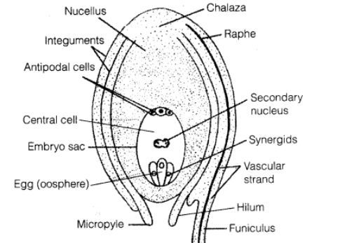
Schematic drawing of the mature eight-nuclei female gametophyte or embryo sac in a typical angiosperm flower ovule. With a neat labelled diagram describe the parts of a typical angiosperm ovule.
With A Neat Labelled Diagram Describe The Parts Of A Typical Angiosperm Ovule Snapsolve
I Funiculus is stalk-like structure by which ovule is attached to the placenta.
Labelled diagram of typical angiosperm ovule. With a neat labelled diagram describe the parts of a typical angiosperm ovule. Each carpel has three parts viz. Well labelled diagram of typical angiospermic ovule showing different parts 2 See answers Brainly User.
Prev Question Next Question 0 votes. Concept Notes Videos 725. Each ovule consists of nucellus surrounded by two integuments and a stalk or funiculus.
Well labelled diagram of typical angiospermic ovule showing different parts Get the answers you need now. The Pistil Megasporangium ovule and Embryo sac. Funicle - It is the stalk of ovule which connects ovule to the placenta.
The diagram of a typical angiosperm ovule is as follows. With a neat labelled diagram describe the parts of a typical angiosperm ovule. With a neat labelled diagram describe the parts of a typical angiosperm ovule.
Asked Mar 12 2020 in Biology by Kasis01 496k points With a. All eight nuclei shown are haploid. Answer An ovule is a female megasporangium where the formation of megaspores takes place.
Sexual Reproduction in Flowering Plants. A typical ovule is composed of the following organs. With a neat labelled diagram describe the parts of a typical angiosperm ovule.
The various parts of an ovule are. With a Neat Labelled Diagram Describe the Parts of a Typical Angiosperm Ovule. Maths Physics Chemistry Biology.
With a neat labelled diagram describe the parts of a typical angiosperm ovule. The various parts of an ovule are 1 Funiculus It is a stalk-like structure which represents the point of attachment of the ovule to the placenta of the ovary. Answers 1 S Sonika.
Our Bootcamp courses are free of charge. An ovule is a female megasporangium where the formation of megaspores takes place. Angiosperm embryo sac diagram.
By Biology experts to help you in doubts scoring excellent marks in Class 12 exams. Ovary style and stigma. CBSE CBSE Science Class 12.
An ovule consists of funicle micropyle integuments nucellus embryo sac hilum chalaza etc. An ovule is a female megasporangium where the formation of megaspores takes place. Mustaqeemareeb687 mustaqeemareeb687 08102020 Biology Secondary School answered Draw a neat and clean.
It is a stalk-like structure by which ovule is attached to the. Step by step video text image solution for With a neat labelled diagram describe the parts of a typical angiosperm ovule. NCERT Class XII Biology - Main Course Book Chapter 2.
Maths Physics Chemistry Biology. Hilum It is the point where the body of the ovule is attached to the funiculus. In double fertilization the egg cell will fuse with a sperm nucleus to form a diploid zygote while the two polar nuclei will fuse with the second sperm nucleus.
With a neat labelled diagram describe the parts of a typical angiosperm ovule. The gynoecium represents the female reproductive whorl of the flower composed of carpels. The various parts of a typical angiospermic ovule are as follows.
With a neat and labelled diagram describe the parts of a typical angiosperms ovule. With a neat and labelled diagram describe the parts of a typical angiosperms ovule. Funicle is short and multicellular.
Funiculus It is a stalk-like structure which represents the point of attachment of the ovule to the placenta of the ovary. Maths Physics Chemistry Biology. 2 Hilum It is the point where the body.
The connecting slender stalk between ovule and placenta is known as Funiculus. With a neat labelled diagram describe the parts of a typical angiosperm ovule. Answer An ovule is a female megasporangium where the formation of megaspores takes place.
Maths Physics Chemistry Biology. Each ovule consists of nucellus surrounded by two integuments and a stalk or funiculus. With a neat labelled diagram describe the parts of a typical angiosperm ovule.
Question Bank Solutions 18579. It is a stalk-like formation which represents the point of the accessory of the ovule to the placenta of the ovary. Get certified as an expert in up to 15 unique STEM subjects this summer.
The source code of this SVG is valid. In which one of the options all the four parts a b c and d are correct.
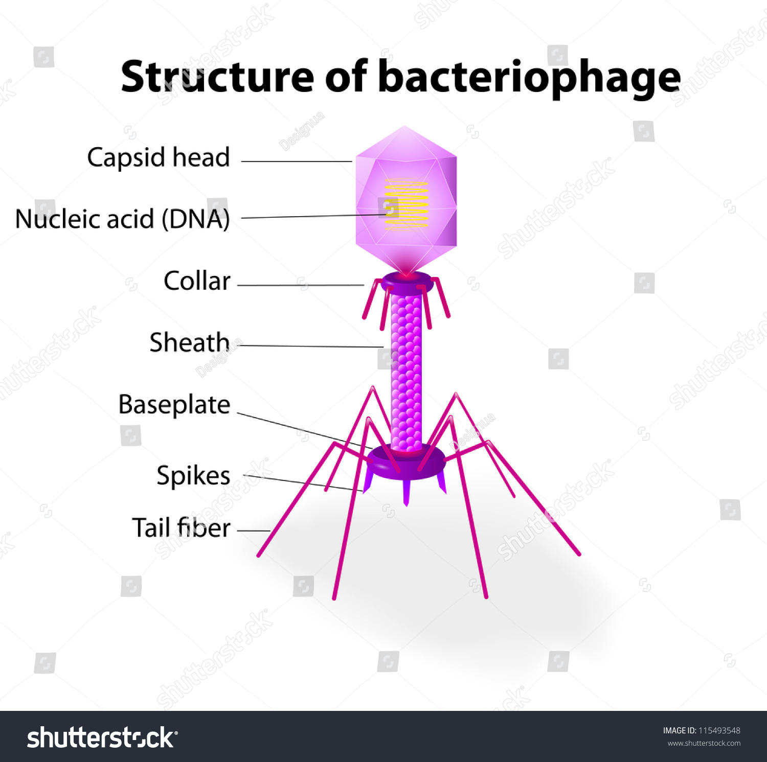
Virus Vector Diagram Typical Tailed Bacteriophage Stock Vector Royalty Free 115493548
Loads of virus pictures sorted by various methods.
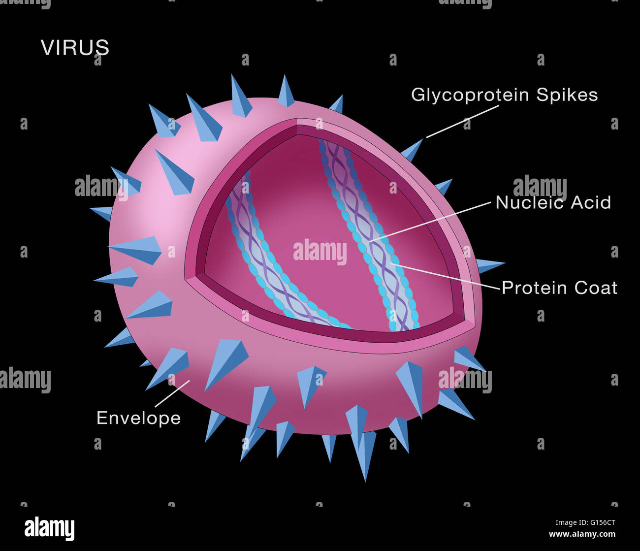
Labelled diagram of a typical virus. The genetic material made from either DNA or RNA. Explore virus structure structure of virus viral structure types and functions of virus structure. Besides these many animal viruses also contain 3 lipid envelope along with some additional parts such as the neck tail sheath tail fibers.
The shape of the capsid may vary from one type of virus to another. A virus is a nonliving particle made of proteins nucleic acids and sometimes lipids. Host cell makes copies of virus parts Biosynthesis Virus attaches to host cell Attachment Virus is released from host cell Release Virus is assembled Assembly DNA from virus enters host cell Penetration 2.
Given is a diagram of a bacteriophage. Well Labelled Diagram Of Bacteria. Labels are a means of identifying a product or container through a piece of fabric paper metal or plastic film onto which information about them is printed.
Structure of Viruses. A virus is an infectious particle that reproduces by commandeering a host cell and using its machinery to make more viruses. The information can be in the form of.
A simple diagram of a bacterium labelled in English. The capsid is made from the proteins that are encoded by viral genes within their genome. Nucleic acid or genome.
Most viruses that have been studied have a diameter between 20 and 300 nanometers. Viruses are much smaller than bacteria. Virus particles have a variety of shapes.
Diagram showing the structure of a typical virus. Virus particles or virions generally consist of two or three parts. A virus is made up of a DNA or RNA genome inside a protein shell called a capsid.
For more anatomy content please follow us and visit our website. Instead they have a core of genetic material surrounded by a protein coat. Check out this page - VIRAL STRUCTURE Look at the pictures of two types of viruses below click on it to.
The largest virus eg poxvirus measures about 250 nm ie as. And in some cases a lipid envelope that su - G156CW from Alamys library of millions of high resolution stock photos illustrations and vectors. A typical virus consists of two basic parts.
A protein coat that protects these genes. Draw a labelled diagram of. Viruses differ widely in terms of size and structure as you can see in the following diagram.
The virions vary widely in size. We hope this picture Label The Diagram Of A Virus can help you study and research. Previous question Next question.
A nonliving particle made up of nucleic acid and a protein coat or nucleic acid and lipid-protein lipoprotein coat. Some filoviruses have a total length of up to 1400 nm. Label the diagram using the descriptions.
Wide collections of all kinds of labels pictures online. Main parts of a virus. A virus is an infectious non-living particle that cannot survive on its own.
This diagram was created with an unknown SVG tool This SVG diagram uses embedded text that can be easily. Replication of a Retrovirus Genentechs Access Excellence Graphics Gallery A simple BW diagram labeled. 100 1 rating 1 The general structure of a virus particle.
Viruses are very diverse. Virus Viroids and Lichens. Some viruses have an external membrane envelope.
They do not have a cellular structure. A virus particle is called virion. In this article we will discuss about the structure of viruses.
Viruses are considered to be nonliving because they do not have the ability to reproduce on their own. The smallest virus measures about 10 mm in diameter eg foot-and-mouth disease virus. It shows the cytoplasm nucleoid cell membrane cell wall mitochondria which bacteria lack plasmids flagella and cell capsule.
Viruses vary in their structure. Diagram of a Retrovirus Genentechs Access Excellence Graphics Gallery A simple BW diagram labeled of a retrovirus. Was this answer helpful.
Make your work easier by using a label. They must be inside a host cell in order to reproduce. Their diameters are only about 80 nm.
A virus particle consists of DNA or RNA within a protective protein coat called a capsid. The life cycle of the virus is a series of steps that enable the virus to infect a host and replicate itself. 2 Viral envelope is a Lipid bilayer membrane acquired from a host cell membrane when virus buds plasma membrane or passes through a mem view the full answer.
We think this is the most useful anatomy picture that you need. Viruses are much smaller than bacteria. I can state the structure and function of a virus.
Draw a labelled diagram of bacteriophage. The capsid and entire virus structure can be mechanically physically probed through atomic force microscopy. Write the stages in the correct order starting with Attachment.
1 nucleic acid genome and 2 protein capsid together called the nucleocapsid. Up to 10 cash back Download this stock image. The shape of the capsid serves as one basis.
The structure and function of synovial joints is our second dash point under the skeletal system. The six types of synovial joints allow the body to move in a variety of ways.

Describe The Structure Of Synovial Joint With The Help Of A Neat Labelled Diagram
Label the friction-reducing structures associated with synovial joints.
Draw a labelled diagram of a typical synovial joint. This activity contains 5 questions. A synovial joint is characterised by the presence of a fluid-filled joint cavity contained within a fibrous capsule. Synovial Joint Diagram Label.
Label the parts of a generalized synovial joint. Branches of a typical spinal nerve. The six types of synovial joints are the pivot hinge saddle plane.
Concept Notes Videos 380. Types of Synovial Joints. Draw a labelled diagram to explain the different parts of a complete flower.
Explain the structure of the rib cage. The morphology of synovial membranes may vary but it. Maharashtra State Board HSC Science General 11th.
The basic structure of a synovial joint is shown in the diagram below. Learn vocabulary terms and more with flashcards games and other study tools. Joints are formed where bones come together.
Learn vocabulary terms and more with flashcards games and other study tools. Start studying Synovial Joint. Synovial fluid is the clear viscid lubricating fluid secreted by synovial membranes.
Synovial joints are the most common type of joint in the body Figure 1. Previous question Next question. Synovial fluid view the full answer.
Question Bank Solutions 5549. Articular capsule o Fibrous layer o Synovial membrane Joint cavity Synovial fluid Articular cartilages Ligaments Yellow bone marrow Periosteum Fibrous. Draw labelled diagram Synovial joint.
The shape of the joint affects the type of movement permitted by the joint Figure 1. List the different types of joints found in the human body. Create healthcare diagrams like this example called Synovial Joint in minutes with SmartDraw.
The main parts of synovial joints are labelled on the synovial joint diagram. A typical synovial joint. Modifications of deep fascia Click here.
This fluid-filled space is the site at which the articulating surfaces of the bones contact each other. On Labelled Diagram Of Synovial Joint. A synovial membrane or synovium is the soft tissue found between the articular capsule joint capsule and the joint cavity of synovial joints.
A key structural characteristic for a synovial joint that is not seen at fibrous or cartilaginous joints is the presence of a joint cavity. It is the most common type of joint found in the human body and contains several structures which are not seen in fibrous or cartilaginous joints. Mention where are they located 5.
Arteries supplying a developing long bone. Allows movement in only one plane. Fibrous connective tissue found in various parts of the body such as the joints.
Synovial joints are the the most common joints present and have a characteristic trait of having the presence of a joint cavity the cavity filled with the synovial fluid. Give one example each for a hinge joint a pivot joint axial skeleton and appendicular skeleton. Elbow joint and knee joint.
Draw a labelled diagram of a synovial joint. Also allows movement in only one plane. Start studying label the synovial joint.
Advertisement Remove all ads. Synovial joints are further classified into six different categories on the basis of the shape and structure of the joint. The skeletal system has a number of different.
Primary movement is a rotation. In this article we shall look at the anatomy of a synovial joint the joint capsule neurovascular structures and clinical. Synovial joints allow for smooth movements between the adjacent bones.
Parts of a developing long bone. The skeletal system has a number of different. These joints can be described as planar hinge pivot condyloid saddle or ball-and-socket joints.
Use specimens in your bone trays illustrations in your textbook atlas to draw label a diagram of a typical synovial joint bellow. Synovial joints are the most movable type of joint found in the human body. A synovial joint is a connection between two bones consisting of a cartilage lined As seen in the above picture the most powerful bite in the world gets its.
A synovial joint or diarthrosis occurs at articulating bones to allow movement. This diagram shows the location of the bursae which are fluid filled sacs in a bone. Synovial joints are subdivided based on the shapes of the articulating surfaces of the bones that form each joint.
For each item below use the pull-down menu to select the letter that labels the correct part of the image. The structure and function of synovial joints is our second dash point under the skeletal system. Draw labelled diagram of the following.
Be sure to include labels of the following structures. The six types of synovial joints are pivot hinge condyloid saddle plane and ball-and socket-joints Figure 943Figure 943 Types of Synovial Joints. SmartDraw includes 1000s of professional healthcare and anatomy chart templates that you can modify and make your own.
Draw a mind map to show the different parts of the Human Skeletal System. Write differences between pectoral and pelvic girdle. Draw labelled diagram Synovial joint.