But water is not the answer. Using Epsom salt and creating a salt soak in the bath can help exfoliate cracked skin even the tough hard skin on heels.

3 Ways To Remove Dead Skin From Feet Wikihow
How to Get Rid of Dry or Dead Skin on Feet Face Lips Hands Natural Tips.

How to get rid of feet skin. You should soak your feet in vinegar solution with half portion vinegar and half portion water. How to Get Rid of Peeling Skin on Your Feet 1. Use warm water to wash your feet in the morning 4Vinegar If you want to remove the thick dead skin from your feet vinegar will not disappoint you.
Just make sure to apply moisturizer later. Steps to get rid of hard skin on feet. Soak the feet in.
Go electric to help you out. How To Get Rid Of Hard Skin On Feet. A pumice stone is made of volcanic rock and is useful for removing dead skin on your body including your feet.
This one is almost everyones favorite remedy against callus because all the ingredients can be found in our very own kitchen. Scrubbing helps in removing the. Wash and dry your feet.
The epsom salts help fluff up the hard skin. Do this for 15 minutes. It will get rid of the dead skin and also moisturize your feet.
Once it gets contacts with your skin it softens and loosens the accumulated dead skin leaving your skin softer and healthier. You can also leave pieces of lemon peel in the water. Prepare a footbath with warm water.
Squeeze in lemon juice from one lemon. Soak your feet for up to 15 minutes. You can make a foot scrub with oatmeal so that you can remove dead skin cells effectively at the same time and you can also keep the skin on your feet moisturized.
Glycerin For Removal Of Calluses. If you decide to use this method. Often used in soaps and skin care products glycerin is a compound that draws moisture through the skin and slows and prevents evaporation and excessive drying.
Just add a whole cup of epsom salt to your hot bath and let it dissolve then soak your skin. You may use a loofa or a pumice stone to scrub the feet with soap and water. Similar to lemon juice vinegar is a multi-purpose product that has been used for skin ailments and other problems for centuries.
Regularly scrubbing the feet helps remove the dead and hard skin that often gets collected and causes dry and cracked soles. How to Get Rid of Dry or Dead Skin on Feet Face Lips Hands Natural Tips - YouTube. They can do this by following the steps below.
Water actually dehydrates your skin. People can use a pumice stone or metal foot file to remove dry skin and calluses from the feet. Warm water can help relax the feet especially if they are soaked in the evening.
You must use oils and moisturizers. This will loosen up dead skin cells that would cause the feet to become dry. This one is like old grandmas scrub of getting rid of callus yet very much useful.
Let your feet soak for several minutes and then rub them with a pumice stone. The key to softening the dry skin on your feet is to moisturize. Scrub the feet well.
Both methods will soften the feet and help reduce calluses. Run a warm bath and add a small amount of Epsom salt to the water. Dryness is the primary cause of peeling skin on the feet so its critical that you keep your skin.
Pumice stone method is a fast way to get rid of the hard skin on feet. Allow the oil to cool rub on the feet and cover overnight with cotton socks. The water epsom salt bath will help to loosen up the dead skin making it easier to remove.
Get a professional to help. Use a foot brush to scrub dead skin off your feet. Many experts recommend first soaking your feet in warm water to soften the skin then using an exfoliating scrub to gently remove dead skin.
A method that a lot of people are now using is going electric and that means checking. In severe cases it may be best to initially seek some professional help from a. If you would like to get rid of dead skin on your feet with an effective remedy oatmeal is the right remedy for you.
You can also soak your feet in a foot bath with epsom salts. 11 Proven Methods 1. Soaking your feet in warm water for a few minutes daily will help loosen accumulated dead skin.
The skin has up to seven layers of ectodermal tissue and guards the underlying muscles bones ligaments and internal organs. Though nearly all human skin is covered with hair follicles it can appear hairless.
Integumentary System Review Key
The skin serves various functions that include acting as the bodys initial barrier against germs UV light chemicals and mechanical injury.

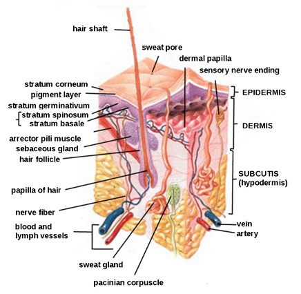
Label the parts of human skin. The epider- mis dermis and subcutaneous tissues Fig. Structure of the Skin. At the boundary between the epidermis and dermis are finger-like projecting structures the dermal papillae that project into the overlying tis- sue the epidermis Fig.
Is a tough coating formed from overlapping layers of dead skin cells. Labelled diagram of human body bones labelled diagram of human body parts labelled diagram of human eye labelled diagram of human heart labelled diagram of human skeleton labelled diagram of the human body labelled diagram of the human body organs Human Anatomy labelled diagram of human. Skin generally consists of a three-layer structure.
25 best ideas about human skeleton labeled on 28 images 25 best ideas about human skeleton labeled on best 25 human skeleton labeled ideas on human 25 best ideas about human skeleton bones on skeleton diagram labeled bones skeleton diagram labeled lesson 2. View Human Skin Worksheetpdf from BIOLOGY MISC at Western University. Human Skin Label the parts of the diagram of human skin Give a description of each.
It contains blood cells sweat glands and hair follicles. The game The Skin is available in the following 4 languages. Epidermis - the outer layer of the skin.
Human skin is similar to most of the other mammals skin and it is very similar to pig skin. An Overview The skin consists of two layersthe outer thinner epidermis and the inner thicker dermis. Webmds skin anatomy page provides a detailed image of the skin and its parts as well as a medical definition.
Papillae--part of the papillary layer Increases the surface area between the epidermis and dermis providing oxygen and nutrients to the outermost layer Nerve cells. WebMDs Skin Anatomy Page provides a detailed image of the skin and its parts as well as a medical definition. From the quiz author.
Learn about the skins function and conditions that may affect the skin. Labeling the parts of the skin. Label the parts of the skin.
Start studying skin parts labels. There is a printable worksheet available for download here so you can take the quiz with pen and paper. The Fitzpatrick scale developed by Thomas B.
The human skin is the outer covering of the body and is the largest organ of the integumentary system. Hair follicle - a tube-shaped sheath that surrounds the part of. Skin makes up the largest part of the integumentary system which.
Human Skinpdf - Read File Online - Report Abuse. Underneath the dermis is the hypodermis also called the subcutaneous layer which is where fat is stored. The first five layers form the epidermis which is the outermost thick layer of the skin.
Human Skinpdf - nimitz126 HUMAN SKIN Name _____ _ Label the following parts of human skin on the diagram below. Labeling the parts of the skin. All seven layers vary significantly in their anatomy and function.
The middle section of the skin is called the dermis. Human Skin Diagram Label - 15 Diagram Of The Skin Anatomy. It was developed as a means to estimate the response of varying skin types to.
Fitzpatrick in 1975 is a numerical classification schema for identifying human skin color accurately. 13 photos of the Labelled Diagram of A Human Skin. The surface layer of the two main layers that make up the skin in animals contains basal cells squamous cells and melanocytes.
Dermis - also called the cutis the layer of the skin just beneath the epidermis. The outer layer of cells on a plant. Diagram of human skin structure.
Learn vocabulary terms and more with flashcards games and other study tools. This is an online quiz called Skin Labeling. From the epidermis to the hypodermis and everything in between when youre learning the anatomy of the skin use this quiz game to make it easy.
Hair shaft the part of the hair that is above the skin.
Find this Pin and more on Dermatologyby Jennifer Bunce-Stone. The deepest layer of the skin.

Human Skin Wikipedia
An easy and convenient way to make label is to generate some ideas first.
Labelled diagram of the skin. Also available for free download. It is designed to withstand repetitive insults in order. Skin Anatomy Song for KidsSkin Song for ChildrenSkin Anatomy.
Skin Structure Diagram - See more about Skin Structure Diagram basic skin structure diagram blank skin structure diagram simple. Skin has two main layers. Finally the stratum corneum is the most superficial layer of the skin.
Skin diagram labeled 1087. The following diagram illustrates the basic structure of the skin labelling key components. The Three Layers of Skin Are.
Sketch The Onion Peel Cell As Seen Under The Microscope. Protection regulation and sen. Find high-quality royalty-free vector images that you wont find anywhere else.
If the damage reaches the dermis and the basal membrane is affected eg. Read the definitions then label the skin anatomy diagram below. Skin has various regeneration and repair mechanisms.
An ulcer then scarring normally occurs. The skin is the largest organ of the body it covers the whole body and they are water resistant. It may be useful preparation for andor revision of courses in bodywork therapies.
Skin diagram labeled 1111. Integumentary System Skin Parts. The basal layer ensures a steady renewal of the epidermis through continual cell division.
We shed more than 500 million skin cells each day. Label the diagram of the skin - heart diagram using Labelled diagram - Skin care - Venn Diagram - Water Cycle Diagram - Ear Diagram Year 4. Blood vessels - Tubes that carry blood as it circulates.
Epidermis - the outer. Labeled Diagram Of An Onion Cell - Example Electrical Wiring Diagram Plant guard cells stoma fully labeled diagram showing open closed 51409771. Skin diagram labeled 1080.
Onion Skin Cell Labeled Diagram. The outermost layer of the skin is called the epidermis. Skin diagram labeled 1082.
Skin structure skin aging stages wrinkle skin structure of the skin the skin anatomy needle skin structure skin collagen infographic skin wrinkles skin glands. 7198 skin diagram stock photos vectors and illustrations are available royalty-free. Veins return oxygen-depleted blood back to the heart and lungs.
The cells are filled with keratin filaments and are devoid of nuclei and organelles. Diagram of the skin is illustrated in detail with neat and clear labelling. Skin Structure Diagram - Anatomy Human.
See skin diagram stock video clips. Arteries bring oxygenated blood from the heart and lungs. Beneath the two layers is a layer of subcutaneous fat which also protects your body and helps you adjust to outside temperatures.
Skin diagram labeled 1097. A Well Labeled Diagram Of An Onion Cell - Explore Schematic Wiring Diagram 09F33069 D38C 4794 AFBA 59DBE1731379. Find Cross Section Human Skin Labels stock images in HD and millions of other royalty-free stock photos illustrations and vectors in the Shutterstock collection.
Skin diagram labeled 1089. The outer layer of the skin. The skin consists of two layers EPIDERMIS and DERMIS.
Here a translucent layer of cells lie above the stratum granulosum and below the stratum corneum. Dermis - also called the cutis the layer of the skin just beneath the epidermis. Thousands of new high-quality pictures added every day.
Label The Parts Such. Skin is the layer of usually soft flexible outer tissue covering the body of a vertebrate animal with three main functions. If an injury is confined to the outermost skin layer the damage known as erosion can heal without scarring.
Try these curated collections. Explore Skin Diagram with BYJUS. Choose from Labeled Skin Diagram stock illustrations from iStock.
Skin diagram labeled 1077. The stratum lucidum is an epidermal layer only appreciated in glabrous skin. The components labelled in the above diagram include the following.
Best viewed on 1280 x 768 px resolution in any modern browser. Label Gallery Get some ideas to make labels for bottles jars packages products boxes or classroom activities for free. Skin Structure Diagram Labelled Diagram Skin Structure - Diagram Body Of Anatomy.
Dead cells are shed continuously from the epidermis as new skin ones take their place New skin. Skin diagram labeled 1075. DRAW AND LABELLED DIAGRAM OF THE SKIN AND DESCRIBE ITS STRUCTURE AND FUNCTION INCLUDE ALL KEY PARTS.
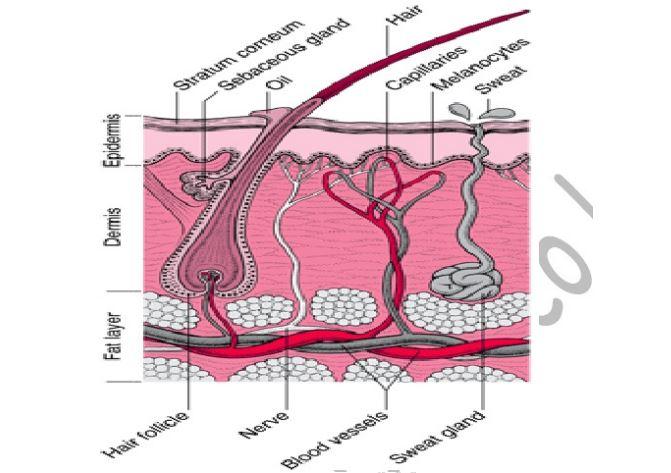
Epidermis Dermis Subcutaneous Layer Hair Stratum Corneum Capillaries Sebaceous Gland Melanocyte Sweat Gland Sweat Pore Hair Follicle Blood Vessel Nerves Arrector Pili Muscle. The part that consists of connective tissue. The Structure of Human Skin Comprises Three Layers.
It gives the skin its tone. Skin Anatomy Diagram Labeled. The Skin - Labelled diagram.
Label the Skin Anatomy Diagram.
Www Ti Com Wed 11 Apr 2018 Tm116w715l Solo 10ao 20 09. Schematic Representation Of Mammalian Skin Structure And Its Cell.
Draw And Label A Mammalian Skin
The skin is an organ that forms a protective barrier against germs and other organisms and keeps the inside of your body inside your body and skin is made up of two layers that cover a third fatty layer.

Labelled diagram of mammalian skin. It is continuous with the mucous membrane in the region of mouth nose urogenital organs and anus. They are 3 layers of skin. The presence of hellical or spiral arrangement of chloroplast makes it from the order Zygnematales of Zygnematacae family of Algae.
Epidermis dermis and hypodermis. Epidermis dermis and hypodermis. Draw a labelled diagram of the generalised vertical section of the mammalian skin.
Epidermis dermis and subcutis. Ii Portion of the nephron between point X and Y. Com an online medical question bank.
Collagen Human Skin Effect. Accessory Structures Of The Skin Anatomy And Physiology I. Labelled Pictures Of Human Skin - A Diagram Of Mammalian Skin Wire Data.
From deep to superficial these layers are the stratum basale stratum spinosum stratum granulosum and stratum corneum. Skin Integumentary System Histology Drawings. Labelled longitudinal section of skin anatomy enchantedlearning com mammalian skin cell biology at the interface between skin diagram and information about your skin how to draw integumentary system skin layers youtube cell cycle wikipedia draw a labelled diagram of the generalized vertical section of the mammalian skin solution e 1.
A Label the parts numbered 1 to 9 b State one main function of each of the following parts. It has three layers. The Structure Of Skin Integumentary System.
The human skin is the outer covering of the body. Andrea Paul a health care provider and main medical officer at Boardvitals. The human skin is the outer covering of the body.
Jun 26 2019 - For the sweet along with savory treat that wont blockage your veins opt for a pot of natural yoghurts which will shield more than just your own heart states that Dr. Skin that has four layers of cells is referred to as thin skin. The diagram below represents a mammalian nephron Solved The diagram below represents a mammalian nephron a Name the istructure labeled P.
Close up damaged old and healthy vector medical skin care cosmetic diagrams. It is continuous with the mucous membrane in the region of mouth nose urogenital organs and anus. A Name the parts labeled E F and G b State two functions in each case of substance secreted by the structures labeled.
B Name the process that takes place at point Q. 12 13 00 GMT Experiment 2 INTRODUCTION Explore Molecule. CreativeMarket Collagen human skin effect 1161237.
Yogurt protects next to gum. Labeled Diagram Of Mammalian Heart Pictures Images. Advanced Labelled Diagram Of The Mammalian Skin.
This is a complicated epithelium consisting of. The human skin and its related structures are known as the integumentary system. The outermost layer of our skin is called the epidermis.
Collagen human skin effect. 1000 Skin Diagram Stock Images Photos Vectors Shutterstock. Most of the skin can be classified as thin skin.
Draw a labelled diagram of the generalised vertical section of the mammalian skin. I H ii I. They are 3 layers of skin.
The diagram below shows a section through the mammalian skin. A well-labelled diagram of spirogyra is attached. Biology 25042020 1612 ImmortalEnigmaYT A well labelled diagram of a mammalian skin.
A well labelled diagram of a mammalian skin. This thin tough layer consists of. Given below is a diagrammatic sketch of the vertical section of human skin.
The diagram below shows a section through the mammalian skin. Thick skin is found only on the palms of the hands and the soles of the feet. THE STRUCTURE OF MAMMALIAN SKIN The diagram above shows a vertical section shows that it is composed of two main parts an outer part called the epidermis and an inner thicker part the dermis.
Skin Gio Vyrjkk Loan. A name the parts labeled e f and g human skin anatomy britannica human skin in human anatomy the covering or integument of the bodyâs surface that both provides protection and receives sensory stimuli from the external environmentthe skin consists. Draw a labelled diagram of the generalized vertical section of the mammalian skin solution e 1 vertical section of the mammalian skin question e 2 given below is a diagrammatic sketch of the vertical section of the human skin a label the parts numbered from 1 to 9 b state one main function of each of the following parts mammalian skin is covered with.
Skin is the outermost covering of mammalian body. Skin Diagram Labeled Simple Written By JupiterZ Saturday June 3 2017 Add Comment Edit. The outer cornified layer The cells in this layer are dead dry flat and horny because.
C Name one substance present at point R but absent at point S in healthy mammal. A spirogyra is a filamentous chlorophyte green algae.