The reticular formation is the medulla pons and midbrain and comprises the brainstem core also known as the tegmentum. The reticular formation is a cluster of nerves within the brainstem that relay sensory and motor signals to and from the spinal cord and the brain.
Neuroscience For Kids The Brain Right Down The Middle
The human brain is an astonishing organ that takes care of each function and action of the body.

Brain diagram labeled reticular formation. Labeled Diagrams of the Human Brain. This may occur either suddenly due to vascular disor. The reticular formation is a set of interconnected nuclei that are located throughout the The human reticular formation is composed of almost brain nuclei and contains many projections into the forebrain brainstem and cerebellum.
It is easier to appreciate the approximate location of these nuclei if they are superimposed on a posterior view of the brainstem with the cerebellum removed. Each of these nuclei performs one of the functions listed above plus a few more. All the functions are carried out without a single glitch and before you even bat an eyelid.
Controlsregulates heartbeat and breathing To and from brain Reticular Formation Helps control arousal responds to change in monotony Thalamus Relays sensory information switchboard between sensory neurons and higher brain regions Deals with sight hearing touch taste. The reticular formation is a neural network located in the medulla that helps control functions such as sleep and attention Reticular formation Important Parts of the Brain Stem. The following are the different regions of the human brain and their functions.
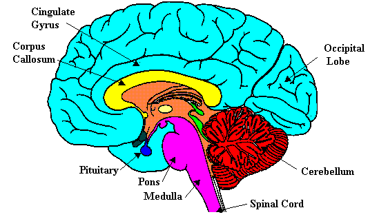
This diagram labels the cranial nerves including olfactory oculomotor trochlear abducens. Central Core The central core consists of the thalamus pons cerebellum reticular formation and medulla. Brain stem Reticular formation.
Download for free from a curated selection of Brain Diagram Reticular Formation for your mobile and desktop screens. It is an arrangement of neurons throughout he brain stem. The thalamus interprets the sensory information and helps determine what is good and bad.
The reticular formation is located in the brain stem. Describe the functions of the reticular formation region of the pons. These five regions are the central areas that regulate breathing pulse arousal balance sleep and early stages of processing sensory information.
These nuclei are the cardiovascular center respiratory rhythmicity center nucleus gracilis and nucleus cuneatus olivary nucleus the reticular formation of the medulla oblongata and the sensory and motor nuclei of the cranial nerves VIII IX X XI and XII. It occupies the anterior portions of medulla pons midbrain hypothalamus and thalamus. The Reticular Formation Descending Reticular Formation Sleep and ArousalNeuronal Basis of Changes in the EEG Sleep Disorders The Ascending Reticular Activating System The brainstem contains many small neural networks that regulate essential functions including the arousal system cardiovascular and respiratory control and the control of somatic muscle tone.
Function and anatomy of parts diagram conditions brain diagram use this interactive 3 d. Cerebellum reticular formation and medulla these five regions are the central areas that regulate breathing pulse arousal balance sleep and early stages of processing sensory information the thalamus interprets the sensory information and helps determine what is good and bad it then. Definition Mass of neurons and nerve fibers extending from the caudal medulla to the rostral midbrain and continuous with the zona incerta of the subthalamus and midline intralaminar and reticular nuclei of the thalamus Organized into definite nuclear groups with known afferent and efferent connections As a whole the reticular formation comprises a neural.
Topographically the nuclei can be. It then sends. The parvocellular neurons receive information relating to arousal.
The reticular formation nuclei are found deep within the brainstem along its length. The descending fibers of the reticular formation as well as the ascending system are critical in gating the sensory inputs and play a critical role in pain modulation mainly by acting on the posterior horn of the spinal cordAll these activities are impaired when a damage affects critical nuclei of the reticular formation. Reticular Formation Anatomy And Clinical Notes Kenhub Pin On Health Maladies Reticular Activating System Diagram In 2019 Reticular Reticular Formation Reticular Formation Brain Anatomy Neuroscience L51 Central Control Of The Ans Neuroscience Goal Setting Disciplined Behavior And Your Brain The Neurotransmitter System And Neural Circuits Governing Sleep Reticular.
It extends throughout the length of the brainstem along the central axis from the spinal cord to the thalamus. Transmits replies from higher brain to cerebellum and medullam Cerebellum Influences memory and learning coordinates. Weve gathered our favorite ideas for Brain Diagram Reticular Formation Explore our list of popular images of Brain Diagram Reticular Formation and Download Every beautiful wallpaper is high resolution and free to use.
Labeled Diagrams of the Human Brain. Our interactive diagram helps you explore the anatomy of the human brain and learn The reticular formation controls muscle tone in the body and acts as the.