Define its magnifying power. I Which lenses should he used as objective and eyepiece.

A Concave Mirror Is Used For Image Formation For Different Positions Of An Object What Inferences Can Be Drawn Science Light Reflection And Refraction 14317813 Meritnation Com
I Which lenses should he used as objective and eyepiece.

Draw a labelled ray diagram to justify your inferences. Nature of image---- real and inverted. Clearly mark angle of incidence angle of refraction angle of emergence and lateral displacement of the ray. Draw A Labelled Ray Diagram Of A Reflecting Type Telescope Write.
It is defined as the ratio of the angle β subtended by the final image on the eye to the angle α subtended by the object on eye. 0 Response to Labelled Refraction Ray Diagram Post a Comment. Share It On Facebook Twitter Email.
Define its magnifying power. Clearly mark angle of incidence angle of refraction angle of emergence and lateral displacement of the ray. I Which lenses should be used as objective and eyepiece.
A concave mirror is used for image formation for different positions of an object. CBSE CBSE English Medium Class 10. B if it hits glass block at an angle other than 90 degrees.
Asked Dec 23 2015 in Class X Science by luckky ramya Expert 18k points 0 votes. Ii Aperture of the objective lens should be. Draw a labelled ray diagram to locate the image of an object fromed by a convex lens of focal length 20 cm when the object is placed 30 cm away from the lens.
Draw a labelled ray diagram to justify your inferences. Draw a ray diagram showing the path of the light ray. B You are given three lenses of power 05 D 4 D and 10 D to design a telescope.
A Draw a labelled ray diagram to obtain the real image formed by an astronomical telescope in normal adjustment position. A Draw a labelled ray diagram to obtain the real image formed by an astronomical telescope in normal adjustment position. Refraction Of Light Science Learning Hub.
Pages 35 This preview shows page 20 - 26 out of 35 pages. Click hereto get an answer to your question 21 Aconcave mirror is used for image formation for different positions of an obiect. Size of the image-- enlarged.
What inferences can be drawn about the following when an object is placed at a distance of 10 cm from the pole of a concave mirror of focal length 15 cm. A if it hits glass block at 90 degrees. Total Internal Reflection Wikipedia.
B You are given three lenses of power 05 D D and 10 D to design a telescope. Draw a Labelled Ray Diagram to Show the Angle of Incidence and the Angle of Refraction for a Refracted Ray of Light. A Position of the image b Size of the image c Nature of the image.
B You are given three lenses of power 05 D 4 D and 10 D to design a telescope. Course Title CHEMISTRY MISC. A ray of light incident at an angle of incidence i passes through an equilateral glass prism.
Answered May 4 2018 by sanjaydas 889k. Question Bank Solutions 20334. Draw a labelled ray diagram to justify your.
Ii Why is the aperture of the objective prefered to be large. Explain why both the objective and the eye piece of a compound microscope must have short focal lengths. How to draw a ray diagram of the convex mirror in an easy way for class 10 beginner students who wants to achieve full mark in boardWatch How To Draw Huma.
Define its magnifying power. Define its magnifying power. A Position of the image b Size of the image c Nature of the image Draw a labelled ray diagram to justify your inferences PIO.
First we draw a ray parallel to principal axis So it passes through focus after refraction We draw another ray which passes through Optical Center So the ray will go through without any deviation Where both refracted rays meet is point A And the image formed is AB This image is formed between beyond 2F 2 We can say that Image is Real. Concept Notes Videos 224. I Image is not free from chromatic aberration and spherical aberration.
Draw a labelled diagram to show how a ray of light passes through a parallel sided glass block. Draw a ray diagram to show the path of the reflected ray corresponding to an incident ray of light parallel to the principal axis of a convex mirror and show the angle of incidence and angle of reflection on it. Draw the labelled ray diagram for the formation of image by a compound microscope.
Post Comments Atom. B You are given three lenses of power 05 D 4 D and 10 D to design a telescope. A Draw a labelled ray diagram to obtain the real image formed by an astronomical telescope in normal adjustment position.
Newer Post Older Post Home. What inferences can be drawn about the following when an object is placed at a distance of from the pole of a concave mirror of focal length. Derive the expression for the total magnification of a compound microscope.
A Position of the image b Size of the image c Nature of the image Draw a labelled ray diagram to justify your inferences. A Draw a labelled ray diagram to obtain the real image formed by an astronomical telescope in normal adjustment position. Draw a labelled ray diagram to justify your inferences 3 Answer Given Position.
Position of image -- 30 cm. Reflection Of Light Visible Light Siyavula. What inferences can be drawn about the following when an object is placed at a distance of 10 cm from the pole of a concave mirror of focal length 15 cm.
Arguably the most important tendon is the Achilles tendon which allows the calf muscles to move the ankle joint. It is often caused by overuse fatigue or improper use of muscles.

Foot Bones Anatomy Conditions And More
The calf muscle may be tight placing stress on the tendons at the top of the foot that pull the foot upward.
What is the muscle on the top of your foot called. The ankle joint or tibiotalar joint is formed where the top of the talus the uppermost bone in the foot and the tibia shin bone and fibula meet. Only two of these muscles are located on the dorsal aspect top of the foot. Extensor tendonitis is a common problem that causes pain across the top of the foot.
If they become inflamed due to overuse or. This is rare and characterized as an inflamed sinus tarsi. The tibialis anterior muscle is the muscle located in the front part of the shin bone of your lower leg.
It flexes and extends the foot ankle and knee. Function of the Tibialis Anterior Muscle. When the muscles tighten contract they pull on the tendons which in turn move the bones.
The muscles at the top of the foot fan out to supply the individual toes. The muscles in the sole of the foot are located in four layers. One of the large muscles of the leg it connects to the heel.
Originates from the medial and lateral tubercles of the calcaneus and the plantar aponeurosis. Bones and Joints of the Foot and Ankle The Ankle Lateral side of the ankle Joint capsule. You can pull any muscle in your body but this is the most common in feet lower back and neck.
Another very common cause of the condition is the foot rubbing against a shoe. This is the muscle that allows the trunk of your body to twist this is controlled by the external oblique muscle on the opposite side of the direction that youre twisting. Collectively they are referred to as the intrinsic muscles of the foot because they are entirely contained within the foot.
A pulled muscle also known as a muscle strain is a result of a torn or overstretched muscle. It is homologous with the abductor digiti minimi of the hand. One of the large muscles of the leg it connects to the heel.
Gastrocnemius calf muscle. You can divide the foot muscles into groups based on their locations either in the sole of the foot the bottom or plantar area or the dorsum on the top of the foot. Extensor tendinitis involves inflammation of the tendons near the middle top of the foot.
Gastrocnemius calf muscle. Most of the muscles are in the sole. This form of foot tendonitis is caused by inflammation or irritation of the tendons that pull the toes up.
The abductor digiti minimi muscle is located on the lateral side of the foot. Adapted screenshot from Kenhub anatomy video CFCF via Wikimedia Commons cc Gluteus medius is responsible for abduction and rotation of the hip. Your external obliques sit on either side of your rectus abdominis and are actually the largest of your abdominal muscles.
This muscle extends from the back of the knee. The extensor tendons located in the top of the foot are needed for flexing or pulling the foot upward. If muscles are overworked or overstressed they can become torn or strained.
Typically tendonitis of the extensor tendons develops due to repeated friction across the top of the foot or excessive pressure from a poorly-fitting shoe. The muscle courses from an area just below your knee down the front of your shin and finally attaches to the top of your foot. Strains usually manifest as pain especially with movement or pressure.
The Muscles of the Foot. The extensor hallucis brevis and the extensor digitorum brevis. What does this mean.
The tendons that run along the top of the foot and pull the foot upwards become inflamed and painful. It flexes and extends the foot ankle and knee. The gluteus medius muscle sits as a deeper layer of muscle beneath the gluteus maximus and is also sometimes referred to as your upper glutes.
The ankle joint is both a synovial joint and a hinge joint. Hinge joints typically allow for only one direction of motion much like a door-hinge. The muscles of the dorsum top of the foot You can learn more about each individual group of muscles in the foot using this table.
This small thin muscle is absent in about. The tendons are thick bands that connect muscles to bones.
It is also known as the cortex and is. Occipital-this bone is on the lower back side of the brain.
:no_upscale()/cdn.vox-cdn.com/uploads/chorus_asset/file/2445172/brain_etymology.0.jpg)
Did You Know There S A Part Of Your Brain Called The Slime Gland Vox
The outer part of the brain is called cerebrumThe outer part located in the back of your head is the cerebellumThe third part of the brain located beneath the cerebrum and in front of the cerebellum is the brain stem.
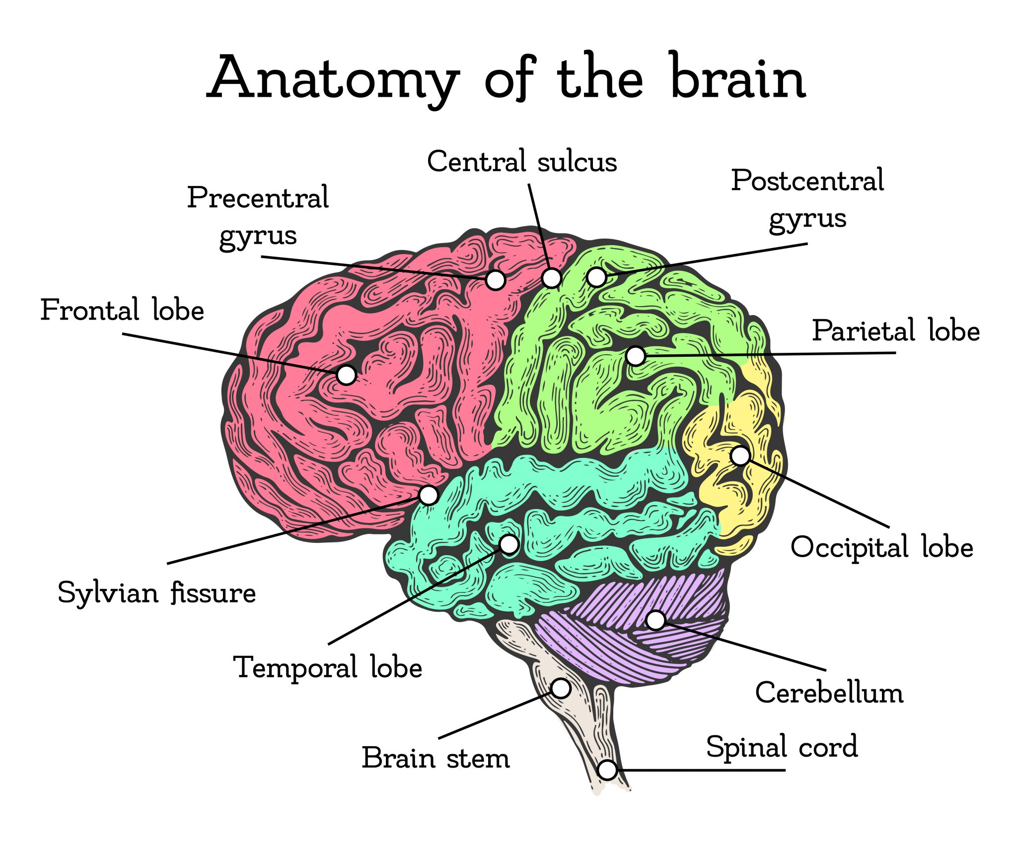
What are the parts of your brain called. This illustration shows three basic parts. Brain Stem Where the spinal cord meets the brain is a bulge called the brain stem. Understanding the Parts of the Brain To understand how emotions relate to thinking and learning you need to understand the parts of the brain.
What Are the Main Parts of the Brain. These parts are the Medulla Pons and the Midbrain. The forebrain is responsible for a number of functions related to thinking perceiving and evaluating sensory information.
The cerebral cortex is the part of the brain that most strongly distinguishes mammals. Its divided into two halves called hemispheres. The forebrain midbrain and hindbrain.
The brainstem is a collection of three areas of the brain. These areas are called lobes. Each lobe controls specific functions.
These three parts control processes in the body including movement memory and thinking. The cerebrum is the largest part of the brain. All the parts of the brain work together but each part has its own special properties.
Each brain hemisphere parts of the cerebrum has four sections called lobes. What Is the Cerebrum. The cerebrum cerebellum and the brain stem.
The cerebrum constitutes the largest part of the human brain. Was I A BeeWikimedia Commons. The forebrain the midbrain and the hindbrain are the three main parts of the brain.
The brainstem is located below the cerebellum and connects the brain to the spinal cord. Each lobe may be divided once again into areas that serve very specific functions. The cerebrum is the largest part of the brain.
The two hemispheres are separated by a groove. Sphenoid-this part of the skull forms the floor of the cranium and part of the eye orbit. Parts of the Brain and Their Functions Cerebrum.
There are three main parts of the brain. One lobe works with your eyes when watching a movie. The limbic system is located centrally and deep within the brain.
The limbic system is involved in emotional memory and mood control. A peek at the lower brain. It has the job of running all the involuntary muscles.
The cerebrum is the largest part of the human brain. The frontal lobe is located in the forward part of the brain extending back to a fissure known as the central sulcus. It consists of several small structures called the hippocampus amygdala thalamus and hypothalamus.
The brain has many different parts. The brain and its parts can be divided into three main categories. In mammals the pallium evolves into a.
The forebrain the midbrain and the hindbrain. Lobes are simply broad regions of the brain. The cerebrum or brain can be divided into pairs of frontal temporal parietal and occipital lobes.
The cerebellum also known as the little brain is located in the back of the brain. Parts of the Brain. It sits just below.
The forebrain has two major parts called the diencephalon and the telencephalon. The brain consists of three main parts. Denise WawrzyniakWikimedia Commons.
Frontal parietal temporal and occipital. The three main regions of the brain are the midbrain hindbrain and forebrain which is broken up into additional sections including the frontal lobe occipital lobe. The basic lower brain consists of the spinal cord brain stem and diencephalon the cerebellum and cortex are also present but will be discussed in later sections.
Each hemisphere has a frontal temporal parietal and occipital lobe. In non-mammalian vertebrates the surface of the cerebrum is lined with a comparatively simple three-layered structure called the pallium. The brain also has specific areas that do certain types of work.
The hindbrain includes the upper part of the spinal cord the brain stem and a wrinkled ball of tissue called the cerebellum 1. It has a rough surface cerebral cortex with gyri and. The cerebellum is known as the little brain and resembles the cerebrum for it has a highly folded surface.
Located in the front and middle part of the brain it accounts for 85 of the brains weight. Structures and Their Functions 1Cerebrum. Lets look at them from bottom to top.
Lobes of the Brain The four lobes of the brain are the frontal parietal temporal and occipital lobes Figure 4. The brain can be divided into three basic units. In turn the brain stem comprises the medulla pons midbrain hypothalamus and thalamus source.
Ethmoid-this bone forms the medial parts of the.
The thinnest part of your foot usually found towards its center is known as the waistline. It contains a lot of moving parts - 26 bones 33 joints and over 100 ligaments.
/footpainfinal-01-d507e82b3e844d068c0089cbb7004d76.png)
Foot Anatomy Physiology And Common Conditions
The area just underneath your toes corresponds to the chest.

Parts of your foot diagram. These arches the medial arch lateral arch and fundamental longitudinal arch are created by the angles of the bones and strengthened by the. Between them the two feet need to balance the weight of the body redistributing it in response to position changes. Anatomically the foot is divided into 3 sections.
There are 14 toe bones two per big toe and three per each of the other four plus five metatarsals. Honeywell Thermostat Wiring Diagram. Leave a Reply Cancel reply.
Like the four other metatarsals it can be divided into three parts. Remove the wrapper obviously and best way to do it is relax stand either feet apart knees slightly bent insert the tampon with thumb and forefinger inside your vagina and gently push up until comfortable. The instuctions will be on the box they come in and a picture diagram but heres what to do.
Such complexity is necessary because the foot is required to do many different activities such as walking running and climbing. Neck and head and the calcaneus. Parts of your foot correlated with the stomach are found above the waistline.
The midfoot contains the rest of the tarsal bones. 2004 F350 Fuse Diagram. The first metatarsal bone is the bone in the foot just behind the big toe.
If you would like to learn all the parts of the foot structure you have come to the right place. Then push the bottom part plastic tube applicator inside the top part holding now the ridgy bit in the middle that. Your email address will not be published.
Additionally the lower leg often refers to the area between the knee and the ankle and this area is critical to the functioning of the foot. The bones of the foot are organized into the tarsal bones metatarsal bones and phalanges. Lewis Dot Diagram For Phosphorus.
List of shoe pattern parts found in the shoe parts diagram drawing and More Picture of the Feet The forefoot Thinnest area of foot is waist and intestine it plays an. The thinnest part of your foot usually found towards its center is known as. This is if youll pardon the.
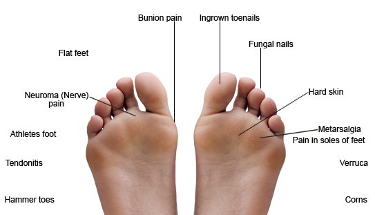
Foot is present at the end of the leg which helps us to stand walk and run. Front Foot Pain Identifier This foot pain diagram shows common problems that cause pain on top of the foot at the front. Posted on April 24 2019 April 24 2019.
Use it to pin point where you are having foot or ankle pain. The talus bone supports the leg bones tibia and fibula forming the ankle. The calcaneus heel bone is the largest bone in the foot.
The hindfoot forms the heel and ankle. Here are the body parts that have reflex points in your foot. Toes of your foot.
The foot diagram has a complex structure made up of bones ligaments muscles and tendons. The foot is located after the long shin bones and it starts from the back of your ankle to your toes. Diagram Of Parts Of The Foot.
The area just above your toes is connected to the chest. 2014 67 Cummins Serpentine Belt Diagram. Hind means posterior so it basically the backward part of the foot.
It may surprise you to know that the foot is one of the most complicated structures of the body. The first metatarsal bone is the shortest and thickest and plays an important role. And the forefoot contains the metatarsals and the phalanges.
Base body and head. Footstep infographic foot anatomy drawing walk infographic nerves leg kids toe infographics footprint feet diseases foot draw health step feet draw. Diagram of normal foot and ankle anatomy.
Parts of foot bones. The first metatarsal bone is the shortest of the metatarsal bones and by far the thickest and strongest of them. The foot diagram has a complex structure made up of bones ligaments muscles and tendons.
The first foot pain diagram looks at the front and top of the foot the second foot pain identifier looks underneath and at the back of the foot. This is the very front part of the foot including the toes or phalanges. The hindfoot midfoot and the forefoot.
Understanding the structure of the foot is best done by looking at a foot diagram where the anatomy has been labeled. Foot Sprain - A foot sprain is an injury to the ligaments and. The hindfoot consists of bone from the leg and the ankle joint.
Parts Of Your Foot Diagram. The insides of your feet correlate to your spine. The insides of your feet correlate to your spine.
In this article we will look at all the different foot components in great detail. The area just underneath your toes corresponds to the chest. The foot is traditionally divided into three regions.
The hindfoot is the posterior part of the foot. Wiring Diagram For Hunter Ceiling Fan With Light. Diagram of The Foot to mainly explain you about every part of bones muscles and tendons in your foot.
The foot begins at the lower end of the tibia and fibula the two bones of. The hindfoot the midfoot and the forefoot Figure 2.
Im one of them. The hindfoot midfoot and the forefoot.
Are Your Feet Aging Part 2 Almawi Limited The Holistic Clinic
Working with a physical therapist to develop a balance program will help reduce your fall risk.

All parts of your feet. The insides of your feet correlate to your spine. If blood vessels are narrow and clogged in the feet theres a good chance theyre narrowing elsewhere tooand that could mean a heart attack or stroke in your future. Tingling in the feet is a common concern.
Numbness in the foot is a common cause of imbalance and can increase your risk of falling. The foot is divided into three sections - the forefoot the midfoot and the hindfoot. Hind means posterior so it basically the backward part of the foot.
The feet are divided into three sections. Wash your feet regularly and dry them thoroughly. This causes numbness in the peripheral parts of the body especially the toes and hands this condition is known as peripheral neuropathy.
Nerves and muscles can wear out as aging occurs causing cramping. Instead of just tinkering with the symptoms. Remember I got gout for the same reasons you own it right now.
The thinnest part of your foot usually found towards its center is known as the waistline. Parts of your foot correlated with the stomach are found above the waistline. Theres now thousands of us who no more endure gout because we tackled it at its cause.
10 Becoming more active as well as. Rinse well and pat your feet dry. 9 Stretching staying active and eating a nutritious diet can help older adults prevent leg cramps.
Soak your feet daily in a mixture of one part white vinegar to two parts warm water for 15 to 20 minutes. Wear socks made of synthetic fiber which wick away moisture better than cotton or wool socks. The area just underneath your toes corresponds to the chest.
Injury overuse or conditions causing inflammation involving any of the bones ligaments or tendons in the foot can cause foot pain. This energy connection should be free-flowing so that all parts of the body - organs muscles nerves glands and blood supply - work in harmony and at. Anatomically the foot is divided into 3 sections.
Now its your turn to understand What Part Of The Foot Does Gout Affect. If your feet become irritated cut down to two or three times. Many people experience a pins and needles sensation in their feet at some point.
In addition the feet may often feel numb and painful. Cold toes numbness discoloration muscle weakness cramping in the legs and other symptoms could all be signs that your body is suffering from a more extensive disease. Foot cramps are more common in older adults.
The midfoot is a pyramid-like collection of bones that form the. People of any age who lead a sedentary lifestyle are also at higher risk for leg and foot cramps. Injury to the nerves of the feet may result in intense burning pain numbness or tingling peripheral neuropathy.
The forefoot contains the five toes phalanges and the five longer bones metatarsals. Dysfunction in the processes that your body uses day-to-day for example diabetes can also affect both the way your nerves function and the diameter of your blood vessels. Arthritis is a common cause of foot pain.
The foot contains a lot of moving parts - 26 bones 33 joints and over 100 ligaments. The hindfoot is the posterior part of the foot.