The membrane over this is 22 times smaller than the tympanic membrane. The middle ear includes.
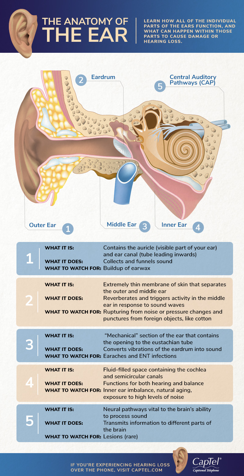
The Anatomy Of The Ear Infographic
This size difference magnifies the vibrations and enables the hearing of low amplitude sounds.
What are the parts of the outer middle and inner ear. The cochlea which is the hearing portion and the semicircular canals is the balance portion. The eustachian tube and the vestibular complex are the important parts of the ear responsible for the balance. The stapes attaches to the membrane over this and transfers the vibrations to the fluid in the inner ear.
But if the ear catches an infection it needs to be treated. This thin membrane vibrates with sounds from the outside and tearing or rupturing of this membrane can. Clearly sound waves travel into funnels through the pinna into the external auditory canal.
Your external auditory canal is just inside your pinna and is also part of your outer ear. Moves in response to air vibrations that have been transmitted from the external acoustic meatus to the auditory ossicles and then to the inner ear. An opening between the middle and inner ear.
The pinna is the part of the ear that sits on the outside of the head. The external outer ear consists of the auricle external auditory canal and eardrum Figure 1 and 2. The inner ear has 3 main parts.
The outer ear middle ear inner ear. The eardrum known scientifically as the tympanic membrane Is a thin piece of tissue that is. The auricle or pinna is a flap of elastic cartilage shaped like the flared end of a trumpet and covered by skin.
The rim of the auricle is the helix. Sound travels through this canal. The cochlea is the auditory area of the inner ear that changes sound waves into nerve signals.
The eustachian tube equalizes the air pressure in the middle ear and maintains the balance. The malleus is attached to the tympanic membrane and the stapes are attached to the oval window of the cochlea. Either of a pair of bones that form part of the side of the skull on each side and enclose the middle and inner ear.
Collects and deflects sound. Divides the external acoustic meatus from the middle ear. Incus or anvil - the bridge bone between the malleus and the stapes.
Ligaments and muscles attach the auricle to the head. The middle ear contains three ear ossicles called malleus hammer incus anvil and stapes stirr-up which are attached to one another in a chain-like fashion. Pinna outer ear Definition.
The pinna is the part of your ear anatomy that collects sounds and protects your middle ear from damage to your ear drum. The outer ear consists of the pinna also called the auricle ear canal and eardrum. The boundary between the inner and outer ear is the tympanic membrane also known as the eardrum.
The vestibular complex contains receptors that maintain body balance. This part of the outer ear collects the sound waves in the air and acts as a deflection device against sounds we do. The peripheral hearing system consists of three parts which are the outer ear the middle ear and the inner ear.
The middle ear is a small air-filled space containing three tiny bones called the malleus incus and stapes but collectively called the ossicles. The malleus is attached to it within the middle ear. The inferior portion is the lobule.
Your ear drum tympanic membrane is what separates your outer ear from your middle ear. The outer ear is made of solid cartilage called the pinna. Cavity also called the tympanic cavity ossicles 3 tiny bones that are attached malleus or hammer - long handle attached to the eardrum.
The inner ear has two main parts. Next to the middle ear in the bone of the skull is a small compartment which contains the hearing and balance apparatus known as the inner ear.
The stratosphere starts just above the troposphere and extends to 50 kilometers 31 miles high. Hinge joints typically allow for only one direction of motion much like a door-hinge.
Foot Pain And Problems Johns Hopkins Medicine

Callus - thick hardened skin usually painless.

Outer parts of the feet. Corns calluses are thick hard raised areas of skin that frequently develop on the side of the feet and toes. Instep The instep has great significance for how a shoe fits. Bones and Joints of the Foot and Ankle The Ankle Lateral side of the ankle Joint capsule.
Supination occurs when weight is placed on the outside of the foot while walking or running. The ozone layer which absorbs and scatters the. The ankle joint or tibiotalar joint is formed where the top of the talus the uppermost bone in the foot and the tibia shin bone and fibula meet.
A doctor may look for swelling deformity pain discoloration or skin changes to help diagnose a foot problem. Other important tendons in the foot include the tibialis posterior posterior tibial tendon which attaches the calf muscle to the bones on the inside of the foot and supports the arch of the foot and the tibialis anterior anterior tibial tendon which runs from the outer tibia to the first metatarsal and surfaces of the median cuneiform tarsal which allows for dorsiflexionbringing the toes toward the shin. The outer part of the ball.
On both feet the lungs are found about an inch below all your toes except the big toe. The sinuses are linked to the tips of each of the toes and the knee is linked to part of the outer border of the sole of the foot. A plain X-ray film of the feet can detect.
This part of the atmosphere is the most dense. The ankle joint is both a synovial joint and a hinge joint. Sprain The overstretching or injury of the ligaments of the ankle Supination The inward turning of the foot the opposite of pronation.
Corns - small circular areas raised skin yellowwhite painful. The number of metatarsals are directly related to the mode of locomotion with many larger animals having their digits reduced to two. The foot contains a lot of moving parts - 26 bones 33 joints and over 100 ligaments.
The foot is divided into three sections - the forefoot the midfoot and the hindfoot. Each part of the body is represented on a certain part of one or both feet. Its main role is to serve as the lateral wall of the ankle mortise Figure 4.
The metatarsals are the bones that make up the main part of the foot in humans and part of the leg in large animals or paw in smaller animals. However the challenge with foot pain is that there are many different potential causes making it difficult at times for even health care professionals to get to the root of your discomfortWhere the pain is and how it feelsthrobbing aching stabbing tender and so oncan offer clues but given all the possible causes symptoms may not be enough to. The inner part of the ball.
Outer ball As above but in reverse that is the outer metatarsal bone meets the bone of the small toe. Almost all weather is in this region. This consists of five long metatarsal bones and five shorter bones that form the toes phalanges.
Its not as simple as drawing a body on your foot instead the size position and scale is altered eg. It might also lead to. In a typical foot the tibia is responsible for supporting about 85 of body weight.
The area just below the waistline is connected to your small intestine on both feet. When the opposite happens and a person shifts their. Toes contain what are referred to as meridian points which are small pressure points correlated to certain body parts.
Foot pain is a very common problem. Talus The talus is one of the major bones that forms the ankle joint. Excessive friction on the skin from tight shoes foot deformities aging.
It throws foot bones out of alignment and can be painful due to pressure or arthritis. Your heels on both feet are connected to your legs. It helps connect the lower leg to the foot and it sits below the tibia and fibula.
Parts of the foot. Supination and pronation are parts of a stride. This bony bump at the base of the big toe causes that toe to veer toward the others.
Inner ball This is where the inner metatarsal bone and the bone of the big toe meet. Tibia and Fibula long bones The foot is connected to the body where the talus articulates with the tibia and fibula. The troposphere starts at the Earths surface and extends 8 to 145 kilometers high 5 to 9 miles.
Walking on the outer side of your feet is called supination You can tell if youre a supinator by checking the soles of your running or walking. The fibula accepts the remaining 15.
Outer ball As above but in reverse that is the outer metatarsal bone meets the bone of the small toe. Learn more about foot bones and foot anatomy here.

A Patient S Guide To Foot Anatomy 2020 Orthonorcal Los Gatos Capitola Morgan Hill Watsonville Ca
In a typical foot the tibia is responsible for supporting about 85 of body weight.

What is the outer part of your foot called. As you walk your weight is transferred from your heel to the ball of your foot and if the weight is unevenly transferred to the metatarsals and over the ball of your foot pain and swelling of the area can occur. The extensor digitorum longus also runs down the leg and through the foot. Lateral Lateral means on the outside or the furthest away from your body so when we talk about lateral ankle ligaments were talking about the ligaments on the outsidefar side of your foot.
Tailors bunions can become painful and inflamed when the enlarged bone gets pressed on by your shoes. The exact location of the pain will depend on which of the many ligaments has sprained torn. If youre starting out with foot reflexology keep your main focus on the sole chart.
Most importantly the fibula bone forms a lateral prominence on the outside of the foot lateral malleolus which is. Causes Symptoms of Peroneal Tendon Injuries. Supination of the foot occurs when your weight rolls onto the outer edges of your feet.
There are eight tarsal bones in the foot. The tibia and fibula are held together by the tibiofibular syndesmosis a collection of 5 ligaments. The outer part of the ball.
For example when it comes to toes the second and third toe after your. Its main role is to serve as the lateral wall of the ankle mortise Figure 4. A midtarsal joint sprain is an injury to the ligaments holding the midtarsal joints together causing pain in the outside middle part of the foot.
Tailors Bunion A Tailors bunion or bunionette is bump on the side of foot at the head of the 5th metatarsal bone just behind the little toe. The muscles tendons and ligaments The muscles are located mainly in the sole of the foot and divided into a central medial group and a group on either side lateral. The tendons are thick bands that connect muscles to bones.
The navicular is on the medial inner side of the foot between the talus and the cuneiform bones in front. The cuboid is on the lateral side of the foot outer foot and sits in front of the calcaneus. The talus forms a joint with four bones.
The muscles at the top of the foot fan out to supply the individual toes. While some of the following conditions can occur anywhere in or on your foot they all belong on a checklist of potential sources of pain on the outside of your foot. The systems vary but once in place they are very secure.
Medial Closer to the central line of the body. One peroneal tendon attaches to the outer part of the midfoot while the other tendon runs under the foot and attaches near the inside of the arch. This is the main cord that allows people to use their big toe and flex the ankle.
When the outer edge of your foot hurts enough to get your attention it can often be difficult to identify the precise cause. They are complex structures with 26 bones. The feet support the human body when standing walking running and more.
It provides slightly more details in regards to which parts of the foot are connected to which parts of the body. The fibula accepts the remaining 15. At the knee the fibula bone articulates with the tibia bone superior tibiofibular then extends downward to ankle and enters the hindfoot.
The main function of the peroneal tendons is to stabilize the foot and ankle and protect them from sprains. In a normal stride your foot should roll inward a bit. Parts of the foot.
This tendon starts in the leg and extends all the way down past the ankle and ends at the big toe. The top of the foot has a major tendon called the extensor hallucis longus. The inner part of the ball.
Another name for supination is underpronation. Tibia fibula calcaneus and navicular. Pole attachment points Most tents have a point at which the end of the pole attaches to either the outer or inner tent depending on the tent design.
The medial ankle ligaments are. Instep The instep has great significance for how a shoe fits. Inner ball This is where the inner metatarsal bone and the bone of the big toe meet.
The ball of the foot is the portion of the foot between your arch and toes where bones called your metatarsals are located.