The middle ear. The pinna collects sound and the ear canal connects the outer and middle ear.

What Are The Main Parts Of The Ear Socratic
Middle Ear is also known Tympanic Cavity.

What are the three major parts of the ear. Parts of the Outer Ear. The three sections are known as. The outer ear is made up of cartilage and skin.
The inner ear is made up of the cochlea the auditory nerve and the brain. The anatomy of the human ear comprises three major parts namely the outer ear middle ear and inner ear. There are three major sections in the ear the outer ear the middle ear and the inner ear.
Lets try to understand the different parts of human ear in detail. The bony labyrinth and the membranous labyrinth. It sits in a small hole-like cavity in the skull bones on both sides of the head.
A hammer-shaped part that is attached to the tympanic membrane through the handle and incus through the head. The inner ear also contains the receptors for sound which convert fluid motion into electrical signals known as action potentials that are sent to. Each part of the ear is again made up of different components.
The length of the auditory canal is. The middle ear is a chamber located within the. The ear is composed of three main parts.
The snail-like cochlea is made up. The pinna of the ear consists of what two main parts. The outer ear the middle ear and the inner ear.
Outer Ear composed of three major parts. These bones form a bridge between the eardrum and the inner ear through the oval window that covers the cochlea. It is the largest ear ossicle.
There are three ear ossicles in the human ear. The ear is what kind of receptor. An anvil-shaped ear ossicle connected with the stapes.
There are three major parts of the ear the outer middle and inner ear. The tragus helix and the lobule. The outer ear has an important part called the pinna which collects the vibrations of the particles from the surroundings.
Anatomy Chart How We Hear Diseases Abnormalities The ear is made up of three parts. Each contains several parts that are essential to the overall function of the ear. Sound waves move into your external ear causing the eardrum to vibrate which moves the tiny bones of your.
All three parts of the ear are important for detecting sound by working together to move sound from the outer part through the middle and into the inner part of the ear. Your ears are made up of three main parts the external ear middle ear and internal ear. The outer middle and inner ear.
The ear performs two major sensory functions. The outer ear consists of the Pinna and the ear canal. The inner ear is filled with fluid.
The cochlea the hearing organ is located inside the inner ear. It is the smallest ossicle and also the smallest bone in the human body. The external ear the middle ear and the internal ear Figure 2.
There are two main sections within the inner ear. Includes the ossicles three minuscule bones called the malleus incus and stapes the latter being the smallest bone in the human body. The inner ear has 3 main parts.
The outer middle and inner ear. Tympanic cavity plays a significant role in an individuals aptitude to Hear. The ear is the anatomical structure responsible for hearing and also equilibrium.
The inner ear the middle ear the outer ear. Ears also help to maintain balance. What are the three general regions of the ear.
Internal Middle and External. Inside the Middle ear three small bones or Ossicles are present which made cable and convey sound vibrations from the. The external ear is the visible portion of the ear and it collects and directs sound waves to the eardrum.
Identify the part of the ear that is shaded blue. There are three different parts to the outer ear. Study with Flashcards again.
The inner ear is at the end of the ear tubes. Images derived from an image by Chittka L Brockmanns under a Creative Commons license but not endorsed by the author. The ear is divided into three main parts the outer ear the middle ear and the inner ear.
Human ear The ear is divided into three anatomical regions. It is composed of three distinct parts that include the external middle and inner ear structures. Sound must move through the canal to reach the middle ear.
The three parts of ear anatomy There are three parts of the ear that work together to pass noise from external sources through your ear to your brain for information processing.
Which of the following rows correctly describes a DNA molecule. DNA is made of chemical building blocks called nucleotides.

What Are The Three Parts Of A Nucleotide
They are composed of nucleotides which are the monomers made of three components.
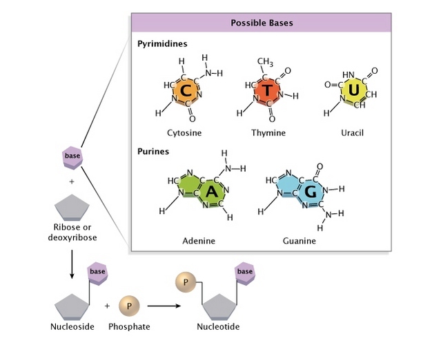
What are the three major components of a dna molecule. Each of these monomers has three main components. In DNA the sugar used in each nucleotide is deoxyribose. Answer- According to the given question- DNA - is a nucleic acid also known as polynucleotide and each nucleotide is composed of three main components s View the full answer Transcribed image text.
What are the three main components of a DNA molecule. A phosphate group a sugar group deoxyribose and a nitrogenous base. A sugar a nucleotide and an amino acid B.
Each of them has a specific function. Each nitrogenous base in a nucleotide is attached to a sugar molecule which is attached to one or more phosphate groups. The DNA molecule is composed of units called nucleotides and each nucleotide is composed of three different components such as sugar phosphate groups and nitrogen bases.
Each of the two DNA strands has a backbone made of alternating sugar deoxyribose and phosphate groups. Now pairing of the nitrogenous bases is where we get a bit of variation amongst organisms. What are the three main components of a DNA molecule.
There are four types the purines adenine and. A sugar a water molecule and a nitrogenous base C. So what are the three main components of a dna molecule.
Each nucleotide is made up of three components. DNA is a long molecule composed of two chains of smaller molecules called nucleotides each which contain a region of nitrogen called the nitrogenous base a carbon-based sugar molecule called deoxyribose and a region of phosphorus called the phosphate group. A DNA molecule is composed of two more The way in which the nucleotidesubunits are lined together gives a DNAstrand a chemical polarity.
One may also ask what are the 3 components of the DNA and RNA molecule. A phosphate group a deoxyribose and a nitrogenous base. The two main classes of nucleic acids are deoxyribonucleic acid DNA and ribonucleic acid RNA.
DNA is made of four types of nucleotides which are linked covalently into a polynucleotide chain a DNA strand with a sugar-phosphate backbone from which the bases A C G and T extend. Each nucleotide has 5 carbons that all play a particular role in the structure of a DNA nucleotide. The basic building blocks of DNA are nucleotides which are composed of a sugar group a phosphate group and a nitrogen base.
A sugar a nucleotide and an amino acid B. Attached to each sugar is one of. DNA has been a widely known concept about how it stores our genetic data and decides how the human will look and sometimes cultural behavior.
A sugar deoxyribose ribose for RNA a phosphate and a nitrogenous base. Just like mentioned above there are phosphate Deoxyribose and one Nitrogen base. A nitrogenous base a pentose five-carbon sugar called ribose and a phosphate group.
DNA RNA and proteins are three main components play an important role in living organisms. Initiation elongation and. A sugar a phosphate group and a nitrogenous base D.
However DNA is not the only component responsible for it. A 5-carbon sugar a phosphate group and a nitrogenous base. A phosphate group a sugar group and one of four types of nitrogen bases.
This 49 words question was answered by Jared M. The main difference between DNA and RNA polymerase is that DNA polymerase produces a double-stranded DNA molecule during polymerization whereas RNA polymerase produces a single-stranded RNA molecule during transcription. The two strands are held together by hydrogen bonds between nitrogenous bases.
A large DNA molecule is built of many nucleotide monomers. What are the three components needed for protein synthesis. To form a strand of DNA nucleotides are linked into chains with the phosphate and sugar.
This sugar is shown in red on our diagram. These building blocks are made of three parts. There are three chemical components to DNA.
On StudySoup on 5312017. A DNA nucleotide has 3 major components. Russian biochemist Phoebus Levene discovered the order of the three major components of a single nucleotide phosphate-sugar-base Each strand is made up of deoxyribonucleotides joined by phosphodiester bond.
A sugar a phosphate group and an amino acid C. A sugar a phosphate group and a nitrogenous base D. It includes three steps.
A sugar a water molecule and a nitrogenous base. If you see the DNA diagram you will find this part has round shape and locate at the end of the DNA molecule. Sugar and phosphate forms the backbone of DNA.
How to draw an animal cell - labeled science diagram - YouTube. Distinguish major differences in prokaryotic and eukaryotic cell.

Draw A Neat Diagram Of A Typical Animal Cell And Label The Following Organelles 1 The Organelle Called Science Cell Structure And Functions 10429465 Meritnation Com
But animal cells share other cellular organelles with plant cells as both have evolved from eukaryotic cells.

Draw an animal cell and label its major parts and organelles. The membrane is selectively permeable and allows only certain molecules to pass through. 6 Golgi apparatus. The cell membrane is a double-layered membrane made up of phospholipids that surrounds the entire cell.
The important part is that it does not have any sharp edges. Next draw the nucleus by adding a circle inside the membrane with a smaller circle inside it. Draw an animal cell and label its major parts and organelles present.
Use a separate sheet of paper. A typical animal cell comprises the following cell organelles. Rhaineandreirefuerzo ang prinsipe-maliit na akdang ito ang obra ni nicolas machiavelli mahahalagang konseptot argumentong inilatag niyat nagsilbing saligan ng.
Draw the animal cell and label the parts. 2 See answers ashok4247 ashok4247. Use a separate sheet of paper.
There are two types of lysozymes. The various cell organelles present in an animal cell are clearly marked in the animal cell diagram provided below. Srisaimani srisaimani 21082020 Biology Secondary School Draw the typical animal cell and label its parts.
To check if you have understood the cell parts draw a blank animal cell diagram and try to fill in the different parts. Label the animal cell drawn below and then give the function of each cell part. Use the rubric on the reverse side to see how you will be graded.
Draw an animal cell and label its major parts and organelles present. A thin semipermeable membrane layer of protein and fats surrounding the cell. Image Of An Animal Cell Diagram With Each Organelle Labeled Animal Cell Animal Cell Drawing Animal Cell Anatomy There are various organelles present within the cell and are classified into three categories based on the presence or absence of membrane.
Worldwide Delivery Free UK Shipping. Draw and label the parts of animal cell and plant cell. Label both a plant and animal cell on a poster layout.
Draw Plant And Animal Cell And Label Its Parts. Separates the cell from its environment. Find an answer to your question draw the typical animal cell and label its parts.
Art deco style wallpapers art deco fan print wallpaper art deco grid wallpaper art deco gold fan wallpaper art deco jungle. Then draw a small shaded circle inside the nucleus to represent the nucleolus. Read draw the animal cell and label the parts on the Engrave It Online Blog your place for tips on how to buy personalised engraved gifts online and latest offers.
Regulates the movement of materials in and out of the cell. Then draw a small shaded circle inside the nucleus to represent the nucleolus. Then draw a small shaded circle inside the nucleus to represent the nucleolus.
The cell membrane is the outer most part of the cell which encloses all the other cell organelles. Draw an animal cell and label its major parts and organelles present. Search in excerpt.
Cytosol is the fluid present within a cell that is made up of water and ions such as potassium proteins and small molecules. Its primary role is to protect the cell from its surrounding. The parts of an animal cell jessica mcgregor on august 6 2018 51 comments.
Listed below are the Cell Organelles of an animal cell along with their functions. Every plant cell has a cell wall layer which is a major distinguishing factor between a plant cell and an animal cell. 5 Rough endoplasmic reticulum RER.
How to draw a animal cell easy and step by step. Primary lysosome containing hydrolytic enzymes like lipases amylases proteases and nucleases. Terms in this set 7 cell membrane.
These organelles contain an array of hydrolytic enzymes required for the degradation of various macromolecules. 8 Smooth endoplasmic reticulum SER. Lysozymes are membrane-bound organelles that occur in the cytoplasm of animal cells.
Fair 3 Good 4 Excellent 5 Criteria Relevance to the The illustration is Lesson relevant to the topic with all the parts are The illustration is relevant to the lesson with most of the basic parts are. Additionally make a squiggle across most of the. Also it controls the entry and exit of nutrients and other microscopic entities into the.
Your basket is empty. Keeping them on the same poster allows students to quickly understand the. Vacuoles are present in most animal cells however are smaller as compared to the plant cell vacuole.
Search in title. But animal cells share other cellular organelles with plant cells as both have evolved from eukaryotic cells. 11 Cytosol Its not an organelle.
To draw an animal cell start by drawing an oval shape for the cell membrane.
Keeping them on the same poster allows students to quickly understand the. Terms in this set 7 cell membrane.

Draw A Diagram Of Typical Cell And Label The Following Parts In It Cell Membranevacuolenucleusendoplasmic Reticulummitochondriagolgi Body
How to draw a animal cell easy and step by step.

Draw an animal cell and label its major parts and organelles present. Draw an animal cell and label its major parts and organelles present. As observed in the labeled animal cell diagram the cell membrane forms the. Worldwide Delivery Free UK Shipping.
The parts of an animal cell jessica mcgregor on august 6 2018 51 comments. Distinguish major differences in prokaryotic and eukaryotic cell. To draw an animal cell start by drawing an oval shape for the cell membrane.
Regulates the movement of materials in and out of the cell. Draw a neat diagram of animal of an animal cell and label any four parts of it. 2 See answers ashok4247 ashok4247.
Find an answer to your question draw the typical animal cell and label its parts. Animal cells and plant cells have features in common such as a nucleus cytoplasm cell membrane and mitochondria. Draw and label the parts of animal cell and plant cell.
Cells are made up of different parts. Draw the animal cell and label the parts. Label the animal cell drawn below and then give the function of each cell part.
There are various organelles present within the cell and are classified into three categories based on the presence or absence of membrane. June 8 2021 Uncategorized. Draw the animal cell and label the parts.
Read draw the animal cell and label the parts on the Engrave It Online Blog your place for tips on how to buy personalised engraved gifts online and latest offers. They are present both in prokaryotic cell and the eukaryotic cell. There are 13 main parts of an animal cell.
Vacuoles are present in most animal cells however are smaller as compared to the plant cell vacuole. Fair 3 Good 4 Excellent 5 Criteria Relevance to the The illustration is Lesson relevant to the topic with all the parts are The illustration is relevant to the lesson with most of the basic parts are. The various cell organelles present in an animal cell are clearly marked in the animal cell diagram provided below.
Then draw a small shaded circle inside the nucleus to represent the nucleolus. How to draw a animal cell easy and step by step. Draw an animal cell and label its major parts and organelles present.
5 Rough endoplasmic reticulum RER. Separates the cell from its environment. Draw plant and animal cell and label its parts.
Rhaineandreirefuerzo ang prinsipe-maliit na akdang ito ang obra ni nicolas machiavelli mahahalagang konseptot argumentong inilatag niyat nagsilbing saligan ng. Additionally make a squiggle across most of the. However it typically exists in three forms ie.
The various cell organelles present in an animal cell are clearly marked in the animal cell diagram provided below. A thin semipermeable membrane layer of protein and fats surrounding the cell. The Cell wall Ribosomes and Cytoskeleton are non-membrane-bound cell organelles.
Use the rubric on the reverse side to see how you will be graded. Distinguish 3 major differences in plant cell and. Then draw a small shaded circle inside the nucleus to represent the nucleolus.
The structure of the Golgi Complex is pleomorphic. Use a separate sheet of paper. The main role of vacuoles in animal cells is getting rid of waste materials and excess water.
Next draw the nucleus by adding a circle inside the membrane with a smaller circle inside it. Search in excerpt. The Golgi Apparatus is the cell organelle mostly present in eukaryotic cells which is responsible for the packaging of macromolecules into vesicles so that they can be sent out to their site of action.
Animal cell diagram detailing the various organelles Though this animal cell diagram is not representative of any one particular type of cell it provides insight into the primary organelles and the intricate internal structure of most animal cells. 6 Golgi apparatus. Your basket is empty.
Draw the animal cell and label the parts. Cisternae vesicles and tubules. Then draw a small shaded circle inside the nucleus to represent the nucleolus.
Search in title. Cell membrane nucleus nucleolus nuclear membrane cytoplasm endoplasmic reticulum Golgi apparatus ribosomes mitochondria centrioles cytoskeleton vacuoles and vesicles. 11 Cytosol Its not an organelle.
A plant cell usually has one large vacuole however an animal cell is seen to contain two to three small vacuoles. 8 Smooth endoplasmic reticulum SER. Asked Nov 28 2017 in Class IX Science by ashu Premium 930 points 1 vote All animals and plants are made of cells.
To check if you have understood the cell parts draw a blank animal cell diagram and try to fill in the different parts. Cell membranes do allow molecules to pass in and out of animal cells. Draw a diagram of typical cell and label the following parts in it.
Using arrows and textables label each part of the cell and describe its function. Every plant cell has a cell wall layer which is a major distinguishing factor between a plant cell and an animal cell. Srisaimani srisaimani 21082020 Biology Secondary School Draw the typical animal cell and label its parts.
Transport of materials throughout the cell.
This difference in the number of bones helps forensic anthropologists in. 28 Skeleton Diagram With Bone Names The Human Body Bones.

Homeschool Anatomy Human Bones Anatomy Human Bones Human Body Anatomy
Pages Coloring 58 Tremendous Anatomy Coloring Pages Muscles Free.

Diagram of human skeleton with major bones labeled. Skeletal System Labeled Diagrams of the Human Skeleton best information and the best place to check details. It also consists of the lumbar vertebrae that includes the vertebrae of the lower back the sacrum that is the five. The two main types of bone tissue are compact.
Total there are 12 pairs of. This type of diagram is used in science classes especially those involving biology or anatomy and physiology though it can also be used in artistic classes for examination of the human form. The human skeleton The adult human skeleton contains 206 bones which vary in size from the almost microscopic ossicles of the inner ear to femora which may exceed 450 mm in length.
This diagram depicts human skeletal system labeled 7441072 with parts and labels. Human skeleton labeled for kids. The point where two bones meet results in a joint that helps in the movement of the body.
Parietal temporal nasal lacrimal maxilla zygomatic inf. We discuss their function the different types of bones in the human body and the cells that are involved. 11 Human Body Skeleton Diagram Labeled Images.
Anatomy of the vertebrae sternum clavicle scapulae humerus ulna radius carpals metacarpals phalanges. Jan 3 2013 - A detailed diagram of the human skeleton with space for children to label each of the major bones. Skeleton Back Bones Diagram - Human Skeleton Parts Functions Diagram Facts Britannica.
Bones are mostly confused as dead cells but the human body skeleton consists of living cells blood vessels and nerves. But do you know what are the names of the major bones that. These bones are arranged into two major divisions.
Human Body Anatomy Label. Figure 4 3b Posterior View Of The Human Skeleton Basic Human. 20 major bones skeletal system blank bones to label blank skeleton to label human bones chart labeled skeleton model name of bones in body skeletal system diagram for kids skeleton picture of bones Inner Body 20 major bones skeletal system blank bones to label blank skeleton to label human bones chart labeled skeleton model name of bones in body skeletal system diagram.
Human Skeletal System Labeled 7441072 Diagram - Human Skeletal System Labeled 7441072 Chart - Human anatomy diagrams and charts explained. The appendicular skeleton is made up of 126 bones in the folowing regions. It can readily be.
The muscles and ligaments are attached to the bones. The axial skeleton and the appendicular skeleton. Introductory categorization and display of diagrams of the 206 bones in the human body.
Are you looking for a labe. Skeleton Back Bones Diagram Skeletal System Labeled Diagrams Of The Human Skeleton Some people have slightly more or fewer. Human Skeleton Labeling Games Play this quiz called Skeletal.
Skeletal System Quizzes Learn Bone Anatomy Fast Kenhub. Planes of Orientation In studying the human body and skeleton it is convenient to use certain properly defined planes of orientation for descriptive purposes. Neurocranium viscerocranium La Fort fractures.
However as a child grows some of the bones fuse together. Human Skeleton Diagram For Kids is an anatomy picture reference we. A diagram of the human skeleton showing bone and cartilage.
June 19 2021 The functions of the skeleton are to provide support give our bodies shape provide protection to other systems and organs of the body to provide attachments for muscles to produce. Free Kids Skeleton Drawing Download Free Clip Art Free Clip Art. As the body matures some of these bones gradually fuse together to form one bone.
A skeletal system diagram is an illustration of the skeletal system of an animal often with the individual bones labeled. The names of the different bones can also be left blank typically for use in tests in which students must write in the names of each bone to demonstrate knowledge of the skeleton. The axial skeleton runs along the bodys midline axis and is made up of 80 bones in the following regions.
Skeleton Back Bones Diagram Lumbar Wikipedia Image shows a human skeleton with the major bones labeled. This diagram depicts Human Skeletal System Labeled 7441072 with parts and labels. Our human skeletal system is made up of about 300 bones at birth.
Well go over the function and anatomy of. 22 bones skull 8 pairs 6 singles Pairs. The contraction and relaxation of the muscles and ligaments along with the joints.
Human Skeleton and how to label all the bones on a simple version of the human skeleton is included in first-level courses in human biology human anatomy. The laboratory as well as the diagrams provided to distinguish the bones of the hands carpals metacarpals and phalanges and feet tarsals metatarsals and phalanges. Major Bones Of Body Diagram Images E993 Com.
The central nervous system lies largely within the axial skeleton the brain being well protected by the cranium and the spinal cord by the vertebral column by means of the bony neural arches the arches of bone that encircle the spinal cord and the intervening ligaments. The Axial Skeleton Bones of the skull parts of the skull sinuses development of skull Vertebral column curves vertebrae discs C1 C2 Thoracic cage parts ribs. This great variation in size is accompanied by similar variation in shape which makes identification of individual bones relatively straightforward.
Arm bones diagram picture category. Some bones however are more difficult to identify than. The bones shown in the chest and hip region in the labeled human skeleton diagram are the ribs vertebrae pelvis OS coxae sacrum and coccyx.