Draw a labelled diagram of. Draw a labelled diagram of a section through ovary.

Draw A Labelled Diagram Of A Section Through Ovary Showing Different Stages Of Follicles Ovulation And Corpus Luteum
To study and identify the transverse section of testes and ovary through permanent slides.

Labelled diagram of a section through ovary. The seminiferous tubules of the testis produce sperm. OR Draw a sectional view of the human ovary showing the different stages of developing follicles corpus luteum and ovulation. Asked Mar 13 2020 in Biology Mar 13 2020 in Biology.
Draw a labelled diagram of a section through ovary. Watch complete video answer for Draw a labeled diagram of a section through ovary. I 12 30 ii 14 49 iii 1535 iv 299 161 Q.
Write two major functions each of testis and ovary. The Leydig cells of testis produce hormones such as androsterone and testosterone together called androgens. How to draw an ovary in exam is the topic.
The ovaries are considered the female gonads. An easy and convenient way to make label is to generate some ideas first. A labelled diagram of a section through the ovary is as follows.
Asked Oct 9. Draw a labelled diagram of a Graafian follicle. This is a well labelled.
Q-With a neat diagram explain the 7-celled 8-nucleate nature of the female gametophyteQ-With a neat labelled diagram describe the parts of a typical angiosperm ovuleQ-Differentiate between a zoospore and a zygote. They have a wall of smooth muscle an inner mucosal lining and an outer layer of loose supporting tissue serosa. TS of Mammalian Testes.
Draw a labelled diagram to show inter-relationship of four accessory ducts in a human male reproductive system. Get FREE solutions to all questions from chapter HUMAN REPRODUCTION. The ovarian fossa is the region that is bounded by the external iliac artery and in front of the ureter and.
Each ovary is whitish in color and located alongside the lateral wall of the uterus in a region called the ovarian fossa. Q-Why is reproduction essential for organismsAnswer. Permanent slides of transverse section of Testes and ovary microscope.
Similar Questions Express each of the following rational numbers in its standard form. Draw a labelled diagram of female reproductive system. Popular Questions of Class Biology.
Draw a labelled diagram of a section through ovary. Draw a labelled diagram of section through ovary. The testes consist of a many seminiferous tubules embedded in the interstitial.
Label Gallery Get some ideas to make labels for bottles jars packages products boxes or classroom activities for free. Q-What is spermatogenesisBriefly describe the process of spermatogenesis. You should make a label that represents your brand and creativity at the same time you shouldnt forget the main purpose.
Q-What is spermatogenesisBriefly describe the process of spermatogenesis. It is the passageway through which the ovum passes from the ovary to the uterine cavity. Popular Questions of Class Biology.
The mammalian testes are covered with a thick fibrous tissue called tunica albuginea. Of Biology Class 12th. This is the diagram of ovary of mice.
The oviducts are part of the genital tract. Draw a sectional view of human ovary and label the different follicular stages ovum and Corpus luteum. This is the well labelled diagram of ovary of mice.
A labelled diagram of a section through the ovary is as follows. Diagrammatic section view of ovary. Draw a labelled diagram of sectional view of a human ovary showing various stages of follicles growing in it.
The oviduct is also known as the fallopian or uterine tube. Q-With a neat diagram explain the 7-celled 8-nucleate nature of the female gametophyteQ-With a neat labelled diagram describe the parts of a typical angiosperm ovuleQ-Differentiate between a. Q-Why is reproduction essential for organismsAnswer.
Diagram of Graffian follicle. The Female Reproductive System.
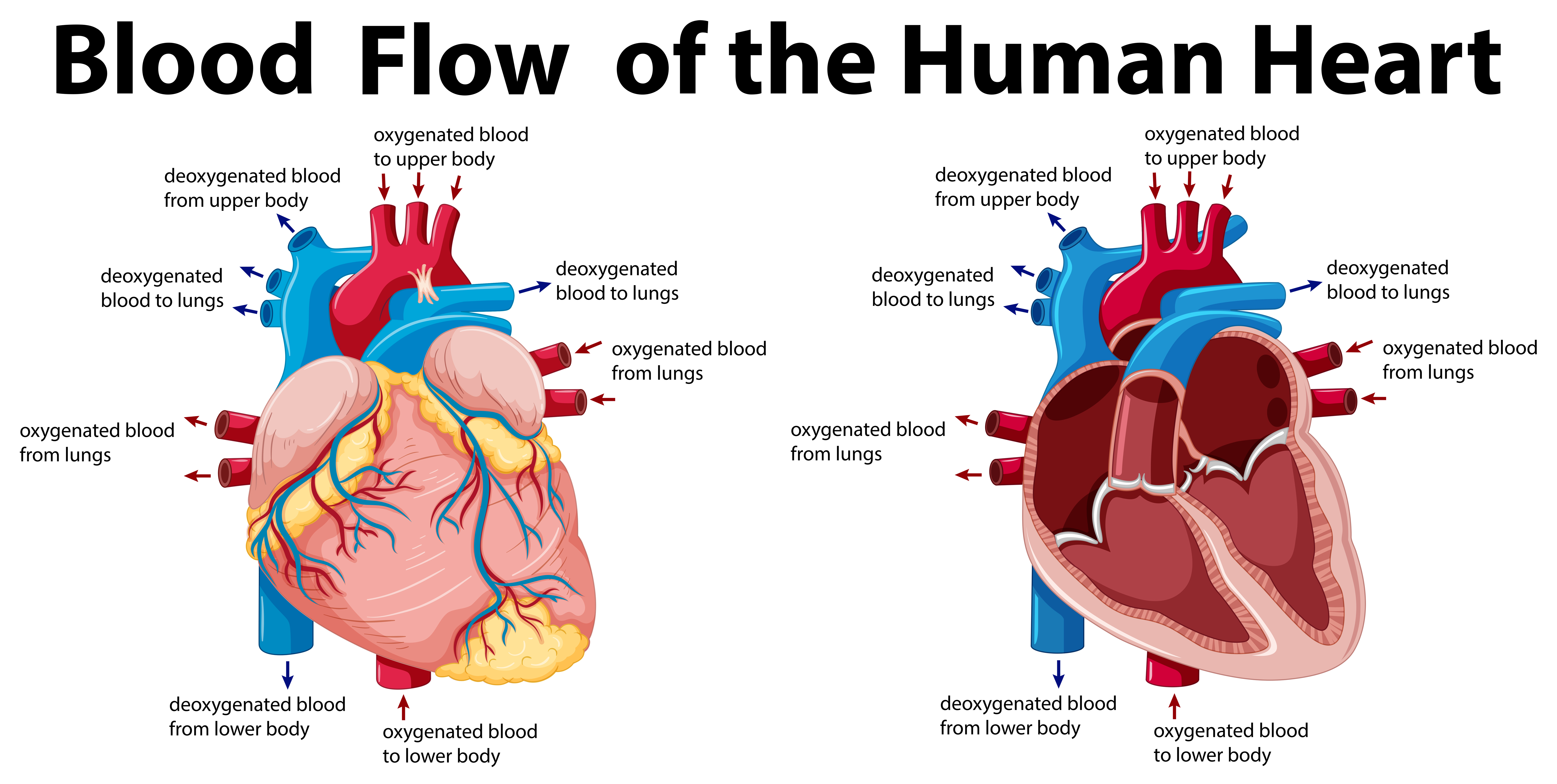
When the ventricle is full the mitral valve shuts to prevent blood from flowing backwards into the atrium. Always remember that it must flow through 6 areas on the right side and then 6 areas on the left side this equals 12 steps.
Blood Flow Of The Human Heart 416577 Vector Art At Vecteezy
The blood then flows through the tricuspid valve into the right ventricle.
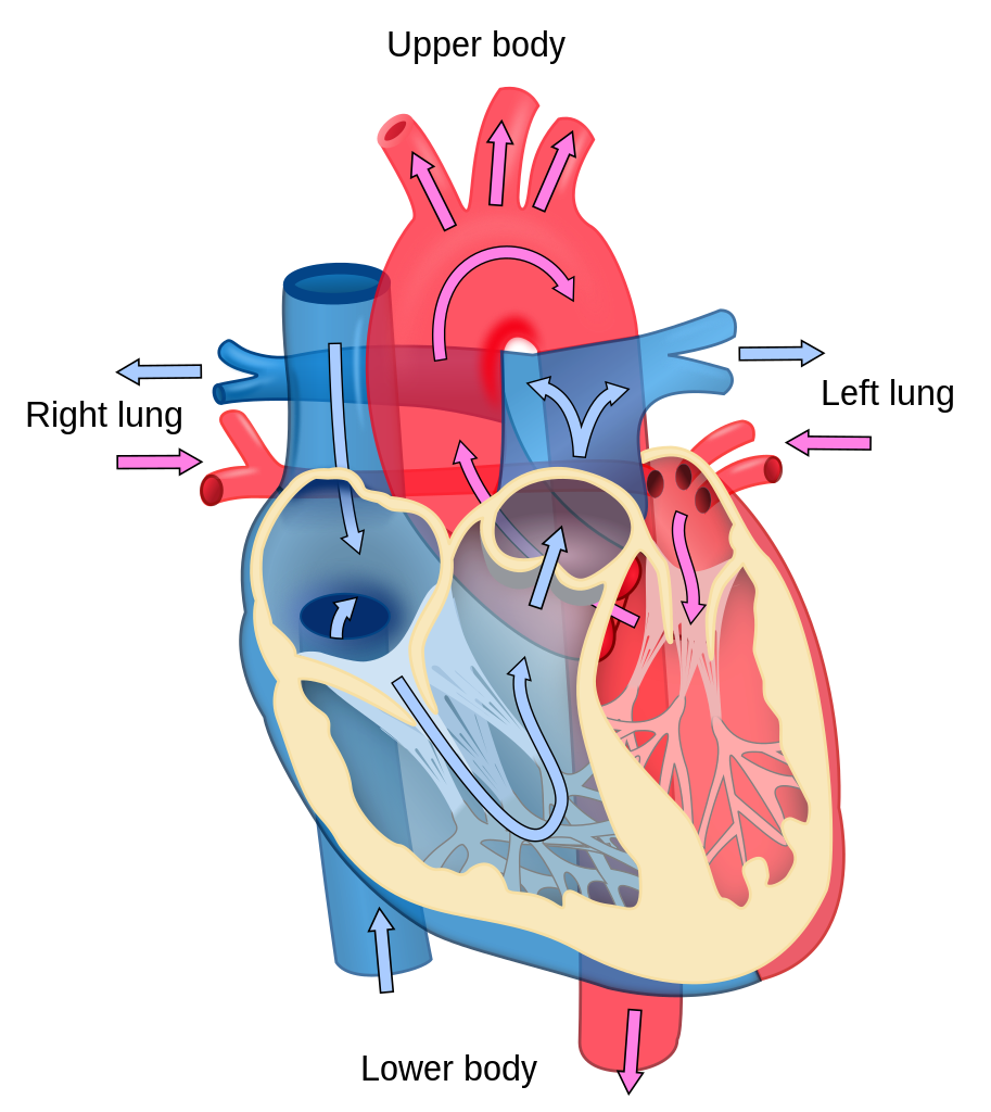
How does blood flow through the heart diagram. An updated version of this vi. The oxygenated blood will flow through the pulmonary veins into the left atrium. Every heart diagram labeledwill clearly show these valves.
Blood leaves the heart through the aortic valve into the aorta and to the rest of the body. Where the atria meet the ventricles there is an area of special cells - called the atrio-ventricular node - which pass the electrical signals throughout your heart muscle by a system of electrical pathways known. The pattern described below is repeated over and over heart rhythm causing blood to flow continuously to the heart lungs and body to supply oxygen and nutrients to the body cells and to deliver waste products to organs that remove them from your body.
Valves to Ensure Unidirectional Blood Flow. How the heart works. For better illustration look at the picture below and note how the.
Task Three - Diagram Assignment The student will create an original diagram of the heart and trace the pathway of the blood through the heart with appropriate labeling of 13 major points of anatomy. A Simple 12 Step Diagram Blood Flow Through the Heart. The right and left sides of the heart work together.
The blood will then become oxygenated as it passes through the pulmonary circulation and lungs. The right and left sides of the heart work together. You are watching a video about the blood flow through the heart.
Blood enters the heart through two large veins the inferior and superior vena cava emptying oxygen-poor blood from the body into the right atrium. The right ventricle pumps the oxygen-poor blood to the lungs through the pulmonary valve. The right atrium receives oxygen-poor blood from the body and pumps it to the right ventricle through the tricuspid valve.
Blood flow through the heart is made easy in this post. How does blood flow through the heart. The right side of the heart pumps blood out into the Pulmonary artery going away from the heart and towards the lungs this blood still has not been oxygenated.
The right atrium receives deoxygenated blood through the superior and inferior vena cavas from the body and pumps it to the right ventricle through the tricuspid valve which opens to allow the blood flow through and closes to prevent blood backing up the atrium. This process is called pulmonary circulation. Blood Flow Through the Heart Blood flows through the heart in 12 easy steps.
These valves allow blood flow in one. Blood flows from the left atrium into the left ventricle through the open mitral valve. Blood flows through your heart and lungs in four steps.
The oxygenated and deoxygenated blood flow through the heart will be clearly distinguished. Anatomy of the Heart. Your heart has four types of valves with primary function of regulating the blood flow through the heart.
The left atrium receives oxygen-rich. The pulmonary artery carries blood to the lungs where it picks up oxygen and then leaves the lungs to return to the heart through the pulmonary vein. The pulmonary vein empties oxygen-rich blood from the lungs into the left atrium.
Blood is pumped through the valves into the ventricles. Animation of blood flow through the heart. From the right ventricle blood will flow through the pulmonic valve and into the pulmonary circulation via the pulmonary trunk and arteries.
Next the blood enters the lungs and performs gas exchange the blood now has high O2 and goes to. How does blood flow through the right and left side of the heart. Blood Flow Through The Heart.
When the heart beats the ventricle pushes blood through the pulmonic valve into the pulmonary artery. The cycle of blood flow and the role of heart valves described. The anatomy of the heart was made easy in a previous EZmed video and post where we learned.
Beginning with the superior and inferior vena cavae and the coronary sinus the flowchart below summarizes the flow of blood through the heart including all arteries veins and valves that are passed along the way. Using a simple diagram to show the. The left atrium receives oxygenated blood through the pulmonary veins from the lungs.
From the pulmonic valve the blood travels to the pulmonary artery into the tiny capillary vessels of the lungs. Blood Flow Through the Heart. The deoxygenated blood from the heart enters the lungs through the pulmonary valve as seen in the human heart diagram.