Step by step video text image solution for Draw a labelled diagram of alimentary canal of a cockroach. How to draw alimentary canal of cockroach alimentary canal of cockroach diagramHello Friends in this video I tell you about how to draw labelled dia.

Draw A Labelled Diagram Of The Alimentary Canal Of A Cockroach Animal Kingdom Biology Class 11
Draw a labelled diagram of alimentary canal of a cockroach.

Well labelled diagram of alimentary canal of cockroach. Prev Question Next Question 0 votes. Draw a well labelled diagram showing the alimentary canal of cockroach and label any four parts. 14 K views 70 people like this Like Share.
Paurometabolous growth is a gradual metamorphosis. Where is it found in human body. Add your answer and earn points.
Around 4600 cockroaches are living aside human habitats. Draw a labelled diagram of the reproductive organs of an earthworm. 325 OR Draw a well labelled diagram of areolar connective tissue and label any four parts.
NCERT NCERT Exemplar NCERT Fingertips Errorless Vol-1 Errorless Vol-2. Alimentary Canal Of Cockroach. What is meant by paurometabolous development in cockroach.
Home NCERT Solution Class Biology Structural Organisation in Animals draw a labelled diagram of alimentary canal of a c. I Give the common name of Periplaneta americana. Well labelled diagram of urinogenital system digestive system in cockroach tutorvista com alimentary canal of cockroach the digestive system in 5 leech drawing well labelled diagram for free download on solved show diagram of side lateral view of cockroach well labelled diagram of open circulatory syestem of dissection of cockroach pdf download.
NCERT P Bahadur IIT-JEE Previous Year Narendra Awasthi MS Chauhan. Cockroaches belong to the category of insects of the Blattodea order. Asked Aug 2 2017 in Biology by Kundan kumar 512k points edited Dec 16 2018 by Vikash Kumar.
Maths Physics Chemistry Biology. Draw a well labelled diagram of alimentary canal of a cockroach. Labelled diagram of alimentary canal 10 labelled diagram of the nervous system nervous digestive system in cockroach tutorvista com labeled male reproductive system human anatomy parts cockroach diagram draw a well diagram of a cockroach pdfsdocuments2 com morphology and anatomy of cockroach biology4isc well labelled diagram of open circulatory syestem of biological.
Find an answer to your question Draw a labelled diagram of alimentary canal of a cockroach nd also write functions ajaynayak4403 ajaynayak4403 05092019 Biology Secondary School answered Draw a labelled diagram of alimentary canal of a cockroach nd also write functions 1 See answer ajaynayak4403 is waiting for your help. Draw a labelled. Q1 Answer in one word or one line.
Draw a labelled diagram of alimentary canal of a cockroach. Prev Question Next Question 1 vote. Draw a labelled diagram of alimentary canal of a cockroach.
Draw a labelled diagram of alimentary canal of a cockroach. Asked Nov 26 2020 in Biology by Maisa 457k points closed Nov 27 2020 by Maisa. Draw a well labelled diagram of alimentary canal of a cockroach.
Draw a labelled diagram of alimentary canal of a cockroach. Click hereto get an answer to your question 26. Q4- Draw a labelled diagram of alimentary canal of a cockroach.
Class 12 Class 11. Draw a labelled diagram of alimentary canal of a cockroach. Draw a labelled diagram of alimentary canal of a cockroach.
Apne doubts clear karein ab Whatsapp par bhi. Ii How many types of nephridia are found in earthworm based on their location. Draw a well labelled.
The digestion of food would take place in the cavities specialized or combined together. By Biology experts to help you in. NCERT RD Sharma Cengage KC Sinha.
Well Labelled Diagram Of A Cockroach draw a labelled diagram of the alimentary canal of a cockroach x get a free home demo of learnnext available for cbse icse and state board syllabus call our learnnext expert on 1800 419 1234 tollfree or submit details below for a call back clear learn keyboard arrow april 2nd 2019 well labelled diagram of human ear well labelled diagram of lice well. How to draw diagram of alimentary canal of cockroach cockroach alimentary canal drawingHello Friends in this video I tell you about how to draw labe. Draw a labelled diagram of alimentary canal of a cockroach.
Watch 1000 concepts tricky questions explained. Maths Physics Chemistry Biology. In this type of development the immature stages resemble small adults and usually.
NCERT DC Pandey Sunil Batra HC Verma Pradeep Errorless. How does it differ from adipose tissue. Loading DoubtNut Solution for you.
- makes the synovial fluid and encloses. - a sheet of cells that lines the joint cavity.
Structure Of Synovial Joint Youtube
Key Structures of a Synovial Joint.

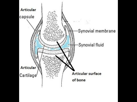
Draw a well labelled diagram of synovial joint. The morphology of synovial membranes may vary but it. Draw labelled diagram Synovial joint. At synovial joints the articular surfaces of bones are covered with smooth articular cartilage.
It consists of two layers. Synovial fluid is the clear viscid lubricating fluid secreted by synovial membranes. The walls of this space are formed by the articular capsule a fibrous connective tissue structure that is attached to each bone just outside the area of the bones articulating surfaceThe bones of the joint articulate with each other within the joint cavity.
Fibrous Capsule outer- provides joint stability. Synovial joints allow for smooth movements between the adjacent bones. This gap allows a free range of motion and space for synovial fluid to lubricate the joint.
Explain the roles of actin and myosin in muscle contraction. Primary movement is a rotation. Give one example each for a hinge joint a pivot joint axial skeleton and appendicular skeleton.
The basic structure of a synovial joint is shown in the diagram below. Movement of the ankle elevating the sole. It is freely movable joint hence known as diarthrosis joint.
Synovial joints are the most common type of articulation and feature a small gap between the bones. Primary movement is a rotation. Reinforcing ligaments Examples of synovial joint-Shoulder jt.
The joint is surrounded by an articular capsule that defines a joint cavity filled with synovial. Examples of this form of articulation are found. Give one example each for a hinge joint a pivot joint axial skeleton and appendicular skeleton.
The main parts of synovial joints are labelled on the synovial joint diagram. Extending the ankle and elevating the heel. Allows movement in only one plane.
Synovial fluid view the full answer. Analyse the electron micrograph for the state of contraction of the muscle fibre. 42 Joint and Movement Type Synovial Joint Movements Circumduction.
Synovial joints are the the most common joints present and have a characteristic trait of having the presence of a joint cavity the cavity filled with the synovial fluid. Also allows movement in only one plane. Draw a labelled diagram of a synovial joint.
Synovial joints are characterized by the presence of a joint cavity. Finally cartilaginous joints are formed. Concept Notes Videos 380.
I articular capsule ii articular cartilage iii synovial fluid. Question Bank Solutions 5549. Some synovial joints are more complicated than others.
Synovial joints allow for smooth movements between the adjacent bones. Draw a labelled diagram of a synovial joint. Allows movement in only one plane.
Elbow joint and knee joint. Fibrous joints exist where bones are very tightly joined and offer little to no movement between the bones. The ball and socket joint or spheroid joint is a type of synovial joint in which the ball-shaped surface of one rounded bone fits into the cup-like depression of another bone.
Elbow joint and knee joint. Also allows movement in only one plane. 19 Draw a well labelled diagram of.
The three main features of a synovial joint are. This gives the bones of a synovial joint the ability to move smoothly against each other allowing for increased joint mobility. Draw a labelled diagram of a synovial joint.
Label the following diagram of the side view of the human elbow joint. Fibrous joints also hold teeth in their bony sockets. Give one example each for a hinge joint a pivot joint axial skeleton and appendicular skeleton.
The distal bone is capable of motion around an indefinite number of axes which have one common center. Figure 941 Synovial Joints. Digging in the heel Plantar flexion.
A metacarpophalangeal finger joint is shown above-rightBBC Bitesize - GCSE Physical Education - Skeletal system - OCR - Revision 3Synovial Joint Labelled Diagram - Anatomy Structure. The structure of a synovial joint is demonstrated by a diagram in which the articulating bones are surrounded by the articular capsule which comprises an exterior fibrous capsule and an interior synovial. Refer to the image for well-labelled diagram of typical synovial joint Synovial joint - A joint between two bones with a cartilage fluid filled cavity in between is called a synovial joint.
This diagram shows the location of the bursae which are fluid filled sacs in a bone. The articular capsule surrounds the joint and is continuous with the periosteum of articulating bones. Synovial Membrane inner- secretes produces synovial fluid for lubrication.
An example of a simple synovial joint eg. Draw a labelled diagram to show the structure of a sarcomere. Draw labelled diagram Synovial joint.
Maharashtra State Board HSC Science General 11th. The main parts of synovial joints are labelled on the synovial joint diagram and described in the table below. A synovial membrane or synovium is the soft tissue found between the articular capsule joint capsule and the joint cavity of synovial joints.
Structural Features of Synovial Joints. It is characterized by. Advertisement Remove all ads.
The Tropic of Cancer 23 1 2 ºN The. The thermal zones of the earth are Tropical Temperate and Polar zones.

Diagram Of Important Latitudes And Heat Zones Class 6 Geography Important Latitudes Drawing Youtube
Question 22 What Is A Circuit Diagram Draw The Labelled Of An Electric Comprising Brainly In Electric Circuit Diagrams Lesson.
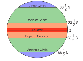
Draw a well labelled diagram of important latitudes and heat zones. The wall of the eyeball is composed of three layers- sclera choroid and retina. This zone lie between the Tropic of Cancer and the Arctic Circle in the Northern Hemisphere and between the Tropic of Capricorn and the Antarctic Circle in the southern hemisphere. This inclination of earths axis helps in marking the following important parallels of latitudes.
The anterior part of this layer is called cornea. There are four important parallels of latitudes. Tropic of Capricorn 235 0 S 23 0 30S or 23 degrees 30 minute south.
Therefore this zone gets the maximum heat from the Sun. The Tropic of cancer is an important parallel of latitude in the northern Hemisphere. D Antarctic Circle 66 12 S.
Therefore the equator is an imaginary circular line and is a very important reference point to locate places on the earth. Draw a labelled diagram of closed electric circuit comprising cell question 22 what is effects cur ncert 7 difference between open and diagrams lesson for label it to. Draw a well labelled diagram of eye.
All parallel circles from the equator up to the poles are called parallels of latitudes. Draw A Labelled Diagram Of Closed Electric Circuit Brainly In Draw The Labelled Diagram Of An Electric Circuit. Name the thermal zones of the earth.
Earthquakes volcanoes geo41 what are the layers of earth 10 h structure of the earth what are the earth s layers earth s internal layers crust mantle. It is the external layer composed of dense connective tissue. Chapter 1 plate tectonics seismic evidence for internal earth draw a well labelled diagram structure the structure of earth marcellus Internal Structure Of The EarthThe Structure Of Earth Earthquakes Discovering GeologyDraw Neat Diagram Label Them And Explain The Interior OfWhat Are The Earth S LayersDraw The Neat Diagram Showing Layers Of Earth Brainly InEarth S Read More.
2 The Tropic of Cancer at 2350 N 3 The Tropic of Capricorn at 2350 S 4 The Arctic Circle at 6650 N 5 The Antarctic Circle at 6650 S. Draw a neat and well labelled diagram of male reproductive system of a frog. This video is related to geographyThis is Latitude and longitude part-2In this video we discussed important Latitudes Heat zones of earth such as Equato.
Draw A Well Labelled Diagram Of Closed Electric Circuit Posted by Margaret Byrd Posted on December 12 2020. The Structure Of Earth Earthquakes Discovering Geology. It is the middle layer bluish in colour and contains many blood vessels.
The adult human eyeball is spherical in shape. Tropic of Cancer 235 0 N 23 0 30N or 23 degrees 30 minute north. Add your answer and earn points.
By measuring the angle of the Pole Star from your place you can know the latitude of your place. Structure Of The Earth Diagram Activity. Draw A Well Labelled Diagram Of Electric Circuit Posted by Margaret Byrd Posted on February 28 2021.
C Arctic Circle 66 12 N. Draw A Well Labelled Diagram To Show The Internal Structure Of Earth. This zone is known as the torrid or the tropical zone.
Important Parallels of Latitudes. 669k 728k 656. The North Pole 90ºN the South Pole 90ºS are important Latitudes since the location of the Equator is obtained from these two fixed points.
Draw a neat labelled diagram of electrolytic cell for the extraction of aluminium. 257k 950k Draw a neat labelled diagram. Internal Structure Of The Earth.
It is at an angular distance of 235 degree 23 degree 30N from the equator. 1 The Equator represents 0 latitude known as Great Circle. 130k 352k Draw a neat labelled diagram to show the structure of the human eye.
By Hilman Rojak July 31 2020. Draw a neat diagram showing pressure belts. There are some other important parallels of latitude which have been given special names.
Important Parallels of Latitude. This zone gets the slanting rays of the Sun as the angle of the Suns rays goes on decreasing towards the Poles. Find an answer to your question Draw a neat and labelled diagram of important latitudes and heat zones of Earth AhmadTaha25 AhmadTaha25 4 weeks ago Geography Secondary School answered Draw a neat and labelled diagram of important latitudes and heat zones of Earth 1 See answer AhmadTaha25 is waiting for your help.
Question 22 what is a circuit diagram electric diagrams lesson for draw schematic labelled of closed and label it to in the shown figure effects cur ncert. Heat zones of the earth will be explained in easy way in this videoanimated video to explain the topic heat zones of the earth for class 5 and class 6Geograp. With the help of degrees name the important lines of latitude.
Structure of PSLV made by ISRO. A Tropic of Cancer 23 12N b Tropic of Capricorn 23 12 S. The equator is the largest possible circle which can be draw around the earth.
Apart from the equator 0 0 and the Poles 90 0. Draw A Well Labelled Diagram Of An Open Circuit Posted by Margaret Byrd Posted on January 18 2021 Labelled diagram of open circuit 7 difference between and a closed short circuits dummies draw their well what is an how with to show. They are as follows.
Draw a well labelled diagram of phloem. The Equator 0º divides the Earth into two hemispheres the Northern Hemisphere and the Southern Hemisphere All the lines of latitude are calculated from it. Important Latitudes and Heat Zones Do you know.
Mosquitoes can live and reproduce inside and outside the home. Aedes life cycle diagram.
Safe Control Of Mosquitos By Steve Tvedten
In the case of Aedes eggs are laid one at a time on a moist surface.

Well labelled diagram of aedes aegypti. Aegypti expressed sequence tags ESTs or expressed orthologs in the Aedes albopictus transcriptome shotgun assembly TSA database. They can survive drying out for up to 8 months. Aegypti host rps17 gene.
285 Revista de la Universidad Industrial de Santander. Pupae develop into adult flying mosquitoes in 2-3 days. Mosquito eggs can even survive a winter in the southern United States.
It is not the same mosquito as the one transmitting malaria females of the Anopheline genus. Lane 1 3 4 6 and 7 represent the dengue-infected Aedes mosquito. Wolbachia density and distribution in the Ae.
Aegypti aegypti is light-colored with white abdominal scales and is found in human-dominated habitats primarily outside Africa. Most eggs hatch into larvae within 48 hours if submerged in water. Aegypti Mosquito Density defined as the likelihood that the average density of.
15 In two previous cases The Netherlands and California we successfully used. 14 17 This pattern of geographic differentiation can be used as population genetic signatures to track the source of novel introductions when compared with a reference panel. Mosquitoes only need a small amount of water to lay eggs.
Aedes aegypti Adult Eggs Larva Pupa The Aedes mosquitoes have 4 life stages. Aegypti formosus is a dark colored mosquito confined to African forests while Ae. Schematic diagram of protein profiles of DENV-2-infected Ae.
Aedes Aegypti biting behavior2 Aedes aegypti bites primarily during the day only female mosquitoes feed on blood in order to lay eggs. Aegypti Figure 3 represents the agarose gel electrophoresis of individual DENV-4-infected Ae. It actively swims in water by wriggling feeds upon aquatic micro-organisms at bottom and grows by undergoing four moults.
Egg 1 Larva 2 Pupa 3 and Adult 4. A similar pattern has been. Aegypti formosus and Ae.
Female mosquitoes lay eggs inside. Albopictus have a white stripe down the middle of the top of the. Aegypti aegypti were formally identified by Mattingly in 1957 7.
Yellow fever mosquito Aedes aegypti Linnaeus Insecta. DISTRIBUTION AND DENSITY OF AEDES AEGYPTI CL AND AEDES ALBOPICTUS SKUSE IN SARAWAK M. Aedes aegypti is the same mosquito that transmits dengue chikungunya and yellow fever.
A Comparison of total Wolbachia density between w Mel w AlbB G18 w Mel w AlbB and w MelPop-CLA infected mosquitoes. Adult female mosquitoes lay eggs on the inner walls of containers with water above the waterline. Culicidae Diseño y construcción de una trampa para el monitoreo.
The mosquito can be recognized by white markings on its legs and a marking in. Aegypti the contents of each vial are transferred to small cups covered with bridal veil secured with a rubber. This species was reported for the first time in S.
The body of larva is distinguished into head thorax and abdomen. If there are other container-inhabiting species in the area besides Ae. The larva of Anopheles however feeds upon the water surface.
Aedes aegypti is the only mosquito vector of these arboviruses so far detected in Cape Verde. Enamel pan from where the pupae from each container are pipetted into a labelled vial. Eggs stick to container walls like glue.
Density is expressed as the mean ratio between the w Mel or w AlbB wsp gene and the Ae. Average numbers per household will be higher than would occur for the naturally occurring. Artículo científico Salud Vol48 No3 Julio - Septiembre de 2016 Design and construction of a trap for the surveillance of Aedes Stegomyia aegypti Diptera.
SCHANG NJUTE SUMMARY A total of 73 localities covering 4894 premises and26 712 breeding habitats were surveyed in 1980 to determine and establish the density and distribution pattern ofAedes aegypti and Aedes albopictus in Sarawak. Vicente Island and later on in all islands since 1964 Ribeiro Ramos Capela Pires 1980. But in the case of Aedes aegypti and Aedes albopictus the.
Their work was based on Carlos Finlays hypothesis that mosquito bites could transmit the organism causing the disease. The entire life cycle from an egg to an adult takes approximately 8-10 days. Culicidae 3 mosquito with a slight difference in size and thorax pat-terns.
In 1901 Walter Reed and colleagues showed that yellow fever was transmitted by Aedes aegypti. Aedes aegypti the yellow fever mosquito is a mosquito that can spread dengue fever chikungunya Zika fever Mayaro and yellow fever viruses and other disease agents. Aedes aegypti is the principal vector of the etiological agents of yellow fever dengue fever and chikungunya fever.
We also excluded mosquito-lineagespecific genes that appear to be members of a multigene family because RNAi knockdown phenotypes may not be immediately obvious because of possible functional redundancy with other gene family members. Two subspecies of Ae aegypti Ae. Aegypti The remaining 25 hazards were all scored as having negligible risk through 8 different combinations of likelihood and consequence.
Egg larva pupa and adult. Aedes aegypti populations are highly genetically differentiated across its range displaying a strong and hierarchical genetic structure. Aegypti executed on 15th day Bandwidth indicates intensity semi-quantitative.
The mosquito goes through four separate and distinct stages of its life cycle. Aedes aegypti adults have white scales on the dorsal top surface of the thorax that form the shape of a violin or lyre while adult Ae. Aegypti after RT-PCR assay.
In the beginning it is about 1 mm.
Draw A Neat And Labelled Diagram Of Female Reproductive System And. Learn vocabulary terms and more with flashcards games and other study tools.
Draw A Labelled Diagram To Explain The Female Reproductive System Biology Topperlearning Com Gi1o8rftt
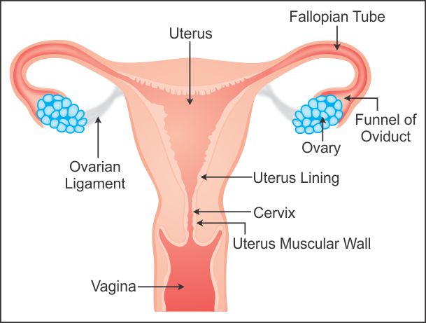
I The Human female reproductive system which produces eggs is Ovary II The human female reproductive system where fusion of egg and sperm takes place is Fallopian tube.

Draw a neat and well labelled diagram of human female reproductive system. Each ovary contains immature ova eggs in follicles. Watch complete video answer for Draw a labelled diagram of female reproductive sys of Biology Class 12th. Q-Why is reproduction essential for organismsAnswer.
Study the same and then answer the questions that follow. A Draw A Sectional View Of Human Female Reproductive System And. Ii Females born with lifetime supply of eggs 250000-400000 in each ovary iii Ovaries release ovum -.
Blank Diagram Of Human Reproductive Systems. Almost all ova degenerate between birth and puberty. Start studying male reproductive system blank.
The human reproductive system includes the male reproductive system which functions to produce and deposit sperm. Following parts constitute the female reproductive system a pair of ovaries fallopian tube uterus cervix vagina and accessory genital glands and a pair of mammary glands. III The human female reproductive system where zygote is implanted is Uterus.
Draw out line of uterus around the triangle as shown. - The human cycle would end. Each ovary is of the shape of unshelled almond and the size is 35 cm long 2 cm wide and 1 cm thick.
Avail 25 off on study pack. Male and female reproductive system blank diagram. I production of female gamete ovum and ii secretion of female hormones estrogen and progesterone.
At puberty these follicles undergo maturation to produce ova. Uterus opens into vagina through narrow cervical canalTwo tube like structures emerge out of uterus on either sides. The female reproductive system is one of the most vital parts of the human reproductive.
Q-What is spermatogenesisBriefly describe the process of spermatogenesis. Each ovary releases one egg cell. Reproductive System - Male Draw neat and well labelled diagram of human femaleDownload this premium vector about human reproductive system vector illustration diagram and discover more than 11 million professional graphic resources on freepik.
A production of gametes b site of fertilization. 400 eggs will be ovulated over womans life. All the best Female Reproductive System Drawing 36 collected on this page.
C site of implantation and. Popular Questions of Class Biology. Draw neat and well labelled diagram of Human female reproductive system - Brainlyin.
The diagram is as follows. It is shaped like an almond size is around 35 cm to 2 cm wide 1 cm thick. Lets start the diagram.
The ovaries perform dual function of. Male And Female Reproductive Systems Harder To. Draw a neat diagram of the female reproductive system and label the parts associated with the following.
It is placed in the abdominal cavity. The ovary is a paired structure located in the upper pelvic cavity. Also write the function of fallopian tube and uterus.
Draw a inverted triangle as shown. Each ovary is composed of ovarian follicles. In human females a pair of ovaries is located in the abdominal cavity near the kidney.
These are called oviductsWe can start with basic shape of Uterus later we go for oviducts and vagina. The female reproductive structure consists of a pair of ovaries uterus fallopian tubes cervix and vagina. Get FREE solutions to all questions from chapter HUMAN REPRODUCTION.
Iii the human female reproductive system where zygote is implanted is uterus. Draw neat and well labelled diagram of Human female. Draw A Well Labelled Diagram Of Female Reproductive System.
Q-With a neat diagram explain the 7-celled 8-nucleate nature of the female gametophyteQ-With a neat labelled diagram describe the parts of a typical angiosperm ovuleQ-Differentiate between a zoospore and a zygote. Draw a neat and well labelled diagram of female reproductive system. 5 Draw A Neat And Well Labelled Diagram Of 1 The Reproductive Human Reproductive System Male And Female Reproductive System Structure Of The Male Reproductive System Men S Health Issues Royalty Free Male Reproductive System Stock Images Photos Human.
Blank Diagram Of Human Reproductive Systems. The reproductive system is a collection of.