Click here to get an answer to your question sketch the reproductive parts of a flower satpalrathi70 satpalrathi70 10032021 Science Primary School answered Sketch the reproductive parts of a flower 2 See answers. A Stamen b Pistil.
Short Answer Question Sketch The Reproductive Parts Of A Flower Snapsolve
Sketch the reproductive parts of a flower Report.

Sketch the reproductive parts of a flower answer. Answer verified by Toppr. Describe the various ways by which seeds are dispersed. CCE 2012 Answerâ.
Click hereto get an answer to your question Sketch the reproductive parts of a flower. Reproductive parts of a Flower. Shilpa2341979 04032020 Science Secondary School 5 pts.
Click hereto get an answer to your question Sketch the reproductive parts of a flower. Sketch the reproductive parts of a flower. Reproductive whorls of a flower are.
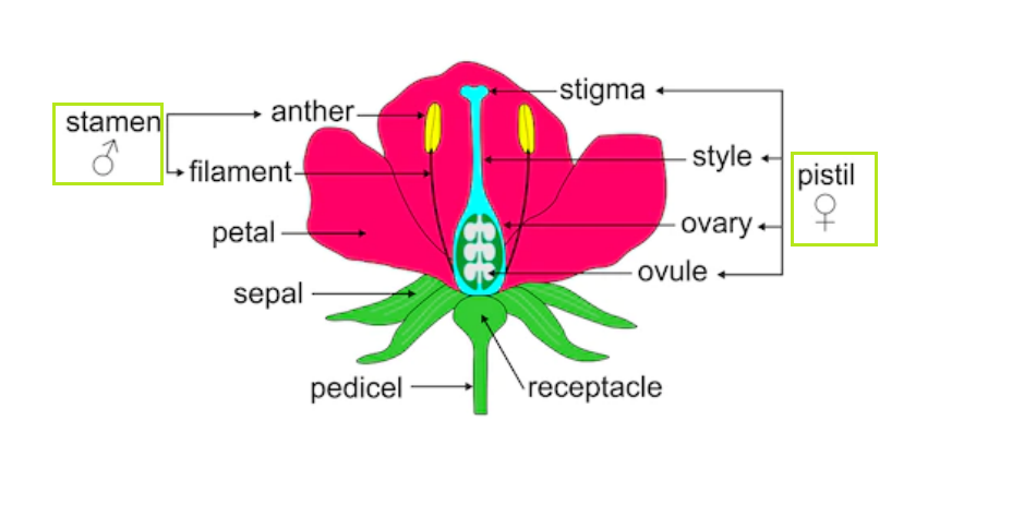
Gaurav Seth 8 months 2 weeks ago. Below well get into what each part does and include some great. Answer 5 Pistil is the female reproductive organ of a plant.
Sketch the reproductive parts of a flower - 33681341 tahseenshariff tahseenshariff 20012021 Science Secondary School answered Sketch the reproductive parts of a flower 2 See answers. Upvote 0 Was this answer helpful. Get Instant Solutions 24x7.
Sexual reproduction involves the fusion of male and. Sketch the reproductive parts of a flower. Apart from these parts a flower includes reproductive parts stamen and pistil.
1 See answer Answers. Also hand out the Flower Dissection worksheet. Flower structure Parts of a flower.
Posted by Piyush Sharma 8 months 2 weeks ago. Advertisement Remove all ads. No Signup required.
The whorls are arranged on the thalamus of a flower in a definite sequence. Upvote 0 Was this answer helpful. The reproductive part of a plant is.
AnswerExplanationthis is the simple structure of the reproductive parts of a flower raj431538 raj431538 04122019 Science Secondary School answered Sketch the reproductive part of a flower 1 See answer raj431538 is waiting for your help. Sketch the reproductive parts of a flower answer. Sketch the reproductive parts of a flower.
1 Answer 1 vote. Add your answer and earn points. Get Instant Solutions 24x7.
Sketch the reproductive parts of a flower. Q5 Sketch the reproductive parts of the flower. Answered Mar 29 2020 by Randhir01 595k points selected Mar 30 2020 by SonaSingh.
Sketch the reproductive parts of a flower. Stamens and carpels 2. It helps in sexual reproduction as it has male parts and female parts.
Sketch the reproductive parts of a flower Answer Question 6 Explain the from MAT 123 at Shivaji University. Can you explain this answer. The transfer of pollen grains from the anther to the stigma of the same or of another flower of the same kind is known as __________.
Explain the difference between self-pollination and cross - pollination. Reproductive parts Prev Question Next. Answer verified by Toppr.
A Stamen b Pistil. Click to find video solution. Sketch the reproductive part of a flower.
Sepals and petals 3. Share It On Facebook Twitter Email. Sketch the reproductive parts of a flower.
CBSE Class 07 Science 1 answers. Reproductive parts of a Flower. Sepals and stamens 4.
The pistil contains the stigma style and ovary. Male and Female reproductive parts of a flower. Correct answer to the question.
The Female Parts of a Flower.
The nephron uses four mechanisms to convert blood into urine. Schematic structure of a transcription unit.
Kidney Wikipedia
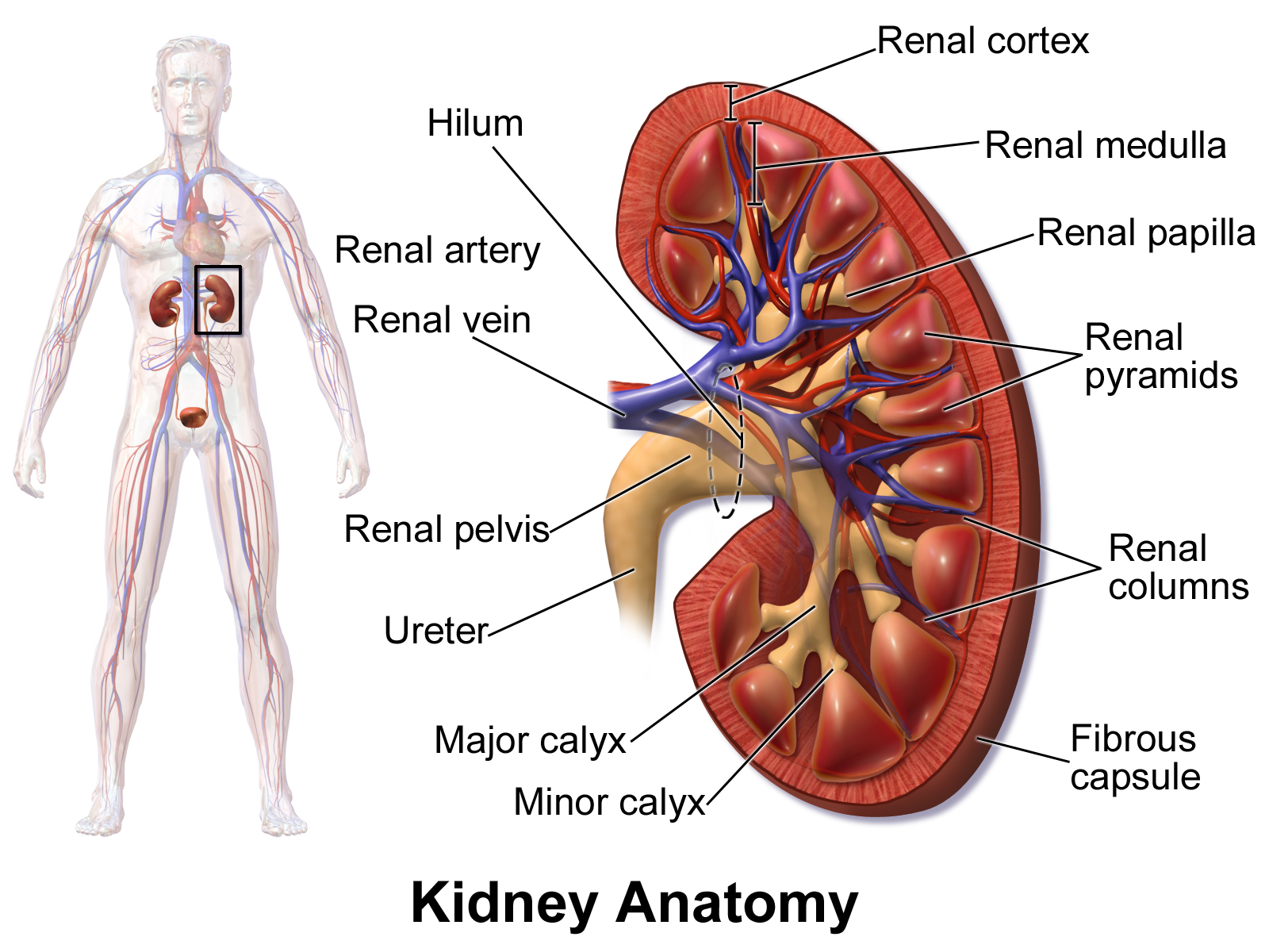
In LS the kidney shows two regions within the capsule.

Explain the structure of kidney with a labelled sketch. The vital structural components of a kidney are enclosed in a smooth but tough fibrous capsule called renal capsule. In the concave side there is a depression in the middle point which is called as hilum or hilus. A solar cell or photovoltaic cell is an electrical device that converts the energy of light directly into electricity by the photovoltaic effect which is a physical and chemical phenomenon.
The kidneys have been bean-shaped appearance. The kidneys are among the most vital organs of the human body. 5437 kidney drawing stock photos vectors and illustrations are available royalty-free.
Each kideney is covered by semi-liquid fatty tissue called adipose capsule. The outer edge of the kidney is convex and the inner concave. Structural organisation in animals.
Filtration reabsorption secretion and excretion of numerous substances. Outer renal cortex and inner renal medulla. Ureter or the duct of the kidney also comes out through this hilum and pass.
It is a cluster of thin-walled blood capillaries. The combination of waste products and excess water constitutes urine. See kidney drawing stock video clips.
The renal medulla comprises a set of 8-18 conical structures called renal pyramids. A healthy adult has 08 to 15 million nephrons in each kidney. Once urine is formed in the kidneys it flows into two thin tube-like organs called ureters.
Gross Structure Of Kidney Kidneys with blood vessels the one on to right is a longitudinal section of the kidney. Medical drawing liver anatomy doodle kidney sketch human organs sketch anatomical kidney drawing internal organs icons digestion sketch stomach illustration kidney bone muscle icon line. It is the outer region of kidney.
The kidneys are responsible for carrying out two very important functions. The average kidneys are about 150 gm in males and 135 gm in females. It codes for enzyme or protein for structural functions.
Renal artery enters and Renal vein comes out of the kidney. Outer covering of this capsule is made up of tough fibrous connective tissue called renal fascia. Malfunction of the kidneys can lead to serious illness or even death.
NCERT Question 11 Class 8. Unlike other organs found in the abdomen the kidneys are located behind the lining peritoneum of the abdominal. Within the kidneys are tiny filtering structures called nephrons.
They are held in position by a mass of mass. Explain with neat diagram construction and working of solar cell. Inside this capsule two distinct regions can be observed.
1 It is a form of photoelectric cell defined as a. The blood vessels ureter and nerves enter the kidneys through the hilum which is a notch at the inner concave surface of the. A kidney is a bean-shaped organ that is reddish-brown in color.
The kidneys have an inner concave structure. Draw a labeled sketch of the human eye. Kidney Structures and Functions Explained with Picture and Video Your kidneys are paired organs found on each side of the back portion of the abdominal cavity.
The structure and function of the epithelial cells lining the lumen change during the course of the nephron and have segments named by their location and which reflects their different functions. Towards the centre of the inner concave surface of the kidney. It has a dimension of 11 cm in length 6 cm in width and 3 cm in thickness.
Kidneys are reddish brown bean shaped structures situated between the levels of last thoracic and third lumbar vertebra close to the dorsal inner wall of the abdominal cavity. It is the region where transcription ends. Please log in or register to add a comment.
Here the blood vessels of the kidney ie. A nephron is the basic filtration unit of the kidney. Draw a labelled diagram of areolar connective tissue.
Humans have two kidneys. Each ureter connects each kidney to the bladder. To flush out harmful and toxic waste products and to maintain balance of water fluids minerals and chemicals ie electrolytes such as sodium potassium etc.
Kidneys are bean-shaped structures located on either side of the backbone and are protected by the ribs and muscles of the back. They have two important functions namely. In humans the excretory system consists of a pair of kidneys one pair of ureters a urinary bladder and a urethra.
Each kidney has a very complex structure and function. A sheath of fibrous tissue. NCERT Question 1 NCERT Question 2 NCERT Question 3 NCERT Question 4 NCERT Question 5 NCERT Question 6.
There are different parts of the nephron in which the formation of urine take place which is the main function of the kidney. Each human adult kidney has a length of 10-12 cm a width of 5-7 cm and weighs around 120-170g. Chapter 16 Class 8 - Light.
The nephrons help eliminate urea other waste products and excess water from the body. The larger left kidney is located a bit higher than the right kidney. It is the binding site for RNA polymerase for initiation of transcription.
Structure of the Human Kidney. A pale outer region called renal cortex and a dark inner portion called renal medulla. As an osmoregulatory organ they regulate the water content of the blood vital for maintaining blood pressure As an excretory organ they excrete the toxic waste products of metabolism such as urea and.
Bowmans capsule and the glomerulus are together called as the glomerular apparatus. Each kidney of an adult human measures 10-12 cm in length 5-7 cm in width 2-3 cm in thickness with an average weight of 120-170 g.