Read the following passage and answer the following questions. Start studying Kidney Anatomy Labeling.
Draw A Well Labelled Diagram Of Ls Of The Human Ki Class 12 Biology Cbse
Here you have to show the full process of digestion.

Diagram of kidney with labelling class 10. Veins of the abdomen and hepatic portal. Draw a well-labeled diagram of the human excretory system. Draw a labeled diagram of the human kidney as seen in a longitudinal section.
Each kidney is about 10 to 13 cm 4- 5 inches in long 6 cm. The diagram given below shows a section of human kidney. 2 ½ inches wide and 3 cm.
On the upper end of each kidney suprarenal glands are situated like a cap. Kidney diagram to label. Human Excretory System.
Label Diagram Of Kidney Wiring Diagram Pipe Activity Pipe Activity Fiocouture It. Draw the diagram of kidney recognize and label the following parts. Kidney diagram class 10 0 comments.
Dehydration a blockage in the urinary tract or kidney damage can cause acute renal failure which may be. A human brain is composed of three main parts-. Current diagram shows a little bit about human excretory system and a detailed diagram of Kidney which is the most important part of Excretory system.
Urinary bladder CBSE 2013 Answer. CBSE Class 10 Physics Important Diagrams. Kidney Diagram Class 10.
Label the parts numbered 1 to 4. Class-10 Welcome to Sarthaks eConnect. Sketch and label the structure of malphigian body.
In males and 135 gms in females. Why does part 2 have a striped appearance. Arteries Of Kidney Great For Anatomy Class Diagnostic Medical.
Q6 Draw A Diagram Of The Human Excretory System And Label Th Lido. Sketch and Label the Diagram. Draw A Well Labelled Diagram Of Human Excretory System And Write Name Of Filtering Unit Of Kidney Brainly In.
The human kidneys house millions of tiny filtration units called nephrons which enable our body to retain the vital nutrients and excrete the unwanted or excess molecules as well as metabolic wastes from the body. Kidney DiagramThe collecting duct labelled in the diagram above is part of the kidney nephron shown much enlarged. It allows small molecules such aswater to.
I Urinary bladder ii Left kidney iii Left ureter. Kidneys are dark brown in colour and are embedded in a mass of fat. What is the fluid that passes down 4.
The average weight of adult kidney is about 150 gms. Draw and label a diagram of the kidney. 101 labeled diagram of the human kidney stock photos vectors and illustrations are available royalty-free.
At September 11 2018 Labels. CBSE Class 10 Chemistry Important Reactions. Draw and label a diagram of the kidney ib biology syllabus.
Click hereto get an answer to your question Draw well labelled diagram of LS. Label the schematic drawing. Learn vocabulary terms and more with flashcards games and other study tools.
1 ½ inch in thickness. Diagram of human excretory system. In this diagram you have to show all the parts of the system which includes two kidneys urethra ureters etc.
Dialysistubing is a semipermeable membrane. The alignment of kidney are. Human Body Organs Diagram From The Back Koibana Info Human Body Organs Human Body Anatomy Body Organs Diagram Students drag labels to the structures on the slide.
You have to mention the mouth liver gallbladder stomach pancreas small and large intestine liver etc. Draw A Well Labelled Diagram Of The L S Of Kidney Label Any. The nephron is one of the most important parts of our body and also one of the smallest functioning units.
Study the diagram carefully and answer the questions that follow. Biology section 3 flashcards the hilus is present on the concave side of the kidney through which the uriters are attached to the kidney the part of the kidney where the renal artery and renal vain enter label the diagram the kidney and nephron below anatomy kidney structure and function the excretory system chegg label the diagram. Students upto class 102 preparing for All Government Exams CBSE Board Exam ICSE Board Exam State Board Exam JEE MainsAdvance and NEET can ask questions from any subject and get quick answers by subject.
Image of a kidney and nephron with the major structures labeled. A unique platform where students can interact with teachersexpertsstudents to get solutions to their queries. Thousands of new high-quality pictures added every day.
Featuring a professional light background with a large amount of vector format various symbols and some other important science elements you wont miss this illustrated kidney diagram template from. Use this interactive 3-D diagram to explore the kidney. Find labeled diagram of the human kidney stock images in HD and millions of other royalty-free stock photos illustrations and vectors in the Shutterstock collection.
1 ½ inch in thickness. Click to find video solution Book a free class. Name the main nitrogenous waste.
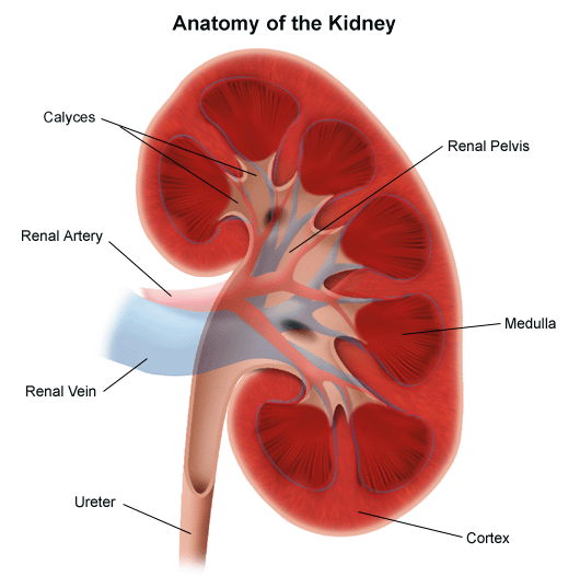
The kidney is one of the primary organs of the human body and the urinary system its evolution until today has been indispensable for. This pair of purplish-brown organs is located below the ribs toward the middle of the back.

25 1 Internal And External Anatomy Of The Kidney Anatomy Physiology
Their function is to remove liquid waste from the blood in the form of urine.

What parts of the human kidney you see. What parts of the human kidney do you see. Science 10122019 1828 kenn14. Kidney and urinary system parts and their functions.
Tamang sagot sa tanong. In this article we are going to analyze the kidney anatomy and the different functions of each of its parts both in humans and animals. Each kidney has about 1 million nephrons.
If blood stops flowing into a kidney part or all of it could die. You could have only 10 of your kidneys working and you may not notice any symptoms or problems. What parts of the human kidney do you see.
Integrated Science 28012020 0828 09389706948. English 08012020 0828 reyquicoy4321 What parts of the human kidney do you see. What parts of the human kidney do you see.
Science 28102019 1445 hajuyanadoy Whats parts of the human kidney do you see. Kidney and urinary system parts and their functions. Each kidney has around a million tiny filters called nephrons.
This pair of purplish-brown organs is located below the ribs toward the middle of the back. Blood containing urea and metabolic waste products enters the kidney from the. What parts of the human kidney do you see.
Tamang sagot sa tanong. 1 Unlock answers. Their function is to remove liquid waste from the blood in the form of urine.
What parts of the human kidney do you see. Keep a stable balance of salts and other. It is the basic structural and functional unit of the kidney and cannot be seen by the naked eye.
What parts of the human kidney do you see. The walls of the nephron are made of a single layer of epithelial cells. What parts of the human kidney do you see.
That can lead to kidney failure. Science 10122019 2128 maledabacuetes What part of the human kidney do you see. - 1774269 beacunanan0701 beacunanan0701 29082018 Science Elementary School What parts of the human kidney do you see.
Integrated Science 28012020 0828 09389706948. What parts of the human kidney do you see. Science 11122019 1028 ShairaGailSanchez What parts of the human kidney do you see.
Integrated Science 28012020 0828 09389706948 What parts of the human kidney do you see. What parts of the human kidney do you see. Keep a stable balance of salts and other.
Biology 02022020 1504 09330399672 What parts of human kidney do you see. The nephron is the filtration unit of the kidney. Refer to Figure 5.
Learn vocabulary terms and more with flashcards games and other study tools. 33 Correctly Label The Following Anatomical Parts Of A Kidney.
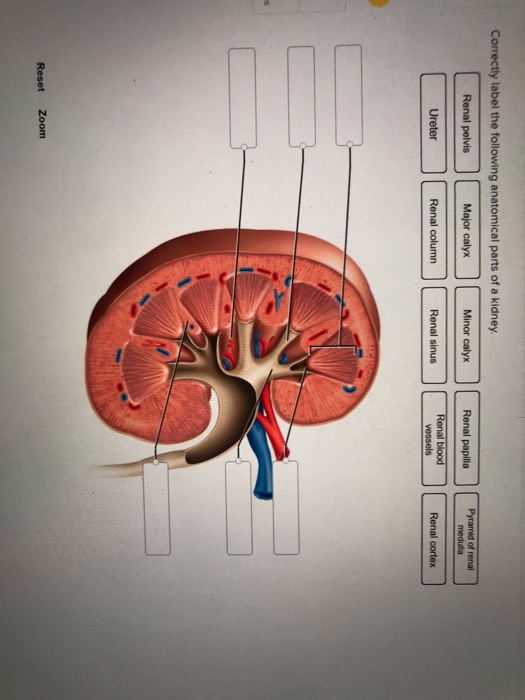
Correctly Label The Following Anatomical Parts Of A Chegg Com
In this article we are going to analyze the kidney anatomy and the different functions of each of its parts both in humans and animals.
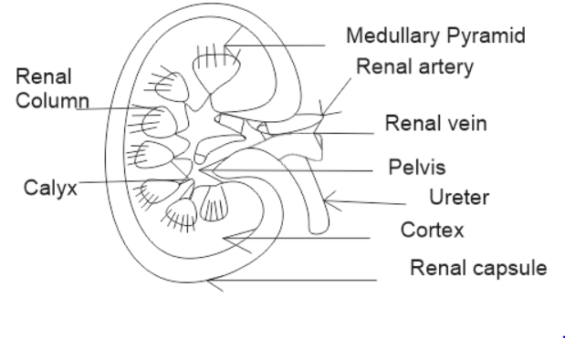
Label anatomical parts of a kidney. Upper portions of the kidneys are somewhat protected by the eleventh and twelfth ribs Figure 2511. Anatomy and Physiology Correctly label the following anatomical parts of a kidney. Ditulis oleh Maria M Beus Sabtu 10 November 2018 Tambah Komentar.
The kidneys are a pair of bean-shaped. Anatomy Human Kidney Main Parts Labeled Stock Vector January 1st 2021 - Find Anatomy Human Kidney Main Parts Labeled stock images in HD and millions of other royalty free stock photos illustrations and vectors in the Shutterstock collection Thousands. Anatomy of the Kidneys Quiz.
The upper parts of the kidneys are partially protected by lower ribs and each whole kidney and adrenal gland are surrounded by two layers of fat. Renal pyramid in renal medulla. Correctly label the following anatomical parts of a kidney Renal vein Renal medulia Renal cortex Ureter Renal sinus Renal artery Renal pyramid Renal pelvis.
Anatomy of the Kidneys Regulation of Urine Concentration Urethra Quiz. Correctly label the following anatomical parts of a kidney. Start studying Correctly label the following anatomical parts of a kidney.
Ureter Renal pelvis Renal blood vessels Renal column Pyramid of renal modulla Major calyx Renal sinus Renal cortex Renal papilla Minor calyx Prey 5 of 20 Next. The basic physiology of a nephron within a kidney. The apex of the pyramid projects medially toward the renal sinus.
There is a printable worksheet available for download here so you can take the quiz with pen and paper. Medulla inner region of the kidney contains 8 12 renal pyramids. The left kidney is located at about the t12 to l3 vertebrae whereas the right is.
The left kidney is located at about the T12 to L3 vertebrae whereas the right is lower due to slight displacement by the liver. Each kidney weighs about 125175 g in males and 115155 g in females. Internal anatomy of the kidney overview The main unit of the medulla is the renal pyramid.
There are 8-18 renal pyramids in each kidney that on the coronal section look like triangles lined next to each other with their bases directed toward the cortex and apex to the hilum. Answer The label is indicated from right View the full answer Transcribed image text. Each kidney weighs about 125175 g in males and 115155 g in females.
This is an online quiz called Label the kidney. Medullary pyramids formed by the collecting ducts inner part of the kidney. Each kidney typically has eight to.
The kidney is one of the primary organs of the human body and the urinary system its evolution until today has been indispensable for. The Male Reproductive System.
Try these curated collections. Bowmans capsule Glomerulus Collecting Duct Renal Artery.

Anatomy Of The Human Kidney Cut To Show Internal Structures Arteries Download Scientific Diagram
The right kidney is placed slightly lower- than the left due to the presence of liver which occupies much space on the right side.

Draw and label the diagram of the human kidney. Biology section 3 flashcards the hilus is present on the concave side of the kidney through which the uriters are attached to the kidney the part of the kidney where the renal artery and renal vain enter label the diagram the kidney and nephron below anatomy kidney structure and function the excretory system chegg label the diagram. A pair of kidneys are present in the human and they are one of the primary excretory systems to remove wastes from the bloodstream and body. Maintains the balance of fluid.
Drag the labels to their appropriate locations on the diagram below. They lie on the posterior abdominal wall. Nephron Diagram And Function.
Function of kidneys-Remove the waste from the body in the form of urine. Label the diagram kidney internal structure. C Write any one function of an artificial kidney.
Bowmans capsule Glomerulus Colleeting duct Renal artery. A Draw a diagram of an excretory unit of a human kidney and label the following. After reading this article you will learn about the structure of the kidney.
Drawing of kidney and label. I i Why does part 2 have striped appearance. The right kidney is placed slightly lower- than the left due to the presence of liver which.
Draw a diagram of the human excretory system and label the following parts. This will also help you to draw the structure and diagram of kidney. Labeled Diagram Of Kidney Nephron Diagram Kidney Anatomy.
Human Body Organs Diagram From The Back Koibana Info Human Body Organs Human Body Anatomy Body Organs Diagram. Label the schematic drawing of a kidney. Search for labeled diagram of the human kidney in these categories.
Draw the LS of kidney and. Kidneys are a pair of bean-shaped organs in the renal system. 113S1 Drawing and labelling of the human kidney Make sure you are able to draw and label the kidney below.
The kidneys are two in number which are situated one on each side of the verteral column and in-front of the last ribs. Types And Grades Kidney Cancer Cancer Research Uk. Kidney Anatomy Nephron Filtration Diagram Photo Kidney Anatomy Basic Anatomy And Physiology Anatomy.
Click hereto get an answer to your question Draw a neat and correct diagram of the of the human kidney and label. Place your mouse pointer on the diagram to view labelled structures of the kidney. Find labeled diagram of the human kidney stock images in HD and millions of other royalty-free stock photos illustrations and vectors in the Shutterstock collection.
Draw and label a diagram of the kidney. 0 Response to Label Kidney Diagram. Start studying Kidney Anatomy Labeling.
113S1 Drawing and labelling of the human kidney Make sure you are able to draw and label the kidney below. Draw a labeled diagram of the human kidney as seen in a longitudinal section. Current diagram shows a little bit about human excretory system and a detailed diagram of Kidney which is the most important part of Excretory system.
Draw A Labeled Diagram Of The Human Kidney As Seen In A Longitudinal Anatomy Of The Urinary System Diagram Of Ureter Slide Wiring Diagram Forward Kidney And Nephron Diagram Westgate Mennonite Collegiate Anatomical Position Labeling Quiz Anatomical Position Labeling Quiz Nephron Definition Function And Structure Biology Dictionary Share this post. Kidney ureter urinary bladder and urethra. Biology 19022020 2210 Firdaus17f Draw and label the diagram of internal structure of human kidney.
Learn vocabulary terms and more with flashcards games and other study tools. A Draw A Diagram Of An Excretory Unit Of A Human Kidney And Label. See labeled diagram of the human kidney stock video clips.
Diagram Of Renal Drug Action Stock Vector Illustration Of Artery. I Label the parts numbered 1 to 4. B Write the important function of the structural and functional unit of kidney.
103 labeled diagram of the human kidney stock photos vectors and illustrations are available royalty-free. Draw a diagram of an excretory unit of a human kidney and label the following. Anatomy of a human kidney-Cortex.
Jul 24 2019 A beautiful drawing of the kidneyAnd it will teach you to draw the kidney very easily. Draw a neat and correct diagram of the of the human kidney and label it. Start studying Kidney Anatomy Labeling.
Draw and label a diagram of the kidney ib biology syllabus. Draw well labelled diagram of LS.
Drawing of kidney and label. Bowmans capsule Glomerulus Colleeting duct Renal artery.

Cross Section Of A Human Kidney Kidney Disease Recipes Kidney Disease Kidney Transplant
The functional units where the kidneys main functions are performed.
Draw and label the parts of a kidney. 1 Renal Artery 2 Renal Vein. 1 Malpighian Body 2 Renal tubule. Each human kidney possesses about 1 -2 millions of nephrons.
Label kidney diagram. 1 Answer 1 vote. I Outer cortex and inner medulla are the two zones in kidney.
1 renal corpuscle 2 renal tubule and 3 collecting tubule with the first two being the main parts and the third an accessory part. BOWMANS CAPSULE-its a part of the kidney which is used to get blood from the nephron. Draw the LS of kidney and.
Renal Pelvis Basin-like area that collects urine from the nephrons the kidneys filtration system it narrows into the upper end of the ureter. Share It On Facebook Twitter Email. Parts of kidney.
8 Branches of the latter vessels in the kidney. It is thin but tough and fibrous. Draw a diagram of an excretory unit of a human kidney and label the following.
Draw the diagram of kidney recognize and label the following parts. C Write any one function of an Artificial Kidney. Each nephron is made up of two main parts.
A pale outer region called renal cortex and a dark inner portion called renal medulla. Draw and lable the parts of kidney. Image of a kidney and nephron with the major structures labeled.
Upgrade to remove ads. There are about a million nephrons in each kidney. The renal medulla comprises a set of 8-18 conical structures called renal pyramids.
Draw a Diagram of the Human Excretory System and Label the Following Parts. Within the kidney both the renal corpuscle and the renal tubule are located in the cortex whereas the collective tubule is found in the medulla. Name the cavity in which the kidney sits.
Health Medicine Endocrine system. A longitudinal section of a kidney. Kidney Anatomy Labeling Diagram Quizlet.
The part of the kidney nephron that collects urine and drains into papillary ducts minor calyx and major calyx and finally into the ureter and urinary bladder. Renal Capsule An outer membrane that surrounds the kidney. Iv Inward extension of cortex between the pyramids is called renal column of Bertini.
Draw the LS of kidney and. Calyx The extension of the renal pelvis. It consists of three parts.
Find an answer to your question a Draw a diagram of kidney and label the following partsi Urinary Bladder ii Aorta iii Urethrab During breathing cycle wh. The vital structural components of a kidney are enclosed in a smooth but tough fibrous capsule called renal capsule. Watch the video and please be kind enough to thumbs up my videos.
DRAW THE STRUCTURE OF A NEPHRON AND LABEL THE FOLLOWING ON IT GLOMERULUSBOWMANS CAPSULE RENAL ARTERY COLLECTING DUCT. Inside this capsule two distinct regions can be observed. Q6 Draw a diagram of the human excretory system and label the following parts Kidney ureter urinary bladder and urethra.
Excretory Products and their Elimination. Draw and label a diagram of the kidney 1133. 113S1 Drawing and labelling of the human kidney Make sure you are able to draw and label the kidney below.
A Draw a diagram of an excretory unit of a human kidney and lable the following Bowmans capsule Glomerulus Collecting duct Renal artery. The location in the kidney where processed filtrate called urine is collected from the renal tubules efferent arteriole carries blood away from the glomerulus. The nephron is one of the most important parts of our body and also one of the smallest functioning units.
Dehydration a blockage in the urinary tract or kidney damage can cause acute renal failure which may be. The two important blood vessels of the kidney are. 1 2 3 Parts of the Kidney.
Answered Dec 28 2020 by Baani 490k points selected Dec 28 2020 by Jaimi. Iii Pyramid projects into calyx. I Label the parts numbered 1 to 4.
Draw the LS of kidney and label the parts. Renal Artery enters into the kidney through hilum. They channel urine from the pyramids.
Kidney Ureter Urinary Bladder and Urethra. B Write the important function of the structural and functional unit of kidney. Ii Medulla is divided into few renal pyramids.