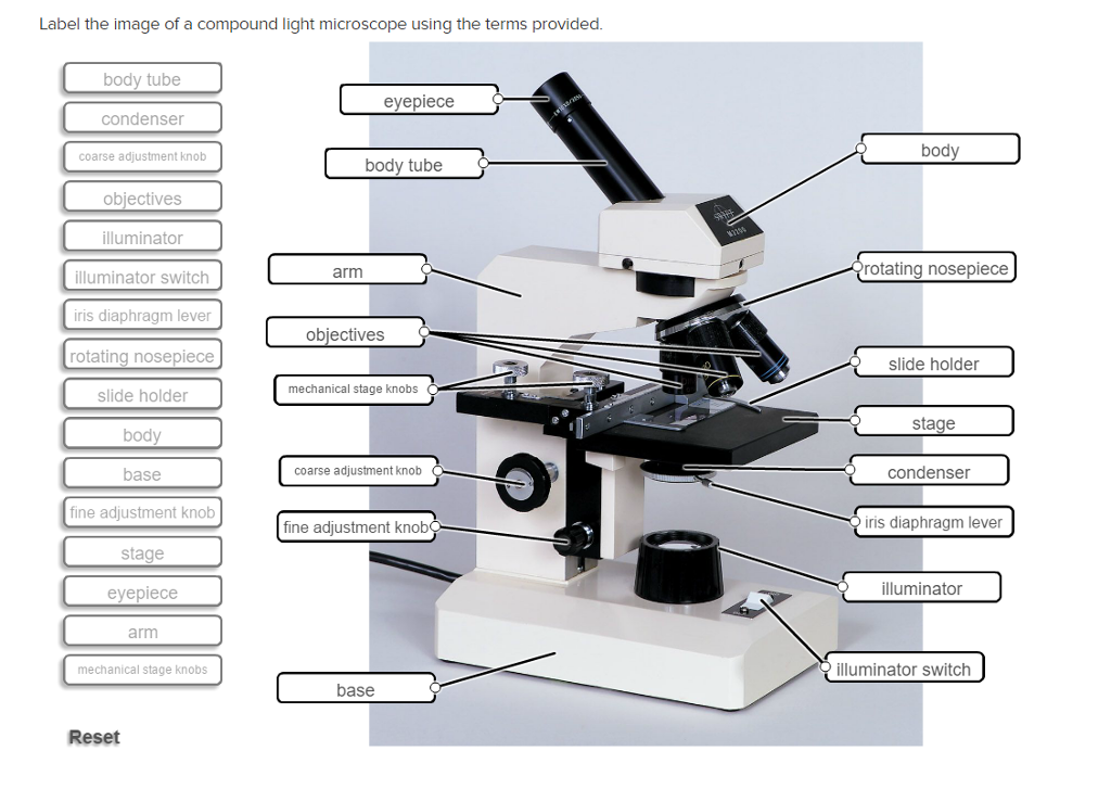
A microscope with a high resolution and uses two sets of lenses providing a 2-dimensional image of the sample. Compound light microscope with label.
Label The Image Of A Compound Light Microscope Using Chegg Com
Transcribed image text.

Labeled picture of compound light microscope. The microscope pictured above is referred to as a compound light microscope. Simple light microscope diagram compound microscope parts blank and compound light microscope parts are three of main things we will present to you based on the gallery title. An easy and convenient way to make label is to generate some ideas first.
Also the compound microscope is one of the types of optical microscopes. You should make a label that represents your brand and creativity at the same time you shouldn. The part that is looked through at the top of the compound microscope.
See 17 Best Images of Microscope Labeling Worksheet. The Best Free Binocular Drawing Images. Labelled Diagram Of Compound Microscope.
First the purpose of a microscope is to magnify a small object or to magnify the fine details of a. Compound deals with the microscope having more than one lensmicroscope is the combination of two words. Its an upright microscope that produces a two-dimensional image and has a higher magnification than a stereoscopic microscope.
Learn vocabulary terms and more with flashcards games and other study tools. Compound Microscope Definitions for Labels Eyepiece ocular lens with or without Pointer. The stage is the flat platform where the slide is placed.
A compound light microscope mostly uses a low voltage bulb as an illuminator. What is a compound light microscope. Compound Microscope Labeled Diagram Quizlet.
In the mean time we talk concerning Light Microscope Diagram Worksheet scroll down to see particular similar images to inform you more. What is Compound Microscope. Compound Light Microscope.
All the best Compound Light Microscope Drawing 36 collected on this page. A compound light microscope is a type of light microscope that uses a compound lens system meaning it operates through two sets of lenses to magnify the image of a specimen. Light Microscope Sketch At Paintingvalley Com Explore Collection.
The term compound refers to the usage of more than one lens in the microscope. Swift Microscope Sw100 Monocular Compound Microscope Amazon Co. Start studying Compound Microscope Labeled.
Nosepiece is a rotating turret that holds the objective lenses. Inspiring Microscope Labeling Worksheet worksheet images. Label the image of a compound light microscope using the terms provided.
Microscope Parts Quiz Worksheet Microscope Parts Blank Diagram Compound Light Microscope Parts Worksheet Microscope Diagram Worksheet Label Microscope Parts Worksheet. Eyepieces typically have a magnification between 5x 30x. Common compound microscope parts include.
The illuminator is the light source for a microscope. Compound Light Microscope Labeled Creative Labels Label Gallery Get some ideas to make labels for bottles jars packages products boxes or classroom activities for free. The viewer spins the nosepiece to select different objective lenses.
A compound microscope is defined as.
A Draw a labelled ray diagram of compound microscope when final image forms at the least distance of distinct vision. Derive the expression for the total magnification of a compound microscope.
A Draw A Ray Diagram To Show The Working Of A Compound Microscope Deduce A Expression For The Total Magnification When The Final Image Is Formed At Th Physics Topperlearning Com
Find an answer to your question draw a labelled ray diagram of a compound microscope and explain its working derive an expression for its magnifying power if.
Draw labelled diagram of compound microscope and write the expression for its magnifying power. Write any one expression for its magnifying power. Draw a labelled ray diagram to show image formation by a compound microscope and write the expression for its resolving power. The focal lengths of the objective and eye-lens of a compoun microscope are 2 cm 625 cm respectively.
Shyam4057 shyam4057 18042019 Physics Secondary School answered draw a labelled ray diagram of a compound microscope and explain its working derive an expression for its magnifying power if final image is. Draw a ray diagram of a compound microscope. How To Determine The Magnifying Power Of Simple Microscope Quora.
The angular magnification of a compound microscope is the ratio of the angle subtended by the final image at the eye to the angle subtended by the object at the eye when both are placed at the least distance of distinct vision. 11i Draw a labelled ray diagram showing the formation of a final image by a compound microscope at least distance of distinct vision ii The total magnification produced by a compound microscope is 20. Draw a labelled ray diagram of an astronomical telescope in the near point position.
Write two important limitations of a refracting telescope over a reflecting type telescope. The ray diagram to show the working of compound microscope is shown in figure. A tiny object AB to be magnified is placed in front of the objective lens just beyond its principal focus fo.
This is the required expression for angular magnification. The object is placed at a distance of 6 cm from the objective lens. Draw a labelled ray diagram of a compound microscope and write an expression for its magnifying power.
Explain why both the objective and the eye piece of a compound microscope must have short focal lengths. Write the expression for its magnifying power. The distance between the lenses is 15cm.
Draw the labelled ray diagram for the formation of image by a compound microscope. Microscopes Geometrical Optics From A Level Physics Tutor. Explain with a neat diagram the working of a compound microscope Obtain an expression for its magnifying power.
All India 2008. In this case the objective lens O of the compound microscope forms a real inverted and. Draw a labelled diagram of an image formed by a compound microscope with the image at.
Click hereto get an answer to your question Draw a labelled diagram of an image formed by a compound microscope with the image at least distance of distinct vision. Notes On Microscope Grade 11 Physics Optical Instruments. I Want To Know About Compound Microscope And Its.
Thus total magnification of the compound microscope M m0 x me Lf0 x Dfe c Aperture and focal length increase or decrease the resolving power of the compound microscope. Magnification Formula For Compound Microscope Physics Ray. Write any one expression for its magnifying power.
Write any one expression for its magnifying power. Draw a labelled ray diagram of a refracting telescope. Magnification power is defined as the ratio of the angle subtended by the final image on the eye to the angle subtended by.
10Draw a ray diagram of compound microscope. B Why is its objective of short focal length and of short aperture compared to its eyepiece. Define its magnifying power and write the expression for it.
The focal length of the objective and eye-lens of a compound microscope are 2cm 625cm respectively. Draw a neat labelled diagram of a compound microscope and give the expression for its overall magnification. Ray diagram of a compound microscopeWhen the final image is formed at the least distance of distinct visionFor the image formed at infinity ue feand By making focal length of the objective small the magnifying power can be increased.
When the final image is formed at the least distance of distinct vision. Write the expression for its magnifying power. Asked Dec 13 2019.
The magnification produced by the eyepiece is 5. Draw a labelled ray diagram of a compound microscope and write an expression for its magnifying power asked May 3 2018 in Physics by paayal 147k points cbse. C The focal length of the objective is 4 cm while that of eyepiece is 10 cm.
Draw a labelled ray diagram of a compound microscope and write an expression for a magnifying power. Derive an expression for its magnification. Write the expression for its magnifying power.
Draw a labelled diagram of an image formed by a compound microscope with the image at least distance of distinct vision. Draw a neat labelled diagram of a compound microscope and explain its working. Draw A Labelled Ray Diagram Of A Compound Microscope And Write An.
The labelled diagram of a refracting telescope is as shown below.
Where L is the distance between second focal point of the objective and first focal point of eyepiece. Name any three optical defects of eye.

Microscope Diagram Labeled Unlabeled And Blank Parts Of A Microscope
Expression of magnifying power of a compound microscope is given by.

Well labelled diagram of compound microscope. Proper Use Of The Light Microscope Kohler Illumination. At the other end of the tube another co-axial smaller and wide tube. Working Principle And Parts Of A Compound Microscope With Diagrams.
These labeled microscope diagrams and the functions of its various parts attempt to simplify the microscope for you. Compound Microscope Labeled Diagram Quizlet. Hence for a small angle of incidence derive the relation d 1 A.
872018 2 Simple Compound Stereoscopic Electron Simple Microscope Similar to a magnifying glass and has only one lens. It consists of a long cylindrical tube containing at one end a convex lens of small aperture and small focal length. Give A Well Labelled Diagram Of Compound Microscope Using Of A Study Of The Microscope And Its Functions With A Labeled Diagram Free Microscope Drawing Download Free Clip Art Free Clip Art On.
Draw a labelled ray diagram of a compound microscope and explain its working. The specimen or object to be examined is usually mounted on a transparent glass slide and positioned on the specimen stage. Download the diagrams and practice labeling the different parts of these fascinating instruments.
This is called the objective lens O. Make your work easier by using a label. Compound microscopes have a combination of lenses that enhances both magnifying powers as well as the resolving power.
Microscope Parts and Functions Microscope One or more lenses that makes an enlarged image of an object. The entire microscope is handled by a strong and curved structure known as the arm. It is a vertical projection.
Microscope With Labels Clip Art At Clker Com Vector Clip Art. M u o v o 1 f e D Where v o is the image distance from the objective lens D is the least distance of distinct vision f e is the focal length of eye lens. Label Diagram Of Compound Microscope And Wrote The Role Of Any.
Light Microscope Labelled Diagram Presentation Name On Emaze 2 1. It is a U-shaped structure and supports the entire weight of the compound microscope. This stands by resting on the base and supports the stage.
Labels are a means of identifying a product or container through a piece of fabric paper metal or plastic film onto which information about them is. Instruments Of Microscopy Microbiology. Learn vocabulary terms and more with flashcards games and other study tools.
The objective lens form image A B near the first focal point ofeyepiece. Diagrammatically show the phenomenon of refraction through a prism. A bright-field or compound microscope is primarily used to enlarge or magnify the image of the object that is being viewed which can not otherwise be seen by the naked eye.
A Mechanical Parts of a Compound Microscope. A Well Labelled Diagram Of A Light Microscope. Here is well labelled diagram of microscope.
Wide collections of all kinds of labels pictures online. Compound microscopes have at least two lenses. 872018 3 Compound Microscope Lets light pass through an object and then through two or more lenses.
Microscope With Labels Clip Art At Clker Com Vector Clip Art. Simple Microscope Drawing At Getdrawings Free Download. Start studying Compound Microscope Labeled.
Define angle of deviation in this case. A Labelled diagram of compound microscope. If the final image A B is formed at the near point.
The earliest microscopes had only one lens and are called simple microscopes. New questions in Biology. Derive an expression for its magnifying power.
However as the saying goes practice makes perfect here is a blank compound microscope diagram and blank electron microscope diagram to label. A compound microscope consists of an objective lens of focal length 2cm an eyepiece of focal length 625 cm separated by a distance of 15cm. Diagram Of A Compound Microscope.
How far from the objective lens should an object be placed in order to obtain the final image at least distance of distinct vision. Light Microscope Drawing At Getdrawings Free Download. Magnification by a microscope is the product of the individual magnifying ability of the oculars and.
Magnification may be defined as the degree of enlargement of the image of an object provided by the microscope.
Head This is also known as the body it carries the optical parts in the upper part of the microscope. Labelled diagram of compound microscope.
Microscope Diagram Labeled Unlabeled And Blank Parts Of A Microscope
Let us take a look at the different parts of a compound microscope and understand each key component.
Compound microscope with labelled diagram. Coarse and fine focus knobs. Draw a labelled diagram of an image formed by a compound microscope with the image at least distance of distinct vision. These labeled microscope diagrams and the functions of its various parts attempt to simplify the microscope for you.
There are more than two lenses in a compound microscope. Binocular microscope drawing at. Compound Microscope Diagram Parts labelled Principle and Uses.
Using sign convention we find that OI 1 v 0 and OO u where v 0 is the image distance due to the objective and u is the object distance for the objective or the compound microscope. Contains the ocular lens. Is used to view samples that are not visible to the naked eye.
A Labelled diagram of compound microscope. This allows the slide to be easily inserted or removed from the microscope. There are three basic parts of a compound microscope as shown in the labeled diagram.
Terms in this set 14 EyepieceOcular lens. 350x453 11 best microscopes. Draw a well labelled diagram of binocular microscope.
However as the saying goes practice makes perfect here is a blank compound microscope diagram and blank electron microscope diagram to label. What is a compound microscope. Monocular or Binocular Head.
I 1 G 1 is negative and OJ is positive. Its main function is to provide the stability to the microscope. The specimen is placed on the glass and a cover slip is placed over the specimen.
Here are the compound microscope parts labeled. One of the most important parts of a compound. Brightfield Microscope Compound Light Microscope USB Microscope- definition principle parts examples uses Cell Organelles- Structure and Functions with labeled diagram.
Eyepieces typically have a magnification between 5x 30x. Sed to focus when using the low power. Compound microscope- definition labeled diagram.
Supports the slide or specimen. As a compound microscope binocular microscopes use two lenses to magnify the image. It forms the base of the microscope.
Common compound microscope parts include. The foot is generally horse shoe-shaped structure Fig. Microscope diagram labeled unlabeled and blank parts of a.
A hollow cylinder that holds the eyepiece. It also allows the specimen to be labeled transported and stored without damage. View Compound microscope- definition labeled diagram parts usesdocx from BIOLOGY 124 at Harvard University.
B Angular magnification of objective lens m0 linear magnification hh where L is the distance between second focal point of the objective and first focal point of eyepiece. Posted on july 18 2019 by admin. Posted on august 18 2019 by admin.
A Draw A Ray Diagram Of A Compound Microscope For The Final Image Free Microscope Drawing Download Free Clip Art Free Clip Art On Draw The Labelled Ray Diagram For The Formation Of Image By A. Major structural parts of a compound microscope. The part that is looked through at the top of the compound microscope.
The objective lens form image A B near the first focal point ofeyepiece. Head base and arm. The eyepiece behaves like a simple microscope.
By Editorial Team Last updated on July 1 2021 As the name suggests a compound microscope uses a combination of lenses coupled with an artificial light source to magnify an object at various zoom levels to study the object. Learn about the working principle parts and uses of a compound microscope along with a labeled diagram here. Part that supports the microscope.
And with the help of the handy microscope diagram and microscope worksheet found on this page youll be an expert on light microscope parts in no time. Compound microscope is a type of optical microscope that is used for obtaining a high-resolution image. The stand is made up of a heavy foot which carries a curved inclinable limb or arm bearing the body tube.
The flat platform where the slide is placed. To find me. Parts of a Compound Microscope - Labeled with diagrams A compound microscope is known as a high-power microscope that enables you to achieve a high level of magnification.
Diagrams have always been of great help in understanding both the structural and functional aspects of entities. Diagram of parts of a microscope There are three structural parts of the microscope ie. 2 which rests on table top or any other surface on which the microscope in kept.
FOOT Piece part of the Compound Microscope. Free microscope drawing download clip art on. Write any one expression for its magnifying power.
Download the diagrams and practice labeling the different parts of these. Structural support that holds connects the eyepieces to the objective lenses. M e 1 Df e.
Compound Microscope Definitions for Labels Eyepiece ocular lens with or without Pointer. Binocular microscope drawing at getdrawings com free for personal. The magnifying power of the eye piece.
The foot is generally heavy in order to increase the. Labeled diagram of a compound microscope. Smaller specimens can be thoroughly viewed using a compound microscope.
Light microscope incorporated in foot piece. The below mentioned article provides a labelled diagram of compound microscope. Optical components of a compound microscope.