The gastrointestinal tract includes the mouth pharynx esophagus stomach small intestine large intestine and anus. Lamina propria comprises loose connective tissue.

Digestive Tract With Labels Pointing To The Liver Stomach Small Intestine Colon Rectum And Anus Media Asset Niddk
The next organ in the gastrointestinal tract is stomach one of the key organs.
Draw and label parts of gastrointestinal tract. The liver and pancreas directly communicate with the GI tract via ducts that join with the duodenum the most anterior segment of. The lower portion of the gastrointestinal tract includes the stomach small intestine and large intestine. Can you label the parts of the human digestive tract.
It is the upper part of the gastrointestinal tract and is mainly involved in delivering the food particles from the mouth to the stomach. Its main function is to digest food and absorb nutrients and fluid. Mouth the first part of the digestive.
The mucosa submucosa muscularis. In stomach houses gastric glands. Digestive system helps in breaking complex food into simpler forms.
Though definitely not the most attractive organs in the body they are certainly among the most important. The upper portion of the gastrointestinal tract includes the oral cavity teeth tongue salivary glands pharynx and esophagus. The gastrointestinal diagram given below represents the different parts of the tract that include the oral cavity oesophagus stomach intestines and the anus.
The end of the small intestine should be towards the bottom of your paper and towards the left side. The wall of the GI tract from the esophagus to the anal canal has four-layer from deep to superficial are the mucosa submucosa muscularis and serosaadventitia. The small intestine has the four layers typical of the other organs of the gastrointestinal tract.
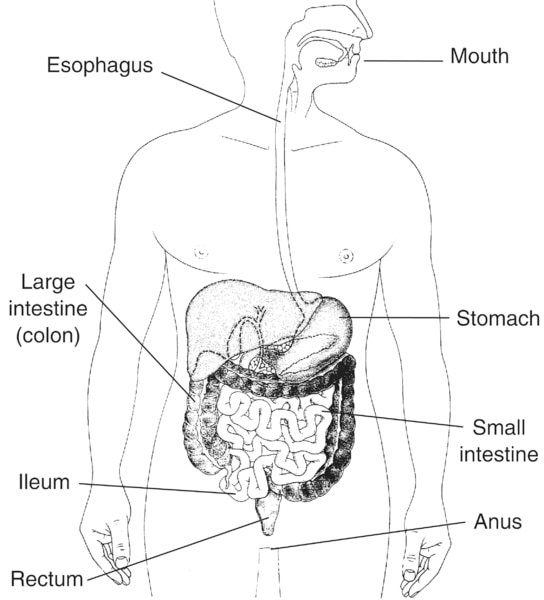
Gastrointestinal Tract Anatomy The gastrointestinal tract in humans begins at the mouth continuing through the esophagus stomach and the small and large intestines. The next part in GI tract is esophagus the food tract located after mouth and extended to the stomach. The mouth esophagus stomach and intestines are all part of the gastrointestinal tract.
Label the parts of the small intestine and color in the various regions and layers of the small intestine. Food taken in through the mouth is digested to extract nutrients and absorb energy and the waste expelled as feces. Identify and label figures in turtle diarys fun online game digestive system labeling.
The GI tract is imperative for our well being and our life-long health. Stomach It is a hollow and muscular organ situated towards the left side of the abdominal cavity and beneath the diaphragm. The digestive system also known as the gastrointestinal tract is made up of the mouth esophagus stomach small intestine large intestine rectum and the anus.
A Mucosa-The mucosa or innermost of the GI tract is a mucous membrane. It is subdivided into 4 regions. 1 esophagus 2 stomach 3 small intestine and 4 large intestine.
The mucosa subdivides into Epithelia comes into contact with contents of GI tract. The gastrointestinal tract is a pathway that begins at the mouth and ends at the anus. The accessory organs include the liver gallbladder and pancreas.
Gastrointestinal is an adjective meaning of or. The gastrointestinal tract is also known as the alimentary tract. Displaying top 8 worksheets found for labeling digestive system.
It receives food consumed in mouth after swallowing through the series of muscular contractions known as peristalsis and its major function is to bring the food to the stomach Gropper and Smith 2012. Terms in this set 14 esophagus. Muscularis mucosae comprises an inner circular and outer longitudinal layer note that it is different than.
Draw it a couple of inches below the stomach because the large intestine will end up going on top of it. Gastrointestinal tract also called digestive tract or alimentary canal pathway by which food enters the body and solid wastes are expelled. The gastrointestinal tract is the tract from the mouth to the anus which includes all the organs of the digestive system in humans and other animals.
Read on to find out more about the digestive system parts and functions. The 30 foot long tube that goes from the mouth to the anus is responsible for the many different body functions which will be reviewed in this chapter. 4 tunics layers of the GI tract From deep to superficial.
Overlooking the gastrointestinal tract GI tract. The gastrointestinal GI tract is a continuous tube beginning at the mouth and ending at the anus. Mucosa Lines the lumen of the GI tract.
The small intestine is a large curving tube that you can draw as squiggly lines beneath the stomach in the center of the body taking up about half of the width of the body. Upper GI tract Draw and label the esophagus diaphragm stomach and duodenum Label these regions of the stomach Fundus Cardia Body Pyloric partregion The small intestine is tricky to draw and label but question 2 on the quiz will ask you to put the regions of the small intestine. It is composed of epithelium connective tissue lamina propria and a layer of smooth muscle muscularis mucosa.
Rumen microbes also produce B vitamins vitamin K and amino acids. Ford Ranger Brake Line Diagram.

Diagram Of The Swine Gastrointestinal Tract With Major Sections Download Scientific Diagram
Digestive system by the diaphragm a thin sheet-like muscle.

A well labelled diagram of the digestive tract of a pig. The digestive system of a pig is well suited for complete concentrate based rations that are typically fed. April 16th 2019 - 11 2 draw label annotate contemporary labeled diagram of digestive system human well label diagram of a pig draw and digestive system human alimentary canal biology notes for igcse 2016 Trending Posts Jazz Piano Chords Diagram Pdf Fishbone Diagram Man Method Machine Digestive System of the Pig Anatomy and Function The. Dissections are best done in specimens preserved Frog dissection manual.
In addition the saliva has an enzyme that helps to begin the digestion of the starch contained in the food. A pigs digestive system is composed of 5 parts namely the mouth esophagus stomach the intestines. This work is licensed under a Creative Commons Attribution-NonCommercial-ShareAlike 40 International License.
The diagram below shows the structure and functions of the human digestive system. Acces PDF Fetal Pig Dissection Diagram Labeled Digestive System Human Anatomy and Physiology Custom Edition Anatomy and Physiology This work has been selected by scholars as being culturally important and is part of the knowledge base of civilization as we know it. C label diagrams of a pigs digestive system.
It comprises mouth buccal cavity pharynx oesophagus stomach small intestine and large intestine which opens to the exterior by cloacal aperture. During a lifetime a person will process between 60000 to 100000 pounds of food. Although this investigation is designed to With scissors remove the skin in the neck and examine the digestive system parts structures from head.
2628 is long tubular and coiled. Traumatic Reticuloperitonitis Digestive System Veterinary. Pig Digestive System Diagram Labeled.
Between the lungs we find the heart swaddled in the pericardial sac. It is the beginning of the digestive tract and the process of digestion begins from the mouth where teeth help by breaking and grinding the food. The fetal pigs that are used for the dissection are from pregnant females that.
The digestive system of pigeon is well developed and includes an alimentary canal and the digestive glands. Carefully lay the pig on one side in your dissecting pan and cut away the skin from the side of the face and upper neck to expose the masseter muscle that. Respiratory system respiratory system the pig site respiratory system in humans with respiratory disorders in cattle respiratory system diagram.
A pig resembles a human both internally and externally in many ways. Cow Respiratory System Diagram. Give the function of each organ or structure listed in Step 1.
Digestive system of the pig anatomy and function the an overview of the pig s digestive system mouth stomach small and large intestines by joel derouchey and colleagues at kansas state university s applied swine nutrition team presented at the swine profitability conference 2009 the digestive system of a pig is well suited for plete concentrate based rations. However anatomy of the human digestive system can be studied by examining the digestive system of a pig an animal similar to a human. If youve Pig Heart Diagram Labeled PDF Download Bobdempsey Org.
Dissect a fetal pig and identify the structures listed in Step 1. Lateral view of pigs head with salivary glands exposed. Is not easy to study the digestive organs of a human.
FETAL PIG DISSECTION OBJECTIVE 1. Fetal Pig Digestive System Diagram. Jan 23 2017 Pictures of Dissection Of Cockroach.
The pigs you will dissect are called fetal pigs. December 27 2018 - by Wandi - Leave a Comment. Just the sight and smell of food begins the digestive.
Let learn the different parts of the human digestive system. Here we can begin to see that the heart is separated into four chambers - the right and left atrium as well as the right and left ventricle. External acoustic meatus make up the external ear in the pig as well as in the human.
Yet this multi-faceted system involves many complex interactive functions. Oct 16 2010 I get my most wanted eBook. The right atrium is the ear-shaped section of the heart labeled 1.
Image of the digestive system has numbers instead of labels it is intended for students of anatomy to practice their knowledge of the system by labeling the various organs and structures. Mouth It includes teeth salivary glands and tongue. The entire digestive tract is relatively simple in terms of the organs involved which are connected in a continuous musculo-membanous tube from mouth to anus.
The alimentary canal of pigeon Fig. Schematic Overview Of The Pathogenesis Bovine Respiratory. PIG DIGESTIVE SYSTEM.
When the pig consumes food it is first chewed in the mouth. Label Diagram Of Cockroachpdf Free Download Here Well Label Diagram Of A Cockroach. The skin is 48 hours to completely digest Try to swallow this The average digestive tract alimentary canal is 27 feet long.
Trace the path of food through the digestive tract of the pig. Some interesting facts about your digestive system. Well Labelled Diagram Of A Pig pig heart drawing at getdrawings com free for personal unlabeled diagram of digestive system diagram body of diagram of the human eye diagram link digestive system labelled diagram anatomy human well labeled diagram of a female pig organ anatomy fetal pig digestive system diagram daytonva150.
The saliva secreted by the mouth helps to soften as well as moisten the food.