The first muscle diagram labeled 2019 above gives you an illustration of the anatomy of the arm muscle. In Chapter 11 the phys-.
Labeled Muscles Of The Human Body Anterior View 3d Rendering Stock Photo Download Image Now Istock

Printable muscle labeling worksheet.

Human body muscles diagram labeled. Muscle diagram labeled front and back muscle system labelling front and back muscular system labeled front and back Human Muscles muscle diagram labeled front and back muscle system labelling front and back muscular system labeled front and back. Muscle-labeled-diagram - Diagram - Chart - Human body anatomy diagrams and charts with labels. The interossei muscles four dorsally and three volarly originating between the metacarpal bones.
There are more than 600 skeletal muscles in the body. The human muscle diagram provided above is the finger muscle diagram. Muscle anatomy quiz for anatomy and physiology.
And the lumbrical muscles. There are around 650 skeletal muscles within the typical human body. Superficial and deep posterior muscles of upper body Anterior and posterior muscles of the upper arm Anterior and posterior muscles of the lower arm Anterior and posterior muscles of upper leg Posterior muscles of lower leg Anterior muscles of lower leg Respiratory muscles.
Use our muscles ks2 labelling activity to learn about key muscles. And together with the scaffolding provided by the skeleton muscles also determine the form and contours of our body. May 28 2016 - The human body muscles are the main contractile tissues of the body involved in movement.
The muscular system consists of all the muscles present in a single body. Check spelling or type a new query. Muscles Labeling Side Body Muscle Anatomy Musculoskeletal System Muscle Body.
Muscle diagrams are a great way to get an overview of all of the muscles within a body region. Muscle Diagrams To Label Localprivate Muscles Muscular System Labeled Muscle Diagram Muscular System. Maybe you would like to learn more about one of these.
This diagram depicts Labeled Muscle Diagram 10241878 with parts and labels. This quiz requires labeling so it will test your knowledge on how to identify these muscles latissimus dorsi trapezius deltoid biceps brachii triceps brachii brachioradialis pectoralis major serratus anterior rectus abdominis etc. Contraction of individual muscle cells is ultimately re-sponsible for purposeful movement.
Human body muscle diagrams. Human Body Muscle Diagram Human body muscles Muscle diagram Human muscular system. Labeling and diagramming is a great way for your children to.
The muscle of the arm is divided by a fascial layer separating the muscles into two osteofascial compartments. Human Body Diagram With Labels Human Body Anatomy With Label. Labeled Muscle Diagram 10241878 Diagram - Labeled Muscle Diagram 10241878 Chart - Human anatomy diagrams and charts explained.
In this image you will find galea aponeurotica frontalis muscle corrugator supercilii muscle levator labii superioris alaeque nasi muscle auricularis muscles superior anterior levator labii superioris muscle zygomaticus minor muscle levator anguli oris musclerisorius muscle. Collectively they constitute 40 to 50 of our body weight. When you are taking anatomy and physiology you will be required to identify major muscles in the human body.
Downloadable pdf anatomy worksheets can be printed labeled and colored to practice your understanding of human anatomy and physiology. The free skeletal system labeling sheet includes a fill in the blanks labeling of the main bones on the body. Human muscle system the muscles of the human body that work the skeletal system that.
Each muscle also has. The anterior and the posterior compartments of the arm. Image Result For Major Muscles Of The Body Worksheet Muscle Diagram Human Body Worksheets Major Muscles.
Black and white muscular system diagram label muscles. We did not find results for. This diagram depicts Muscle Labeled DiagramHuman anatomy diagrams show internal organs cells systems conditions symptoms and sickness information andor tips for healthy living.
Studying these is an ideal first step before moving onto the more advanced practices of muscle labeling and quizzes. Nevertheless the exact number is difficult to define. If youre looking for a speedy way to learn muscle anatomy look no further than our anatomy crash courses.
Using The Word Bank Label The Back Muscles In This Free Worksheet The Answers Are Below Human Body. Each of these muscles is a discrete organ constructed of skeletal muscle tissue blood vessels tendons and nerves. There are around 650 skeletal muscles within the typical human body.
15 Human Body Muscle Diagram Labeled Pictures. The sartorius l the sartorius runs from the outside of the hip down and across to the. Almost every muscle constitutes one part of a pair of identical bilateral muscles found on both sides resulting in approximately 320 pairs of muscles as presented in this article.
The fascia merges with the periosteum outer bone layer of the humerus. The intrinsic muscle groups in finger muscle are the thenar thumb and hypothenar little finger muscles. They cause motion and produce a force that the body uses to move and manipulate the body.
Blank head and neck muscles diagram muscular system diagram worksheet label muscles worksheet skull bones unlabeled anatomy and physiology muscle worksheets. This is a table of skeletal muscles of the human anatomy.
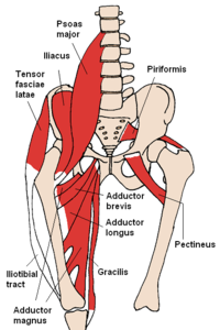
Diagram Labelled Of The Hip Muscles 6 photos of the Diagram Labelled of The Hip Muscles back muscles diagram body muscles diagram labeled diagram of hip muscles and ligaments hip anatomy diagram hip muscles pain thigh muscles diagram Human Muscles back muscles diagram body muscles diagram labeled diagram of hip muscles and ligaments hip anatomy diagram hip muscles pain thigh muscles diagram. The anterior muscles are located on the front of the hip while the posterior muscles are located on the back of the hip.
Muscles Of The Hip Wikipedia
The fascia merges with the periosteum outer bone layer of the humerus.

Labeled diagram of hip muscles. These are often divided into four groups according to their orientation around the hip joint. The anterior and the posterior compartments of the arm. And the iliopsoas group.
The sartorius serves to flex and rotate your hip and bend your knee. This deep muscle begins in the low back and pelvis and connects on the inside edge of the upper femur. The sartorius muscle is a long muscle located in the front of your thigh.
The four groups are the anterior group the posterior group adductor group and finally the abductor group. Diagram Of Hip Muscles And Ligaments. It is controlled by the axillary nerve.
The anterior muscle group features muscles that flex bend the thigh at the hip. A complete list of muscular system quizzes. Anterior muscles front posterior back and medial inside.
Muscles Of The Lower Back And Hip Diagram. We are pleased to provide you with the picture named Human Body Muscle System Diagram With Detailed LabelsWe hope this picture Human Body Muscle System Diagram With Detailed Labels can help you study and research. Its origin is on the ilium of the coxal bone and it inserts part-way down the shaft of the femur.
The muscles of the anterior thigh consist of the quadriceps or quads. Another large hip flexor is the rectus femoris. The muscles that flex the hip are in front of the hip joint.
The quads make up about 70 of the thighs muscle mass. Anatomy Hip Muscles Diagram Learn All Muscles With Quizzes And Labeled Diagrams Kenhub The iliopsoas is a fused muscle composed of two muscles the iliacus and the psoas major. Large and superficial muscles which mainly abduct and extend the thigh at the hip joint.
Push down carefully leaning just as far as you can without overextending your hips. The muscles of the hip can be divided into three different groups. Superficial and deep anterior muscles of upper body.
These are the gluteus maximus gluteus medius gluteus minimus and tensor fasciae latae. The first muscle diagram labeled 2019 above gives you an illustration of the anatomy of the arm muscle. For more anatomy content.
Our LATEST youtube film is ready to run. It courses from your hip and crosses the front of your thigh inserting near the inner part of your knee. There are three layers of gluteal muscles on the posterior hips just like there are three layers of muscles in the abdominal trunk.
Most will label a diagram of muscle with its. In human anatomy the muscles of the hip joint are those muscles that cause movement in the hip. The quadricep group of muscles is the muscles found on the anterior portion of the thigh and they are the rectus femoris the lateralis the vastus medialis and.
1 hip anatomy function and common problemsApproaching the muscular anatomy around the hip can be undertaken in a number of ways. Small and deep muscles which mainly externally rotate the thigh at the hip joint and stabilize the pelvis. In human anatomy the muscles of the hip joint are those muscles that cause movement in the hip.
It is a 3-part muscle with anterior front middle and posterior back heads. Muscle Charts of the Human Body For your reference value these charts show the major superficial and deep muscles of the human body. Labeled diagram View the muscles of the upper and lower extremity in the diagrams below.
The largest of them is the most superficial muscle the gluteus maximus. For a more detailed analysis of the muscles used refer to the biomechanics of rowing. You can pull your toes up at the exact same time to add another measurement to the stretch.
Use the location shape and surrounding structures to help you memorize each muscle. The hip joint is one of the most flexible joints in the entire human body. The hip muscles are divided up into three basic groups based on their location.
The muscle of the arm is divided by a fascial layer separating the muscles into two osteofascial compartments. The lateral rotator group. Most modern anatomists define 17 of these muscles although some additional muscles may sometimes be considered.
The medial muscles are located on the outside of the hip. For a much deeper release in the hips location your elbows on your legs as you lean forward. These include the iliopsoas muscle.
Vastus medialis intermedius lateralis and rectus femoris muscles. 12 photos of the Muscles Of The Lower Back And Hip Diagram. It is the longest muscle in the human body.
These muscles can be grouped based upon their location and function. The hamstring muscle group extends across the posterior surface of the thigh from the ischium of the pelvis to the tibia of the lower leg. The front fibers flex the arm and the middle fibers help abduct the arm bring the arm away from the body.
Just need a glimpse leave your valuable advice let us know and subscribe us. Diagram Of Hip Muscles And Ligaments. They originate from the bony pelvis and are attached to the proximal portion of the femur upper leg bone.
Posterior back fibers help to extend the arm. Oct 29 2020 anterolateral trunk muscles diagram. The many muscles of the hip provide movement strength and stability to the hip joint and the bones of the hip and thigh.