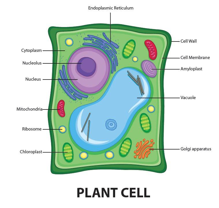
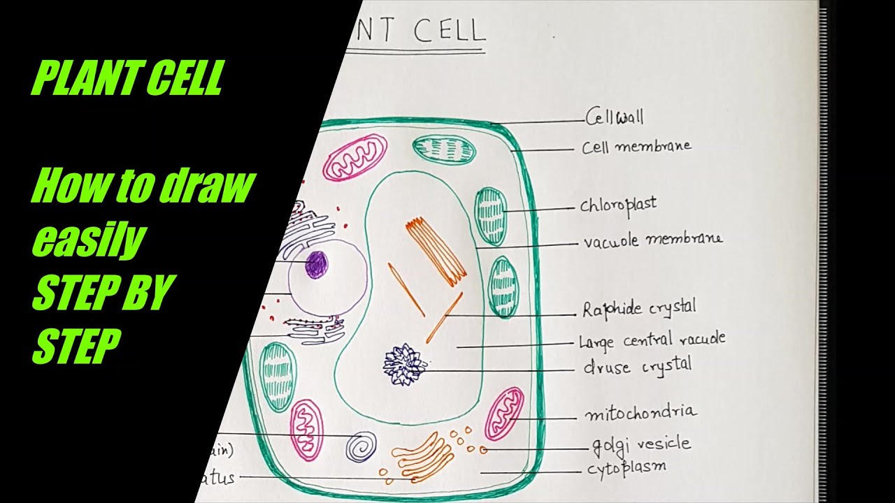
Draw a neat diagram of plant cell and label any three parts which differentiate it from animal cell. Get Draw A Neat Labelled Diagram Of Plant Cell And Label Its Parts BackgroundThe nitrates absorbed by the plant roots are converted into various types of amino acids about 20 types which sequences respectively to form polypeptide chains of various protein molecules in the ribosomes.
Draw A Welllabelled Diagram Of A Plant Cell Class 11 Biology Cbse
Answered Feb 6 2020 by KumariJuly 536k points selected Feb 6 2020 by.
Neat labelled diagram of plant cell class 11. How to draw animal cell labelled diagram Animal cell diagram for class 910 and 11 - YouTube. Compare The Location Of Nucleus In Animal Cell And Plant Cell Draw A. Draw a labelled diagram of a plant cell.
Labelled diagram of a plant cell. Find an answer to your question 1 Draw a neat labelled diagram of plant cell and list the function of each organalle. Energy is produced in the form of ATP in the process.
Compare Plant cell and Animal cell 4 Why Animal cell do not have cell wall for class 8 plz answer fast. The diagram given below represents a plant cell after being placed in a strong sugar solution. Link of our facebook page is given in sidebar.
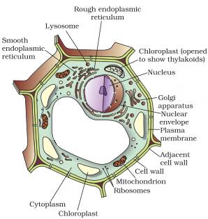
If playback doesnt begin. Photosynthesis occurs in the chloroplasts of the plant cell. Draw a neat labelled diagram of animal cell.
Define plant cell and animal cell. Draw A Neat Diagram Of Plant Cell And Label Any Three Parts Which. ExplanationDiagram of plant cell and thier three parts Diagram of plant cell and thier three parts.
Study the diagram and answer the questions that follow. Answer verified by Toppr. Photosynthesis is the major function performed by plant cells.
Get Instant Solutions 24x7. 1 Answer 1 vote. Aaryansharmasml aaryansharmasml 20112020 English Secondary School answered Draw a neat labelled diagram of a plant cell.
The membrane is selectively permeable and allows only certain molecules to pass through. It is the process of preparing food by the plants by utilizing sunlight carbon dioxide and water. Ncert Solutions For Class 8 Science Chapter 8 Cell Structure And.
Cells under the microscope. Share It On Facebook Twitter Email. Rajubasak49041 rajubasak49041 28042020 Biology.
Class 11 Oscillations Redox. Cell Structure And Functions Class 11 Notes Biology Mycbseguide. Draw a labelled diagram of a plant cell.
Draw a neat diagram of the structure of chromosome and label the parts. A Centromere b p-arm. Plant and animal cell diagram class 8 NCERTHi friends In this video we will learn how to draw diagram of plant and animal cell this diagram is.
Draw a neat labelled diagram of the following cell organelles and explain their structure. Draw a neat and labelled diagram of a Plant cell and animal cell. It is very thin delicate elastic and selectively permeable membrane.
Class-11 Welcome to Sarthaks eConnect. Further plant cells are green in color due to the presence of special pigments that aid in photosynthesis. 13 Plant Cell Diagram Class 9Th PicturesThe class students can check below the diagram of plant cell and animal cell which can help them in understanding how to draw a cell diagram concept.
1st lab exam material biology 440 with dr. The plant cell is rectangular and comparatively larger than the animal cell. The cell membrane is a double-layered membrane made up of phospholipids that surrounds the entire cell.
Plant cells kept in hypertonic solution will get. Draw a neat diagram of plant cell and label any three parts which differentiate it form animal cell. The unit of life.
A Mitochondri b Centrioles. Students upto class 102 preparing for All Government Exams CBSE Board Exam ICSE Board Exam State Board Exam JEE MainsAdvance and NEET can ask questions from any subject and get quick answers by subject teachers. Draw a neat labelled diagram of an animal cell.
Class 8th 9th 10th 11th and 12th can use this diagram. Upvote 0 Was this answer helpful. 1 Draw the concept Map for the Cell 2.
How to draw a animal cell easy and step by step. Cytosol is the fluid present within a cell that is made up of water and. A unique platform where students can interact with teachersexpertsstudents to get solutions to their queries.
The cell wall is made up of cellulose which provides support to the plant. It is responsible for coordinating many of the important cellular activities such as protein synthesis cell division growth and a host of other important functions.
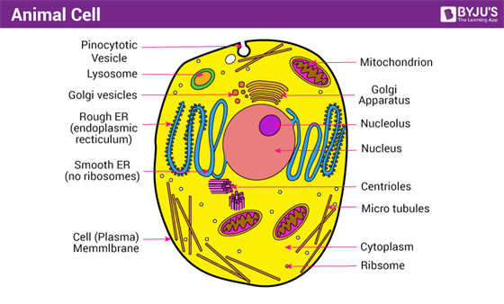
A Well Labelled Diagram Of Animal Cell With Explanation
Class 9 ScienceHome Page.

Labelled diagram of eukaryotic cell class 9. Draw well-labeled diagrams of any one. They are present in eukaryotic cell but absent in prokaryotic cells. A Draw a neat and labelled diagram of a prokaryotic cell.
Question Bank Solutions 7801. 9th Grade Prokaryotic Cell Diagram Class 9. Genetic material enclosed in nuclear envelope Genetic material not enclosed in nuclear envelope 3 Nucleolus and one or more chromosomes present.
Topic 1 2 Ultra Structure Of Cells Amazing World Of Science With. Viruses are smaller than prokaryotic cells. Name the kingdom in which they are placed.
Example plant cellanimal cell. Explain the structure and function of the nucleus. V Draw a labelled diagram of a prokaryotic cell.
These neat and well labelled diagram will ma. Draw a neat diagram of plant cell and label any three parts which differentiate it from animal cell. B Differentiate between a prokaryotic and eukaryotic cell any 4 points of difference.
A diagram of an eukaryotic nucleus is as given. Prokaryotic Cells Types Of Prokaryotic Cells Cell. B Differentiate between plant cell and animal cell.
It is responsible for storing the cells hereditary material or the DNA. Compare and contrast distinguishing characteristics of all these cell types only on the basis of diagrams. A well labeled diagram of mitochondria Return to CBSE Class 9 Chapter Wise Notes Structure and Fundamental Unit of Life - Cell Posted on February 14 2018 by Manoj Saxena Posted in No Comments A well-labelled diagram of mitochondria is given below for your better understanding of the structure.
A Well Labelled Diagram Of Animal Cell With Explanation. Significantly bigger than the prokaryotic cells eukaryotic cells have diameter ranging from 10µm -100µm. Start studying 9th grade.
A labeled diagram of the plant cell and functions of its organelles we are aware that all life stems from a single cell and that the cell is the most basic unit of all living organisms. Chioroplast and a large centrally placed vacuole. You can draw flowcharts wherever necessary.
Genetic material in the form of single chromosome. Example plant cellanimal cell. Eukaryotic cells are exclusively found in plants animals fungi protozoa and other complex organisms.
Draw a labelled diagram of mitochondria. Long Answer Type 1. Advertisement Remove all ads.
Found in eukaryotic cell. Neither of the two. Explain the structure and functions of the chloroplast.
The examples of eukaryotic cells are mentioned below. Draw a well labelled diagram of mitochondria class 9. Cell wall is the non-living protective layer outside the plasma membrane in the plant cells bacteria fungi and algae.
ICSE Class 9 Biology Sample Question Paper 6 with Answers. They are the building block or smallest unit of life of organisms as simple as amoeba and protozoa to the most complicated plants and animals. Test your Knowledge on Nucleus -.
2018 in Class IX Science by saurav24 Expert 14k points the fundamental unit of life 1 vote. Found in prokaryotic cell. Draw a labelled diagram of a animal cell and Plant cell.
Genetic material in the form of single chromosome. Genetic material enclosed in nuclear envelope Genetic material not enclosed in nuclear envelope 3 Nucleolus and one or more chromosomes present. Prokaryotic and Eukaryotic Cell.
CBSE CBSE English Medium Class 9. Download free cbse sample paper for class 9 biology. How can you differentiate between prokaryotic cell and eukaryotic cell.
Draw a well labelled diagram of typical prokaryotic cell. Inside it are various cell organelles which performs individual functions. Write the functions of mitochondria.
8th Grade Science Class. Discuss the main components of a typical cell. Class 9 Cell Basic Unit of Life Long Questions.
Large collection of high quality biology pictures photos images illustrations diagrams and posters on marine biology cell biology microbiology. Animal Cell Diagram For Class 10In pairs discuss the different organs in. 4 Cell divides by mitosis and meiosis.
A bacteria diagram basically facilitates us to profit more approximately this single cell organisms that have neither membrane. The nucleus has 2 primary functions. Important Science Diagrams From All Chapters For CBSE Class 8.
Draw a neat diagram of plant cell and label any three parts which differentiate it from animal cell. Eukaryotic cell are the developed advanced and complex forms of cells. Ncert Solutions For Class 9 Science Chapter 5 The Fundamental Unit.
Comparison of chart of all class mates can be done. Found in eukaryotic cell. Draw a neat well-labelled diagram of an animal cell.
Well Labelled Diagram Of An Animal Cell. Sketch Plant Cell Diagram For Class 9. Draw a labelled diagram of mitochondria.
A list any two structural differences and two similarities between animal cell and plant cell. A Draw a well labelled diagram of a plant cell and label any 4 parts. 4 Cell divides by mitosis and meiosis.
Prokaryotic and Eukaryotic Cell video tutorial 000715. It initiates and regulates cell division. Draw a neat labelled diagram of an animal cell.
I It is plant cell as it contains cell wall. Asked Feb 5. Prokaryotic And Eukaryotic Cells Read Biology Ck 12 Foundation.
Found in prokaryotic cell. Also Read Different between Plant Cell and Animal Cell Though this animal cell diagram is not representative of any one particular type of cell it provides insight into the primary organelles and the intricate internal structure of most animal cells.
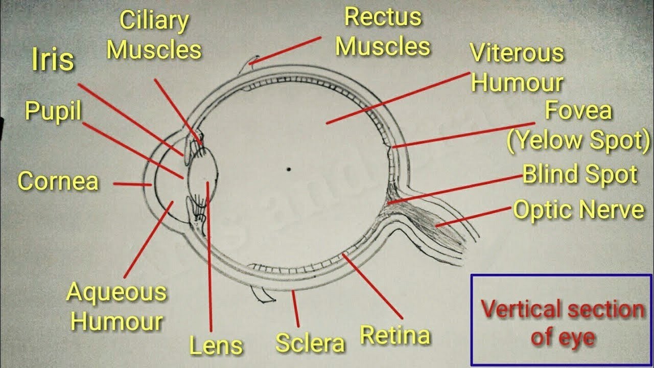
It is the coloured diaphragm between the cornea and lens. Human Eye is the most important optical instrument gifted to us by God.

Human Eye And Colourful World Class 10 Notes Science Chapter 11 Learn Cbse
Following are the main structures in the human eye.
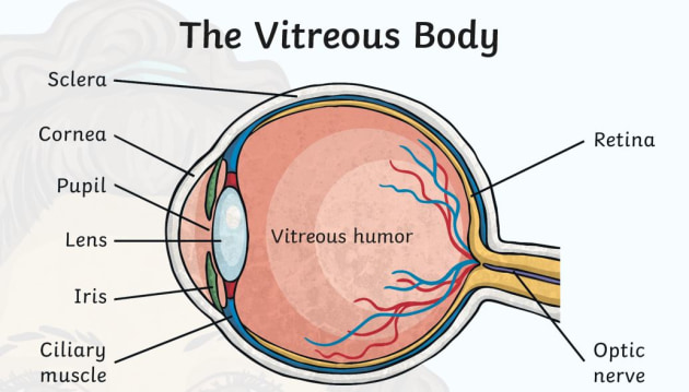
Diagram of human eye class 8. It is enclosed within the eye sockets in the skull and is anchored down by muscles within the sockets. It is the transparent spherical membrane covering the front of the eye. The iris and the choroid are connected by the ciliary body.
The human eye is almost spherical in shape. A simplified anatomy of the eye is shown in the diagram below. Diagram Of Human Eye With Lable Class 8 Brainlyin Rbse Solutions For Class 8 Science Chapter 14 Refraction Of Light How The Human Eye Works Cornea Layersrole Light Rays Explain Parts Of Human Eye And Its Functions Science The Human Cross Section Of The Eye Main Lesson Book Page From Waldorf Today Cbse.
2 The eye-lens is a convex lens so it converges the light rays and produces a real and inverted image of the object on the retina. This can be written with the help of Braille slate and stylus. Pupil regulates and controls.
Human Eye Teachoo Human Eye Diagram For Class 8 Hd Png Human Eye And Colourful World Class 10 Notes Science Chapter 11 Eye Lessons Tes Teach Kids Health Topics Eyes How Your Eyes Work Ncert Solutions For Class 8 Science Chapter 16 Light Learn Cbse The Human Eye And Its Defects Draw It. Human eye is one of the sense organs in human body that reacts to light and helps in seeing objects. Hi friends in this video we will learn how to draw diagram of human eye this diagram is based on class 8 ncert textbook1.
Human Eye Diagram Image will be Uploaded Soon The structure of the human eye is shown above in the image Parts of the Human Eye. It is the small hole in the iris. The pupil is a small opening in the iris.
Human eye diagramhuman eye diagram for class 8construction of human eye diagramdrawing human eye drawing easy stephuman. The iris controls the size of the pupil. The anterior chamber of the eye is the space between the cornea and the iris and is filled with a lubricating fluid aqueous humour.
FranchiseePartner Enquiry North 8356912811. 5284 74 543 966 إحجز الأن. Download Human Eye Diagram Class 8 Ncert PNGHuman eye is spherical about 25 cm in diameter.
It controls the amount of light entering the eye. Thanks for watching our Channel. The outer covering of the eye is called sclera.
The Diagram Given Below Represents The Cross Section Of The Human Label Eye Diagram Wiring Diagram Centre Light Diagram Science Convex Mirror Light Reflection Class 8 Diagram Of Human Eye With Lable Class 8 Brainlyin Class 8 Science Structure Working Of Human Eye Cbse Board Cbse Physics 8th Physics Light Reflection Refraction Eyes Defect. This layer is transparent on the front. The human eye is a large spherical ball which consists of Crystalline lens Aqueous Humour Pupil Iris Cornea Ciliary Muscles Retina Optic Nerve Vitreous Humour.
Can Lease for only 1599. Jul 232021 - Well diagram of human eye. The pupils function is to adjust the amount of light entering the eye.
It regulates the amount of light entering the eyes. Optical aids can help a person who is partially visually challenged. The eyeball is covered with a tough layer.
EduRev Class 8 Question is disucussed on EduRev Study Group by 417 Class 8 Students. It works like the shutter of a camera. You can identify a flower by smelling and touching it but you cant identify the color without the sense of vision.
The human eye structure and function makes it easy to distinguish the objects based on colour size and other parameters. Iris is a thin circular structure. The eyeball is covered with a tough layer.
This transparent portion is called cornea. Anatomically the eye comprises two components fused into. This document is highly rated by Class.
The vascular layer of the eye known as the choroid contains the connective tissue. Human eye diagram for class 8. 1 When we look at an object then light rays coming from the object enter the pupil of the eye and fall on the eye-lens.
Cataract is treated by surgery. Well-Labelled Diagram of Eye. نشر في يناير 12 2021 بواسطة.
Holiday Cottages Peak District. The human eye is a roughly spherical organ responsible for perceiving visual stimuli. This article is helpful for Class 10 Science.
A detailed explanation of the human eye along with its structural representation covering the important parts is mentioned in the chapter.
A group of cells having similar structure origin and functions is called a tissue. Eukaryotic cells are one which have organised nucleus with a nuclear membrane and genetic material is organised into chromosomes.

Draw The Diagrams Of Plant Cell And Animal Cell Label Any Five Organelles Common In Plant And Animal Brainly In
Draw A Neat Labelled Diagram Of An Animal Cell Cbse Class 9 How To Draw Plant Cell In Easy Steps Fundamental Unit Of Life Draw A Labelled Diagram Of A Plant Cell How To Draw Animal Cell Step By Step Tutorial For Beginners Youtube Draw Labelled Diagrams Of Animal And Plant Cells To Show Their Plant Cell Diagram For Class 8 Ncert Bookfanatic89 Printable Cell Diagrams Plant Cell Diagram.
Draw a neat labelled diagram of plant cell class 9. Observes leaf cells using a microscope. Avia Salon Day Spa 5500 Grossmont Ctr Dr Suite 285 La Mesa CA 91942. How To Draw Plant Cell For Class 9 To 12 Step By Step Demonstration Simple And Easy Bio Diag Youtube This document is highly.
Are solved by group of students and teacher of Class 9 which is also the largest student community of Class 9. The cell being the smallest unit of life is akin to a tiny room which houses several organs. Asked Dec 29 2018 in Class X Science by muskan15 -3980 points cell division.
NCERT syllabus plays an i. Label the Plant Cell Worksheets SB11867 SparkleBox. Plant cell diagram for class 8 image information.
Draw a labelled diagram of a animal cell and Plant cell. Draw a labelled. Draw a labelled.
ExplanationDiagram of plant cell and thier three parts Diagram of plant cell and thier three parts. Cell organelle is a specialized entity present inside a particular type of cell that performs a specific function. Diagram of plant cell.
Hello friends I know first picture is more clear but heshe have not drawn it and I. 2 See answers. The most important structures of plant and animal cells are shown in the diagrams below which provide a clear illustration of how much these cells have in common.
The Questions and Answers of Draw neat and labelled diagram of plant and animal cell. Draw a neat labelled diagram of an animal cell. Sunday 14 April 2013.
Ways To Make Models Some Edible Of Plant And Animal Cells And. Diagram of plant cell wall. You can see some here it is the labeled diagram of plant and animal cell class 9 notes edurev sample questions with examples at the bottom of this page.
Plant and animal cells are different from each other as plant cell. The cell theory was further expanded by a german physiologist rudolf virchow 1855. Is done on EduRev Study Group by Class 9 Students.
Both plant and animal cells belong to eukaryotic cells. The cell structure is also explained in this chapter. Topic starter Draw a neat labelled diagram of plant cell and label its parts.
NCERT Solutions CBSE Sample papers and Lattest CBSE Syllabus For Class 9 to 12 All Subjects Maths Science Physics Chemistry. Labelled diagram of plant and animal cell are as follow---. Schahat6836 schahat6836 18042020 Biology Secondary School answered Draw A Neat Labelled Diagram Of Plant Cell 2 See answers at176396 at176396 Answer.
Plants cells are usually eukaryotic cells. Draw a labelled diagram of a animal cell. Draw A Neat Labelled Diagram Of Plant Cell Get the answers you need now.
Draw a neat diagram of plant cell and label any three parts which differentiate it from animal cell. Aaryansharmasml aaryansharmasml 20112020 English Secondary School answered Draw a neat labelled diagram of a plant cell. Plant cells can be larger than animal cells.
Hand drawn labelled diagram of plant cell class 9. This discussion on Draw neat and labelled diagram of plant and animal cell. Plant cell diagram 9th class.
Trisha Dave trisha-dave Noble Member. Draw a neat labelled diagram of plant cell and label its parts. Draw a neat labeled diagram to show the metaphase stage of mitosis in an animal cell having 6 chromosome.
Time may be required prior to lesson to read background. 36 Well Labelled Diagram Of Plant Cell For Class 9 Pictures. Draw a neat labelle.
Notifications Clear all Draw a neat labelled diagram of plant cell and label its parts.
The cornea is the clear outer part of the eyes focusing system located at the front of the eye. Use this Label the Eye worksheet to teach your class all about the different parts of the human eye.
Eye Diagram With Easy Steps How To Draw Human Eye Youtube
Diagram Of Human Eye With Labelling.
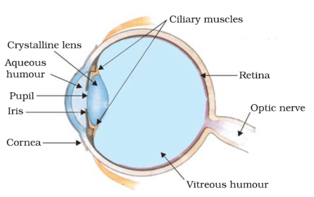
Diagram of eye with labelling class 10. Eyediagramxnperiodoffsetplotstring â Filename. Human Eye Diagram Class 10 How To Draw Human Eye Diagram Easy Eye Diagram By Firkin Human Eye Diagram Diagram Of The Eye Eye Labelled Diagram Of Human Eye Transparent Png Download Eye Diagram Structure And Function Of The Human Eye Your Eyes For Kids Nemours Kidshealth The Human Eye 1000 Human Eye Diagram Stock Images Photos Vectors Shutterstock The Eye Diagram Label 2 Wiring Schematic Diagram. Figure 24a shows a perfect eye diagram.
Four Human Biology Diagrams to Label - Heart Lungs. Printable Human Eye Diagrams. Human Eye Origami Organelles.
The diagram of eye is beneficial for Class 10 and 12 and is frequently asked in the examinations. Anatomy Eye Diagram To Label Health Class Eye Anatomy Eye The Eye Diagram To Label By Canonuk Teaching Resources Label Eye Printout Enchantedlearningcom Anatomy Vision Label Parts Of The Human Eye Label Eye Diagram Wiring Diagram Centre Draw A Neat And Label Diagram Of Human Eye And Explain Functions Of The Eye Diagram Label Worksheets Differentiated By Zmzb Teaching Eye Diagram Without Labels. Label The Eye Teaching Resources.
Iklan Tengah Artikel 2. Draw It Neat How To Draw Lungs Diagram. Iklan Tengah Artikel 1.
People say that the eyes are the. A detailed explanation for the labelling of diagrams is also provided here. Human Eye Diagram Class 10 How To Draw Human Eye Diagram Easy Step Anatomy And Structure Of The Eye Brightfocus Foundation Draw A Simple Diagram Of The Human Eye And Label Clearly The Cornea How To Draw Human Eye Diagram Step By Step 10th Physics Science Eye Diagram Basic Wiring Diagram Human Eye Anatomy Quiz Eyes Anatomy Overview Parts And Functions Biology Dictionary Label Eye.
Diagram Of The Eye Lions Eye Institute. Human Eye Definition Structure Function Parts Diagram. It is made up of dense connective tissue and protects the inner parts.
The human eye diagram displayed at right shows all of the major features of human ocular anatomy. It lines the sclera and is made up of stratified squamous epithelium. Just behind the iris and pupil lies.
Human Eye Diagram Images Stock Photos Vectors Shutterstock. Check important biology diagrams for CBSE Class 10 Science. The image is taken from above the left eye.
The Human Eye Primary Science Worksheets Teaching Resources. It is a white visible portion. Eye Diagram With Labels And Functions.
In biology human biology physics and practical courses in medicine nursing and therapies. Eye Diagram Label Purposegames How To Draw Human Eye Diagram Step By Step For Beginners Youtube 1000 Human Eye Diagram Stock Images Photos Vectors Shutterstock Draw A Labeled Diagram Of Human Eye Write The Functions Of Cornea External Anatomy Of The Human Eye With Labels Stock Photo Drawing Of Heart With Labels At Paintingvalley Com Explore How The Eyes Work National Eye. How To Draw Human Eye Diagram Eye Diagram Step By Step Class 10 Science Physics NCERT.
In this resource youll find a 2-page PDF that is easy to download print out and use immediately with your class. Human eyes are camera-type eyes which means they work like camera lenses focusing light onto film. Iklan Bawah Artikel.
Simple Anatomy Of The Retina By Helga Kolb Webvision. The External Structure of an Eye. Human Ear Diagram For Children Human Ear Diagram For Children.
The Eye Science Quiz. The parts of the eye that are visible externally include the following-. Trace period specified as a positive scalar.
Read the definitions then label the eye anatomy diagram below. The eye eye diagram how we see eye year 6 light parts of the eye light light year 6 eyes human body What is this Human Eye Labelling Activity useful for. An unregistered player played the game 1 day ago.
Next light passes through the lens a clear inner part of the eye. The lens is a clear part of the eye. Schematic diagram of the Structure of the Human Eye.
Start studying labelling the human eye. Diagram of 1986 nissan maxima 3 0 engine 1992 dodge dakota fuse diagram 110 outlet wiring diagram diagram of a eye 2008 saab 9 3 fuse diagram 2008 ford e350 fuse panel diagram. A brief description of the eye along with a well-labelled diagram is given below for reference.
In the class today we covered parts of the eye and what changes in them should be alarming. The first page is a labelling exercise with two diagrams of the human eye. Diagram Eye Label Diagram Labels Label Gallery Get some ideas to make labels for bottles jars packages products boxes or classroom activities for free.
The most common eye diseases include myopia hypermetropia glaucoma and cataract. Label diagram of eye. Here are descriptions of some of the main parts of the eye.
One is a view from the outside and the. Human Eye Diagram Class 10 How To Draw Human Eye Diagram Easy. FileDiagram of human eye without labelssvg.
Choose a web site to get translated content where available and see local events and offers. Great for science classes. It is found in different colors like brown black blue green etc.
Smith Lab Investigating Vascular Eye Diseases Boston. Newer Post Older Post Home. Fileschematic Diagram Of The Human Eye Ensvg Wikimedia.
Inges Mild Myopia The Questionable Value Of A. Easy to download and print out. The iris is the colored part of the eye that regulates the amount of light entering the eye.