Brain stem works together to regulate essential life functions including body temperature breathing heartbeat and blood pressure. Main Parts of the Brain and Their Functions Cerebrum.

Found On Bing From Www Pinterest Com Brain Diagram Brain Anatomy Brain Anatomy And Function
The brain role as part of the Central Nervous System is to regulate most functions of human body including vital functions such as heart rate or breathing basic functions like being hungry sleeping or sexual instinct also complex functions like speaking thinking remembering etc.

Human brain labeled and functions. The amygdala is a cluster of nuclei located close to the base of the brain that is primarily involved in functions including memory emotion and the bodys fight-or-flight response. The Seat of Consciousness. It is made up of more than 100 billion nerves that communicate in trillions of connections called synapses.
The brain is one of the most complex and magnificent organs in the human body. The human brain is the command center for the human nervous system. It receives signals from the bodys sensory organs and outputs information to the.
It includes the cerebellum reticular formation and brain stem which are responsible for some of the most basic autonomic functions of life such as breathing and movement. 15 kilograms The human brain contains close to 86 billion nerve cells neurons the grey matter The human brain has billions of nerve fibers axons and dendrites the white matter These neurons are connected by trillions of connections or synapses. 7 The structure processes external stimuli and then relays that information to the hippocampus which can then prompt a response to deal with outside threats.
Psychology Brain StructureAnatomy and Function BRAIN FACTS Composition of the brain. The function of this structurally and functionally specialized region of the brain is not just limited to auditory processing. The midbrain also called the mesencephalon connects the hindbrain and the forebrain.
Human Brain Divisions and their Functions Amygdala. The brain is the most complex part of the human body. Lying in its bony shell and washed by protective fluid the brain is the source of all the qualities that define our humanity.
It is also responsible for planned voluntary muscle movements throwing a ball walking chewing etc and for taking in and interpreting sensory information such as vision hearing smell touch and pain. They coordinate specific functions including visual memory such as facial recognition verbal memory such as understanding language and interpreting the emotions and reactions of others. The human brain just like most other mammals has the same basic structure but it is better developed than any other mammalian brain.
The cerebrum is the largest brain structure and part of the forebrain or prosencephalon. The cerebral hemispheres control reasoning thought emotion and language. The human brain weighs about 33 lbs.
It functions by receiving and sending signals via neurons to different parts of the body. The parietal lobe is located on top of the brain and its primary functions include the perception of stimuli as well as the recognition of objects movement and orientation. This region of the brain helps in balancing movement maintaining.
The amygdala is situated deep within the limbic center in the area where emotions are perceived. Working on the principle of division of labour different parts of brain are specialized for only specific tasks. In addition the brain stem coordinates the fine movement of the face and limbs.
The brain is composed of the complex network of billions of neurons that are arranged in a specific pattern which is vital to the essential functioning of this organ. Functions of this area include sneezing vomiting swallowing and movement of the eyes and mouth. 78 water 12 lipids 8 protein 1 carbs 2 soluble organics and 1 salt 10 seconds is the amount of time until unconsciousness after the loss of blood supply to the brain.
A summary of the function of brain. The brain is one of the largest and most complex organs in the human body. The forebrain is responsible for a number of functions related to thinking perceiving and evaluating sensory information.
The brainstem middle of brain connects the cerebrum with the spinal cord. The human brain controls nearly every aspect of the human body ranging from physiological functions to cognitive abilities. It is associated with motor functions and auditory and visual responses.
It controls our muscle movements the secretions of our. The brain stem contains the pons and medulla oblongata. High Intellectual Functions Occur in the Cerebrum.
The hindbrain contains both the metencephalon and the myelencephalon. The cerebellum lies below the cerebrum. Our brain gives us awareness of ourselves and of our environment processing a constant stream of sensory data.
It is also involved in producing emotional attitudes storing new memories processing sensory output and the retention of visual memories. The cerebellum adjusts body movements speech coordination and balance while the brain stem relays signals from the spinal cord and directs basic internal functions and reflexes. This three-pound organ is the seat of intelligence interpreter of the senses initiator of body movement and controller of behavior.
The cerebrum front of brain comprises gray matter the cerebral cortex and white matter at its center.
The cerebrum can be divided into four parts or an oblongata medulla locates in the lower portion of the brain is the medulla oblongata. Divided into two hemispheres the cerebrum is the largest region of the human brain the two hemispheres together account for 85 of total brain mass.
The Medulla Oblongata Internal Structure Vasculature Teachmeanatomy
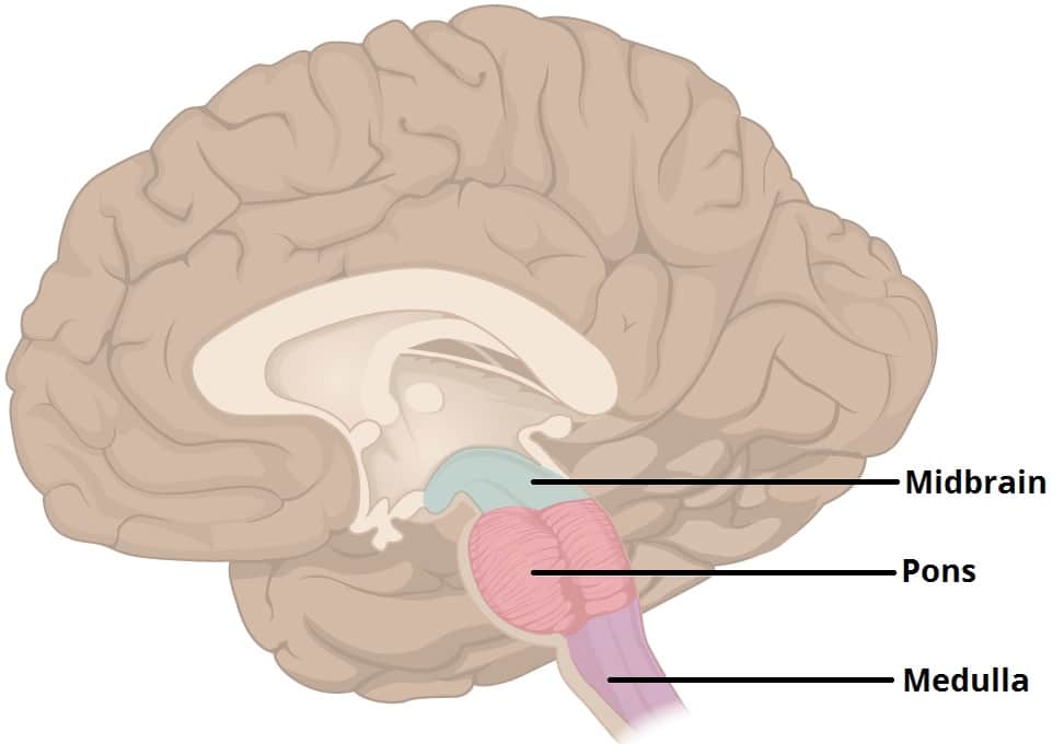
The image on the left is a side view of the outside of the brain showing the major lobes frontal parietal temporal and occipital and the brain stem structures pons medulla oblongata and cerebellum.

Brain diagram labeled medulla. The brainstem is made of three regions. It is the transition from the spinal cord to the brain. The portion which encloses the brain differs in several essential particulars from that which surrounds the medulla spinalis.
At the bottom of the brainstem the medulla is where the brain meets the spinal cord. The image on the right is a side view showing the location of the limbic system inside the brain. The medulla contains the vital autonomic cardi.
It is an extremely important part as it deals with vital and basic activities of the human body as it contains the cardiac respiratory and vasomotor centers. Split into two hemispheres and is highly. The cerebrum forms the superior part of the brain covering and obscuring the diencephalon and brain stem similar.
Connecting the brain to the spinal cord the brainstem is the most inferior portion of our brain. We arrive at everyones favorite part of the brain the medulla oblongata. A net-like structure of mixed gray and white matter known as the.
Medulla Oblongata - the lowest section of the brainstem at the top end of the spinal cord. The limbic system consists of a number of. Quite literally the medulla oblongata regulates all the functions we need to live.
46 Brain Diagram Labeled Medulla Pictures. Medulla medulla oblongata The functions of these areas are. It controls automatic functions including heartbeat breathing etc.
Anatomy and Syndromes The anatomy of the brainstem is complex. The medulla is essential to survival with functions that regulate many bodily activities including heart rhythm breathing blood flow and oxygen and carbon dioxide levels. These five regions are the central areas that regulate breathing pulse arousal balance sleep and early stages.
Labeled diagram showing the main parts of the brain Blank brain diagram free download Use the arrows and fill-in-the-blank spaces on this diagram to write the name of each brain structure. The upper thicker region of the medulla is connected to the fourth ventricle while the lower portion of the. It contains numerous cranial nerve nuclei and is traversed by multiple tracts between the brain and spinal cord.
Besides being a fun term to say the medulla oblongata is known for its critical role in autonomic functions that keep us alive including respiration digestion and swallowing control of the heart rate constriction and relaxation of blood vessels and sneezing. The brain contains the following main areas. Many of the most basic survival functions of the brain are controlled by the brainstem.
The medulla oblongata forms the lower half of the brain stem. The medulla oblongata frequently just called the medulla is the lowest portion of the brainstem and it is also thought to be the oldest part of the brain. Improved MRI resolution now allows the radiologist to identify a higher level of anatomic detail but an un-derstanding of functional anatomy is crucial for correct interpreta-tion of disease.
Midbrain Pons and Medulla. The medulla is responsible for the control of heart rate sneezing breathing and other automatic functions of the body. There are four main areas in the brain.
The medulla oblongata or simply the medulla is the most caudal part of the brainstem between the pons superiorly and spinal cord inferiorly. Thus it is responsible for functions such as breathing maintaining a steady heart rate and blood pressure inciting regurgitation vomiting swallowing urination. The medulla oblongata the pons and the midbrain.
Learn about the brains anatomy including the limbic system and the role and function of different lobes of. Medulla Regulates breathing and heart rate hanging a person works bc if done correctly it breaks this in half Pons Involved in sleeping waking and dreaming Cerebellum The lesser brain coordinates balance and coordination Thalamus Relays all sensory information to specific perception areas of the brain with the exception of smell Hypothalamus Part of the old brain it. The structure of the brain.
Use this interactive 3-D diagram to explore the brain. Lets use a common method and divide the brain into three main regions based on embryonic development. Occipital Lobe of the Cerebrum - the region at the back of each cerebral hemisphere that contains the centers of vision and reading ability located at the back of the head.
The medulla contains the vital autonomic cardiovascular and respiratory centers controlling heart rate blood pressure and breathing. The brain contains the following main areas. General anatomy of the human brain Marieb Hoehn Human Anatomy and Physiology 9th ed Figure 122 CEREBRUM.
The plant cell contains a large central vacuole and a protective outer covering called the cell wall. The middle lamella acts as the strengthening layer between the primary walls of the neighboring cells.

A Labeled Diagram Of The Plant Cell And Functions Of Its Organelles Plant Cell Diagram Cell Diagram Plant Cell
Amusing Human Cell Coloring Page Animal Cell Worksheet Labeling.

Parts of a plant cell labeled. Their distinctive features include primary cell walls containing cellulose hemicelluloses and pectin the presence of plastids with the capability to perform photosynthesis and store starch a large vacuole that regulates turgor pressure the absence of flagella or centrioles except in the gametes and a unique method of cell division involving the formation of a cell plate or phragmoplast that separates the new daughter cells. For more information about the mostparts of plant cell game. You may also like.
Cell membrane also called plasma membrane is present inside the cell wall and surrounds the cytoplasm. Cell wall peroxisome vacuole cytoplasm cell membrane Golgi apparatus nucleolus nucleus ribosome mitochondrion endoplasmic reticulum chloroplast Parts of a Plant Cell. Inspiring Labeled Plant Cell Parts Worksheet worksheet images.
The Parts of a Plant Cell and an Animal Cell. Both animal and plant cells are eukaryotic cells which means they have complex structures enclosed within membranes. Oct 4 2019 - Label a plant cell and match the parts with its definition.
Samsung ipad 2 price in india Or cells with organelles and that the cell game plant names. The parts of a plant cell include the cell wall the cell membrane the cytoskeleton or cytoplasm the nucleus the Golgi body the mitochondria the peroxisomes the vacuoles ribosomes and the endoplasmic reticulum. Made of cellulose and strengthens the cell.
A digital version of this resource is also included and is compatible with Google Classroom. A plant cell has a rigid cell wall which is the outermost of the cell. Different Parts of a Plant Cell Cell wall.
The plant cell will store water in the central vacuole which expands the vacuole into the sides of the cell. Identify and label each part of the plant cell. Components of a Plant Cell Plant Cell Wall.
Plant cell labeled parts and functions Further information about the mostparts of a glossary of learn. Centrosome The microtubule organizing centre A small body located near the nucleus - it has a dense center and radiating tubules. Where protein synthesis occurs.
It is the semipermeable membrane also called the plasma membrane that is present within the cell. The cell wall is made up of two layers a middle lamella and a primary cell wall and sometimes a secondary cell wall. Nucleus cell wall and chloroplast.
It determines the function and development of the cell. It is the. It is a site for many biochemical.
It packages materials coming from the endoplasmic reticulum. It is a rigid layer that is composed of cellulose glycoproteins lignin pectin and hemicellulose. The centrosomes is where microtubules are made.
During cell division mitosis the centrosome divides and the two parts move to opposite sides of the dividing cell. Plant Cell Definition Functions And Structure Biology Dictionary Plant Cell Labeled Parts And Functions Animal Cell Diagram Labeled Pencil Wiring Diagrams Cross section of a leaf showing various plant cell. The other components of a plant cell the cell wall and central vacuole work together to give the cell rigidity.
Prokaryotic Cell Coloring Page Label Animal Cell Diagram Worksheet Plant and Animal Cell Worksheet Plant Cell Diagram without Labels Plant Cell Structure and Function Worksheet. Contains chlorophyll and it is where photosynthesis happens. The cell wall then pushes against the walls of other cells creating a force known as turgor pressure.
It is made up of chitin lignin. There are two major types of cells namely prokaryotic and eukaryotic cells. Jan 5 2021 - Parts Of A Plant Cell Labeling Matching Plant Cell Labeled Plant Cell Plant Cells Worksheet.
Featured in this printable worksheet are the diagrams of the plant and animal cells with parts labeled vividly. The cell wall also bonds with other cell walls to form the structure of the plant. It provides resistance to microbes to withstand hypotonic external media without bursting.
Where the majority of the activities take place. Cell wall is the outermost tough and rigid layer which comprises cellulose hemicellulose pectin and at. Contains DNA and controls the functions of the cell.
This enhanced visual instructional tool assists in grasping and retaining the names of the cell parts like mitochondrion vacuole nucleus and more with ease. See 14 Best Images of Labeled Plant Cell Parts Worksheet. The plant cell can be drawn as.
Parts of an Animal Cell - Labeling MatchingParts of an Animal Cell - Organelle. Where the majority of respiration takes place. Every plant cell has a cell wall layer which is a major distinguishing factor between a plant cell and an animal cell.
Plant Cell Parts 1. This is also called a plasma membrane and is present adjacent to the cell wall. This is a fluid matrix.
Answer keys are included for easy grading. Compare the parts and structure of a plant cell and an animal cell using this labeled diagram.
A nucleotide is composed of a phosphate a deoxyribose sugar molecule and a nitrogen-containing base A T C or G. DNA Paper Model Homework Sheet 9.
Dna Structure And Replication Review Article Khan Academy
335 Draw and label a simple diagram of the molecular structure of DNAHere I demonstrate drawing the structure of DNA.
Labeled parts of a dna model. ThearaBbox includes genes on the DNA which work together in. CLOSURE 5 minutes Targeted Resources Quickly review the components of DNA. You dont need to be an artist its.
Labeling a DNA structure with appropriate fonts and font sizes is the last step in a DNA molecule project. Diagram And Label A Section Of Dna. In 1953 two scientists James D.
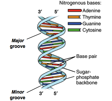
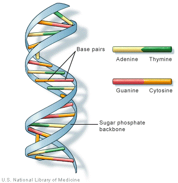
The two strands of DNA are held together with the help of base pairing. DNA has a double-stranded helical structure. Guanine and cytosine or adenine and thymine connect with hydrogen bonds to form the nitrogenous base pairs or rungs.
Dna model worksheet labeled dna model worksheet and dna paper model activity are three main things. The building blocks of DNA are nucleotides which are made up of three parts. Label Gallery Get some ideas to make labels for bottles jars packages products boxes or classroom activities for free.
The two nucleotide chain is arranged as stacks connected by. It is made up of nitrogenous base deoxyribose sugar and phosphate. Using research from many sources including chemically accurate models.
Watson and Crick 1953 have proposed a model for the structure of DNA molecule which is now usually accepted by all. DNA - Deoxyribonucleic acid a macro molecule which has all the hereditary data or information in many living beings. An easy and convenient way to make label is to generate some ideas first.
Answer The above model represents an operon which consists of genes that function in the same process. DNA replication is the process in which the parent DNA molecule produces its identical copy during cell division This is a necessary step because each newly formed cell has to receive a copy of DNA material so that parental characteristics may be. It was given by Watson and Crick model.
There are four types of nitrogenous bases in DNA. They described the molecule as a double helix two spiraling strands composed of nucleotides. Now lets consider the structure of the two types of nucleic acids deoxyribonucleic acid DNA and ribonucleic acid RNA.
By the way about DNA Model Cut Out Worksheets below we can see various similar pictures to inform you more. Go around the room one student at a time and have them tell which base would be. Adenine always bonds with thymine with the help of 2 hydrogen bonds and guanine always bonds with cytosine with the help of three hydrogen bonds.
Nitrogen base of one strand makes a hydrogen bond with the nitrogenous base of the second strand. The instructions carried on DNA are used to make proteins from amino acids in the cytoplasm. Figure 2 below shows the structural formula of DNA in greater detail.
DNA to its function in the cell. This time we will show you some awesome pictures we have collected in case you need more example today we will see more concerning DNA Model Cut Out Worksheets. About Press Copyright Contact us Creators Advertise Developers Terms Privacy Policy Safety How YouTube works Test new features Press Copyright Contact us Creators.
The nitrogenous base deoxyribose sugar and phosphate link together forms the nucleotide chain. 1 Bases 2 Deoxyribose sugar molecules 3 Phosphate molecules Put up the Half-a-helix transparency on the overhead. DNA is made up of six smaller molecules -- a five carbon sugar called deoxyribose a phosphate molecule and four different nitrogenous bases adenine thymine cytosine and guanine.
Watson and Francis H. It is also known as B form of DNA. The basic unit of structure for a DNA molecule is the nucleotide.
Phosphate and deoxyribose molecules form the backbone or sides of the model. Components- Deoxyribose sugar A phosphate A nitrogenous base adenine guanine thymine and cytosine. Function- DNA plays an important role in the production of proteins along with the metabolism and reproduction of the cells.
Alternating sugar and phosphate units form the two sides of a ladder-shaped arrangement with the rungs or steps each formed by a pair of nucleotide bases. The nitrogen bases are ring compounds with their carbon and nitrogen atoms arranged in single or double rings. According to this model called as Watson Crick Model the DNA molecule is a double helix structure consisting of two long polynucleotide chains coiled round each other around an imaginary axis and running opposite to each other.
A deoxyribose 5-carbon sugar a phosphate group and a nitrogenous base Figure 93. Free 3D dna models for download files in 3ds max c4d maya blend obj fbx with low poly animated rigged game and VR options. DNA replication simple definition.
Crick proposed a model for the structure of DNA. Human chromosomes are structures composed of two long strands of deoxyribonucleic acid DNA combined with proteins.
Brassica rapa can be purchased under the trade name Wisconsin Fast Plants and used in this activity. The pistil is considered the female part of a flower because it produces seeds.

Nature Cultural And Travel Photography Blog Brassica Flower Parts
Thursday December 14 2017.

Labeled diagram of brassica flower. Rapa Turnips - Plantinfo. The vegetative shoot shows unlimited growth whereas the flower shows the limited growth. Plants primary teaching resources and printables sparklebox.
Make 10 MCQs draw a labeled diagram of brassica flower also collect flower and dry it. Find flower anatomy diagram lesson plans and teaching resources. 12 When a terminal flower is depicted the axis is not present and therefore cannot be shown.
One especially fast-bolting bolt means to set flowersusually making the leaves bitter variety of brassica was developed by the University of Washington for research and education and has a life-cycle of just one month. They are not only involved in reproduction but are also a source of food for other living. Brassica Campestris Flower Diagram.
Plant Flower Anatomy - Anatomy Drawing Diagram. Diagrams are usually depicted with the subtending bract below and the axis above the flower itself both in the median line. Parts of a plant.
Flower develops on the mother axis stem in the form of floral bud. Napus from other brassicas. The anatomy and morphology of stamens and carpels of cruciferous flower bears testimony to a papaverous ancestory.
Opal Diagram Brassica Flower Youtube. The leaves of turnip Brassica campestris left grasp the stalk completely. For this plant observation lesson students examine a diagram of a Brassica plant and identify its anatomy before planting their own seeds and viewing the changes.
Spring is almost here and flowers are blooming its the perfect time to make a flower anatomy 3D project. Wild Turnip Brassica rapa - Flowers - NatureGate. Diagram of a flowering plant with label.
The reproductive parts of the flower that are necessary for seed production are the stamen the male organ and carpel the female organ. Flower diagram not labeledEach female and male organs are discovered on the identical flower. Shown on the plant cell diagram labeled above that plant cells are surrounded by a thick rigid cell wall.
Labeled hibiscus labeled parts of flower. Flower diagram not labeled. The extraordinary diversity of Brassica oleracea Plant.
A flower is a attribute characteristic of flowering vegetation and is definitely an extension of the shoot meant for copyflowers are enticing and seem in numerous colors and shapes to draw pollinators who assist in pollen switch. Bracteoles if they are present are usually drawn on the sides of the diagram. Education Chart Of Biology For Anatomy Of Hibiscus Flower Diagram Grass Features And Structure Draw A Well Labeled Diagram Of Flower To Show Its Parts Gbzhcrb00.
Diagram Flower Mature Free Vector Graphic On Pixabay Parts Of A Flower Lovetoknow Flower Diagram To Label Wiring Diagram Blog. They are not only involved in reproduction but are also a source of food for other living organisms. The axis corresponds to the position of the main stem relative to a lateral flower.
Quickly find that inspire student learning. Parts and function of the ovary in the flower. Flower diagram not labeled.
Paste it Write definition of set also types of sets i Empty set ii Finite set iii infinite sets iv equal set v disjoint set vi over lapping set Do Ex 11 2nd Learn WriteVerb. U 1935 Office of the Gene Technology Regulator 2008 Figure iv. Flower diagram labeled flower diagram labeled and functions flower diagram labeled parts labeled diagram of brassica flower.
Draw neat and labelled diagram of l s of hibiscus flower. Parts of a flower. The leaves of canola Brassica napus centre only half-grasp the stalk.
But in Brassicaceae the stamens are tetradynamous and not in Papaveraceae. Comparison of floral diagram indicates that Brassicaceae is closely allied to Capparidaceae. Brassica Flower Parts Labeled images similar and related articles aggregated throughout the Internet.
Typical floral diagram of a Brassicaceae Erysimum Bowles Mauve Flowers may be arranged in racemes panicles or corymbs with pedicels sometimes in the axil of a bract and few species have flowers that sit individually on flower stems that spring from the axils of rosette leaves. Life cycle of a plant content classconnect the structure of a flowering plant brassica rapa parts of a flower diagram ks2 beautiful. Flower anatomy flowers consist of reproductive parts and modified leaves.
Its made up of the following parts. By constructing a three-dimensional flower and labeling all the parts children will learn a lot about flower anatomy. Showing posts with label labeled diagram of brassica flower.
Getting an A for outstanding achievement in science by following this step by step 3-D flower project is within your reach. How to draw diagrams on Biology Practical copy Punjab Board Lahore by Naveed Akhtar Uppal calligrapher artist Jhelum Pakistan. Get Free Access See Review.
A bird rape plant Brassica campestris with an associated.