There is no distinct branch from the cephalic vein to the basilic vein. Heart diagram using - Labelled diagram.
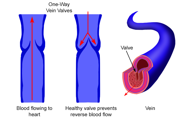
Labelled Diagram Of Vein Valves And Vein Artery Circuit
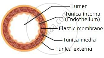
Lining the core of each is a thin layer of endothelium and covering each is a sheath of connective tissue but an artery has thick intermediate layers of elastic and muscular fiber while in the vein these are much thinner and less developed.
Labelled vein diagram. Right Atrium Right Ventricle Left Atrium Left Ventricle Tricuspid Valve Mitral Valve Pulmonary Valve Aortic Valve Superior Vena Cava Inferior Vena Cava Pulmonary Trunk Right Pulmonary Artery Left Pulmonary Artery Pulmonary Veins. Diagram Iv Vein Diagram Full Version Hd Quality Vein Diagram. Free Artery Vein Diagram Templates.
State how each of these are adapted for its functions. Aorta Pulmonary artery Pulmonary vein Right atrium Left atrium Right ventricle. The veins carry the blood towards the heart which consists of the waste.
Human Body Vein Diagram In Detail. Both arteries and veins are types of blood vessels in the cardiovascular system. 20 5 Circulatory Pathways Anatomy And Physiology.
In this image you will find a facial vein internal jugular vein superior vena cava hepatic vein renal vein gonadal vein inferior vena cava in human body vein diagram in detail. Fully Labeled Arteries Diagram Jpg Superficial Temp Oral Artery. The vein is the blood vessel which carries the deoxygenated blood from different parts of the body back to heart.
Want to learn more about it. Labeled Diagram Of Arm Veins Diagram Labels Label Gallery Get some ideas to make labels for bottles jars packages products boxes or classroom activities for free. Venipuncture And Arterial Puncture Policy.
A labeled diagram of the human heart you really need to see. Labels are usually small in size so you should carefully choose the font of the texts to make sure it is readable. Arteries and veins have the same layers of tissues in their walls but the proportions of these layers differ.
Labeled diagram of arm veins. Labelled Diagram Of Vein Valves And Vein Artery Circuit. There are three veins most commonly used in venipuncture or phlebotomy.
The major nerves and veins start in your neck and run the length of your arms often into your hands. Try to label the following structures yourself and then check to see if you are correct. It is the outermost coat which is formed of connective tissues.
The placement of these three veins forms a letter H as in the diagram below. Draw neat labelled diagram of a cross section of an artery and a vein. Tunica externa is also called tunica adventitia.
Anatomy Label Major Arteries And Veins Major Arteries Of The Head And Neck Labelled Medical Stock Images Company - Related posts of anatomy veins arteries diagram. Labeled Arteries And Veins Diagram Written By JupiterZ Tuesday July 18 2017 Add Comment Edit. Arteries dont have valves in them with the exception of the semilunar valves found in the pulmonary artery and the aorta.
These three veins are found in the antecubital area. Major arteries veins and nerves of the body. This diagram shows the major veins in human body venas.
They are the cephalic median cubital and basilic veins. Arteries and veins diagram 205 circulatory pathways anatomy and physiology. As you can see there are also have a common iliac vein internal iliac vein external iliac vein deep femoral vein femoral vein.
With the help of labelled diagrams describe the structure of an artery a vein and a capillary. Arterial And Venous Network In The Forearm Download Scientific. Lab Practicum 2 Circulatory System Anatomy.
You can also put your logo at the top or bottom corner of the label. How to draw labelled diagram of TS of artery vein and capillaries step by stepHello Friends in this video I tell you about how to draw labelled diag. A Simple Schematic Of The Layers Within An Artery.
Below is a blank diagram followed by the labeled diagram with the answers. Venous system diagram Use this interactive 3-D diagram. Circulatory Pathways Anatomy And Physiology Ii.
It is a middle coat which is chiefly formed of elastic connective tissue and smooth muscle fibres. Valves in these veins allow blood to flow from the superficial veins to your deep veins but not the other way. Most of the times we put the labels to show some specific information.
Veins have valves or more precisely non return valves preventing reverse blood flow. Diagram Showing The Venous Anatomy Of The Leg Vascular. Arteries Of The Body Diagram Untpikapps.
This diagram shows the major veins in the human body. Cephalic Vein Anatomy And Clinical Points Kenhub. The wall of an artery and a vein consists of three coats.