Examples from our community 10000 results for labelled diagram eyes Photosynthesis Diagram Labelled diagram. Ii within an arc of 45.

Labeled Eye Diagram Human Eye Diagram Eye Anatomy Human Anatomy Drawing
Well Labeled Diagram Of Eye Ditulis JupiterZ Minggu 04 Oktober 2020 Tulis Komentar Edit.
Well labelled diagram normal eyes vs jaundice. Well labeled diagram of eye well labeled diagram of human eye. Octopuses see below for variants relates to approximately 300 species of soft bodied eight limbed molluscs of the order Octopoda ɒ k ˈ t ɒ p ə d ə ok TO pə dəThe order is grouped within the class Cephalopoda with squids cuttlefish and nautiloidsLike other cephalopods. 5 Angle closure glaucoma is a well-known complication of scleral buckling and it is of particular interest when it occurs in eyes with previously normal angles.
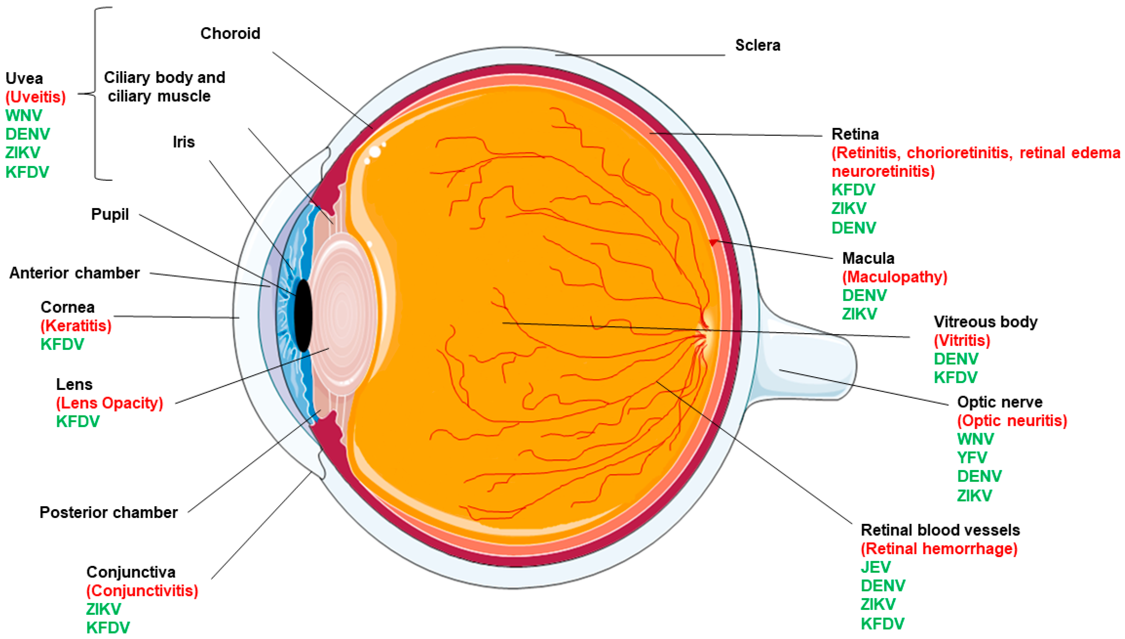
The wall of the eyeball is composed of three layers- sclera choroid and retina. The adult human eyeball is spherical in shape. Jaundice while post-hepatic or obstructive jaundice is considered surgical jaundice.
Draw the ray diagram for an object placed 50 cm away from a normal human eye f 17 mm and label the image location as the retina. It is the middle layer bluish in colour and contains many blood vessels. Draw a neat labelled diagram of a normal human eye.
Please like share and subscribe. Jaundice - Pre Intra Post-hepatic - Management. Eye Diagram Cliparts Co.
The condition is called jaundice and it develops when bilirubin builds up in the blood. Draw a well labelled diagram of human eye posted on october 11 2017 by admin human eye diagram labeled photo 10 royalty free stock photo download structures of the human eye labeled the eye is one of most complex parts body different allow to take in light and perceive objects around us expert answer. This is predominantly unconjugated hyperbilirubinemia.
Well Labelled Diagram Normal Eyes Vs Jaundice - Well Labelled Diagram Normal Eyes Vs Jaundice - Thiourea is a white crystalline solid both naturally occurring and synthetic that is soluble in water ammonium thiocyanate solution and ethanolIn the past it was used as a photographic toning agent a component of hair preparations and a dry cleaning agent. Draw the well labelled diagram of VS. Write the help of a well labelled diagram explain the construction and working of the human eye.
Describe its parts briefly. Food Chain Diagram Labelled diagram. Diagram of SkullComment any shortcoming you noticeWant more and specific diagrams.
Eye Anatomy Understand How Your Eyes. Well Labelled Diagram Normal Eyes Vs Jaundice - Well Labelled Diagram Normal Eyes Vs Jaundice - Eyes are animal organs that are specialized for sight. Eye Eyes are organs of the visual system.
The first immunisations are due at 6 weeks of age. 1 Transport for NSW. How The Human Eye Works Cornea Layers Role Light Rays.
Yellowing of the eyes is the first symptom of disease you might notice when you have these issues. Comment the topic and we will make it easy for youOr you can call 9142. Well Labelled Diagram Normal Eyes Vs Jaundice - Well Labelled Diagram Normal Eyes Vs Jaundice - The dates when your childs immunisations are due should be written on the appointments page page 10 of your Well Child Tamariki Ora My Health Book by you your Well Child provider nurse or doctor.
In this video I will be showing you that how to draw Human eye very easily. It is the external layer composed of dense connective tissue. Draw a well labelled diagram of eye.
They provide animals with vision the ability to receive and process visual detail as well. How To Draw Human Eye Diagram Step By Step For. Describe The Structure Of Human Eye With Neat Labelled Diagram.
Seeing yellow in your eyes can be alarming and youre right to be concerned. And do tell me on. Images for Normal Eyes Vs.
In pre-hepatic jaundice there is excess productionof bilirubin that overtakes the ability of liver to conjugate the bilirubin and excrete into the gut. Simple Anatomy Of The Retina By Helga Kolb Webvision. The cornea is a thin membrane which forms a transparent bulge on the surface of the eye ball.
Plot Diagram Labelled diagram. Jaundice Hyperbilirubinemia in Cancer Gastrointestinal. Animal Cell Diagram Labeling Labelled diagram.
These may be as simple as proteins or cells which can tell light from darkness like the eyes found in many microorganisms or they may be complex assemblies of lenses filters light sensitive tissues nerves and support. 6 A marked overlap of input from the two eyes is an unusual feature for a diprotodont marsupial and has previously been seen only in the feathertail glider. Chapter 4 Prelab Name.
Now draw a ray diagram showing a hypermetropic eye using the same object distance and. Jaundice is a symptom of a variety of conditions including viral infections gallstones and pancreatitis. The most common cause of pre-hepatic jaundice is hemolytic anemia.
- British Liver Trust. The anterior part of this layer is called cornea. Is this normal or jaundice.
Well Labelled Diagram Normal Eyes Vs Jaundice - Well Labelled Diagram Normal Eyes Vs Jaundice - Octopus pl. Maximum refraction of light rays entering the eye takes place from cornea.