Describe the structure and give the name of four cell organelles. The tabernacle consisted of a tent-like structure the tabernacle proper covered by rug-like coverings for a roof and an external courtyard 150 feet by 75 feet.

Draw A Labelled Diagram Of The Basic Body Plan Of Chordates Brainly In
Click hereto get an answer to your question Draw a labelled diagram of the structure of mature dicot embryo.

Draw a labelled diagram of the basic body plan of chordates. 167953095 stock photos online. New users enjoy 60 OFF. What are the 5 major body plan characteristics of the Phylum Chordata Draw a from BIO 102 at Santa Barbara City College.
Draw a labelled diagram of the experimental set-up to study the latent heat of vaporisation of water. Soft iron core PrincipleTransformer is based on the principle of electromagnetic mutual inductionWhen the current flowing through the primary coil changes an emf is induced in the secondary coil due to the change in magnetic flux linked with the primary coil. The total surface area of the cerebral cortex is about 2500 cm2 and when stretched it will cover the area of a night table.
The fence was made of linen hangings held by pillars. It contains 8 proteins 1 carbohydrates 2 soluble organics and 1 insoluble salts. Draw a neat labelled diagram showing the LS of a typical flower.
Quizlet flashcards activities and games help you improve your grades. A flower is a seed-bearing part of a plant consisting of reproductive organs stamens and carpels that are typically surrounded by a brightly coloured corolla petals and a green calyx sepalsFlowers are attractive and appear in different colours and shapes to. All vertebrates are built along the basic chordate body plan.
Draw a labelled diagram of the basic body plan of chordates. A stiff rod running through the length of the animal vertebral column with a hollow tube of nervous tissue the spinal cord above it and the gastrointestinal tract below. I Labelled diagram of a step-down transformer.
Basic chordate body plan study guide by Venabiezer includes 8 questions covering vocabulary terms and more. The brain is composed of 77 to 78 water and 10 to 12 lipids. The worksheet has two pages.
The whole compound was surrounded by a high fence about 7 feet in height. Drawing Anatomy Diagram Anatomy Of Human Body. Download 782 Fertilization Diagram Stock Illustrations Vectors Clipart for FREE or amazingly low rates.
Asked May 21 2020 in Structure of Living Organisms by Rukmani 511k points. Your lungs human body human body systems match column a with. This course covers the NCERT syllabus for Chordates.
It has a diagram of the human body to label and a diagram of the human face to label on the same page. The first page of. The diagram below shows the experiment set up to study the latent heat of vaporisation of water.
I have created a simple body and face worksheet for the study of basic body parts. This course is all about Phylum Chordata and their detailed classification their general features including examples on a systematic basis. Ii Turn ratio in terms of voltage isn iii For an ideal transformer according to.
Draw a labelled diagram of an animal cell. Diagrams of the Tabernacle and Basic Layout. Advertisement Remove all ads.
Of the spinal cord of man. Wiring Diagram 10 Major Muscles In Human Body A Draw A Labelled Diagram Of The Human Digestive System Skeletal System Function And Components Amazon Com Internal Organs Of The Human Body Anatomical The Human Organ Systems Human A. Correct answer to the question.
Click hereto get an answer to your question Draw a labelled diagram of the TS. Atent heat of vaporisation.
With the help of a clean spatula or a toothpick the inner side of the cheek is gently scrapped. They are found in the brain spinal cord and the peripheral nerves.

Solved Draw A Labelled Diagram Of Human Cheek Cells 3 Marks Self Study 365
Name the parts labelled 1 2 and 3.

Draw a labelled diagram of human cell. Name the stage prior to this stage and draw a diagram to represent the same. Label any three parts and write their functions. Labelled diagram of plant and animal cell are as follow---Both plant and animal cells belong to eukaryotic cells.
The cell membrane is semipermeable and flexible. Eukaryotes are sophisticated cells with a well defined nucleus and cell organelles. Unicellular to multicellular in nature and evolved 1 billion years ago.
This structure contains enzymes used for penetrating the female egg. It is a double-layered membrane composed of proteins and lipids. Identify the above stage and give a reason to support your answer.
The middle piece has a central filamentous core with many mitochondria. The cell organelles are membrane bound present within the cells. The sperm cells are the haploid gametes which are produced in the male.
462 378 pixels. Draw a diagram of the microscopic structure of human sperm. Diagram of the human cell illustrating the different parts of the cell.
Size of this PNG preview of this SVG file. FileDiagram human cell nucleussvg. The above diagram is of the sperm cell.
The structure of a neuron varies with their shape and size and it mainly depends upon their functions and their location. V Egg is the largest human cell. Mention the type of cells in our body where this type of cell division occurs.
A neuron is also known as the nerve cell. These cells reproduce both asexually and sexually. Contains the genetic material that the sperm has to pass on a haploid genome because it contains only one copy of each chromosome.
Vi Ovaries are located lower abdomen. There is the genetic material which is a haploid genome because it contains only one copy of each chromosome. The scrapped material is transferred into a drop of water and taken on a clean slide.
Draw A Labelled Diagram Of A Animal Cell And Plant Cell Ncert. State the functions of the two parts labelled. Draw a Neat and Labelled Diagram of the Human Ear.
With the Help of this Diagram Explain the Construction and Working of the Human Ear. Label the following parts in it and write their functions. This is a file from the Wikimedia Commons.
Fallopian tubes i Two thin tubes attached to the upper sides of uterus ii Tubes terminate near the ovaries but are not attached iii Fimbriae are finger-like structures on the end of each tube. Posted by Unknown at 2209. A neuron is a specialized cell primarily involved in transmitting information through electrical and chemical signals.
Solution For Draw a well labelled diagram of eye. Draw a diagram of a mature human sperm. The cells are comparatively larger in size 10-100 μm.
There are different parts of the sperm cell. 293 240 pixels 587 480 pixels 733 600 pixels 939 768 pixels 1252 1024 pixels 2503 2048 pixels. Information from its description page there is shown below.
Iii In what two ways is mitotic division in an animal cell different from the mitotic division in a plant cell. Lakhmir Singh Science Class 8 Solutions Chapter 8 Cell Structure And. Cell Membrane The cell membrane is the outer coating of the cell and contains the cytoplasm substances within it and the organelle.
It contains enzymes used for penetrating the female egg. 1 left and 1 on the right. With the help of a needle the material is uniformly spread.
Eukaryotic cells are one which have organised nucleus with a nuclear membrane and genetic material is organised into chromosomes. It is the inner layer and consists of three sub layers- ganglional cells bipolar cells and photoreceptor cellsThe innermost ganglionic cells give rise to optic nerve fibre that forms optic nerve in each eye and is connected with the brain. Draw a labelled diagram of the embryonic stage that gets implanted in the human uterus.
Human Cell Diagram Parts Pictures Structure and Functions parts of a human cell Diagram of the human cell illustrating the different parts of the cell.
- makes the synovial fluid and encloses. - a sheet of cells that lines the joint cavity.
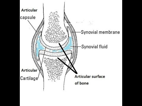
Structure Of Synovial Joint Youtube
Key Structures of a Synovial Joint.

Draw a well labelled diagram of synovial joint. The morphology of synovial membranes may vary but it. Draw labelled diagram Synovial joint. At synovial joints the articular surfaces of bones are covered with smooth articular cartilage.
It consists of two layers. Synovial fluid is the clear viscid lubricating fluid secreted by synovial membranes. The walls of this space are formed by the articular capsule a fibrous connective tissue structure that is attached to each bone just outside the area of the bones articulating surfaceThe bones of the joint articulate with each other within the joint cavity.
Fibrous Capsule outer- provides joint stability. Synovial joints allow for smooth movements between the adjacent bones. This gap allows a free range of motion and space for synovial fluid to lubricate the joint.
Explain the roles of actin and myosin in muscle contraction. Primary movement is a rotation. Give one example each for a hinge joint a pivot joint axial skeleton and appendicular skeleton.
The basic structure of a synovial joint is shown in the diagram below. Movement of the ankle elevating the sole. It is freely movable joint hence known as diarthrosis joint.
Synovial joints are the most common type of articulation and feature a small gap between the bones. Primary movement is a rotation. Reinforcing ligaments Examples of synovial joint-Shoulder jt.
The joint is surrounded by an articular capsule that defines a joint cavity filled with synovial. Examples of this form of articulation are found. Give one example each for a hinge joint a pivot joint axial skeleton and appendicular skeleton.
The main parts of synovial joints are labelled on the synovial joint diagram. Extending the ankle and elevating the heel. Allows movement in only one plane.
Synovial fluid view the full answer. Analyse the electron micrograph for the state of contraction of the muscle fibre. 42 Joint and Movement Type Synovial Joint Movements Circumduction.
Synovial joints are the the most common joints present and have a characteristic trait of having the presence of a joint cavity the cavity filled with the synovial fluid. Also allows movement in only one plane. Draw a labelled diagram of a synovial joint.
Synovial joints are characterized by the presence of a joint cavity. Finally cartilaginous joints are formed. Concept Notes Videos 380.
I articular capsule ii articular cartilage iii synovial fluid. Question Bank Solutions 5549. Some synovial joints are more complicated than others.
Synovial joints allow for smooth movements between the adjacent bones. Draw a labelled diagram of a synovial joint. Allows movement in only one plane.
Elbow joint and knee joint. Fibrous joints exist where bones are very tightly joined and offer little to no movement between the bones. The ball and socket joint or spheroid joint is a type of synovial joint in which the ball-shaped surface of one rounded bone fits into the cup-like depression of another bone.
Elbow joint and knee joint. Also allows movement in only one plane. 19 Draw a well labelled diagram of.
The three main features of a synovial joint are. This gives the bones of a synovial joint the ability to move smoothly against each other allowing for increased joint mobility. Draw a labelled diagram of a synovial joint.
Label the following diagram of the side view of the human elbow joint. Fibrous joints also hold teeth in their bony sockets. Give one example each for a hinge joint a pivot joint axial skeleton and appendicular skeleton.
The distal bone is capable of motion around an indefinite number of axes which have one common center. Figure 941 Synovial Joints. Digging in the heel Plantar flexion.
A metacarpophalangeal finger joint is shown above-rightBBC Bitesize - GCSE Physical Education - Skeletal system - OCR - Revision 3Synovial Joint Labelled Diagram - Anatomy Structure. The structure of a synovial joint is demonstrated by a diagram in which the articulating bones are surrounded by the articular capsule which comprises an exterior fibrous capsule and an interior synovial. Refer to the image for well-labelled diagram of typical synovial joint Synovial joint - A joint between two bones with a cartilage fluid filled cavity in between is called a synovial joint.
This diagram shows the location of the bursae which are fluid filled sacs in a bone. The articular capsule surrounds the joint and is continuous with the periosteum of articulating bones. Synovial Membrane inner- secretes produces synovial fluid for lubrication.
An example of a simple synovial joint eg. Draw a labelled diagram to show the structure of a sarcomere. Draw labelled diagram Synovial joint.
Maharashtra State Board HSC Science General 11th. The main parts of synovial joints are labelled on the synovial joint diagram and described in the table below. A synovial membrane or synovium is the soft tissue found between the articular capsule joint capsule and the joint cavity of synovial joints.
Structural Features of Synovial Joints. It is characterized by. Advertisement Remove all ads.
Define its magnifying power. I Which lenses should he used as objective and eyepiece.

A Concave Mirror Is Used For Image Formation For Different Positions Of An Object What Inferences Can Be Drawn Science Light Reflection And Refraction 14317813 Meritnation Com
I Which lenses should he used as objective and eyepiece.

Draw a labelled ray diagram to justify your inferences. Nature of image---- real and inverted. Clearly mark angle of incidence angle of refraction angle of emergence and lateral displacement of the ray. Draw A Labelled Ray Diagram Of A Reflecting Type Telescope Write.
It is defined as the ratio of the angle β subtended by the final image on the eye to the angle α subtended by the object on eye. 0 Response to Labelled Refraction Ray Diagram Post a Comment. Share It On Facebook Twitter Email.
Define its magnifying power. Clearly mark angle of incidence angle of refraction angle of emergence and lateral displacement of the ray. I Which lenses should be used as objective and eyepiece.
A concave mirror is used for image formation for different positions of an object. CBSE CBSE English Medium Class 10. B if it hits glass block at an angle other than 90 degrees.
Asked Dec 23 2015 in Class X Science by luckky ramya Expert 18k points 0 votes. Ii Aperture of the objective lens should be. Draw a labelled ray diagram to locate the image of an object fromed by a convex lens of focal length 20 cm when the object is placed 30 cm away from the lens.
Draw a labelled ray diagram to justify your inferences. Draw a ray diagram showing the path of the light ray. B You are given three lenses of power 05 D 4 D and 10 D to design a telescope.
A Draw a labelled ray diagram to obtain the real image formed by an astronomical telescope in normal adjustment position. A Draw a labelled ray diagram to obtain the real image formed by an astronomical telescope in normal adjustment position. Refraction Of Light Science Learning Hub.
Pages 35 This preview shows page 20 - 26 out of 35 pages. Click hereto get an answer to your question 21 Aconcave mirror is used for image formation for different positions of an obiect. Size of the image-- enlarged.
What inferences can be drawn about the following when an object is placed at a distance of 10 cm from the pole of a concave mirror of focal length 15 cm. A if it hits glass block at 90 degrees. Total Internal Reflection Wikipedia.
B You are given three lenses of power 05 D D and 10 D to design a telescope. Draw a Labelled Ray Diagram to Show the Angle of Incidence and the Angle of Refraction for a Refracted Ray of Light. A Position of the image b Size of the image c Nature of the image.
B You are given three lenses of power 05 D 4 D and 10 D to design a telescope. Course Title CHEMISTRY MISC. A ray of light incident at an angle of incidence i passes through an equilateral glass prism.
Answered May 4 2018 by sanjaydas 889k. Question Bank Solutions 20334. Draw a labelled ray diagram to justify your.
Ii Why is the aperture of the objective prefered to be large. Explain why both the objective and the eye piece of a compound microscope must have short focal lengths. How to draw a ray diagram of the convex mirror in an easy way for class 10 beginner students who wants to achieve full mark in boardWatch How To Draw Huma.
Define its magnifying power. Define its magnifying power. A Position of the image b Size of the image c Nature of the image Draw a labelled ray diagram to justify your inferences PIO.
First we draw a ray parallel to principal axis So it passes through focus after refraction We draw another ray which passes through Optical Center So the ray will go through without any deviation Where both refracted rays meet is point A And the image formed is AB This image is formed between beyond 2F 2 We can say that Image is Real. Concept Notes Videos 224. I Image is not free from chromatic aberration and spherical aberration.
Draw a labelled diagram to show how a ray of light passes through a parallel sided glass block. Draw a ray diagram to show the path of the reflected ray corresponding to an incident ray of light parallel to the principal axis of a convex mirror and show the angle of incidence and angle of reflection on it. Draw the labelled ray diagram for the formation of image by a compound microscope.
Post Comments Atom. B You are given three lenses of power 05 D 4 D and 10 D to design a telescope. A Draw a labelled ray diagram to obtain the real image formed by an astronomical telescope in normal adjustment position.
Newer Post Older Post Home. What inferences can be drawn about the following when an object is placed at a distance of from the pole of a concave mirror of focal length. Derive the expression for the total magnification of a compound microscope.
A Position of the image b Size of the image c Nature of the image Draw a labelled ray diagram to justify your inferences. A Draw a labelled ray diagram to obtain the real image formed by an astronomical telescope in normal adjustment position. Draw a labelled ray diagram to justify your inferences 3 Answer Given Position.
Position of image -- 30 cm. Reflection Of Light Visible Light Siyavula. What inferences can be drawn about the following when an object is placed at a distance of 10 cm from the pole of a concave mirror of focal length 15 cm.
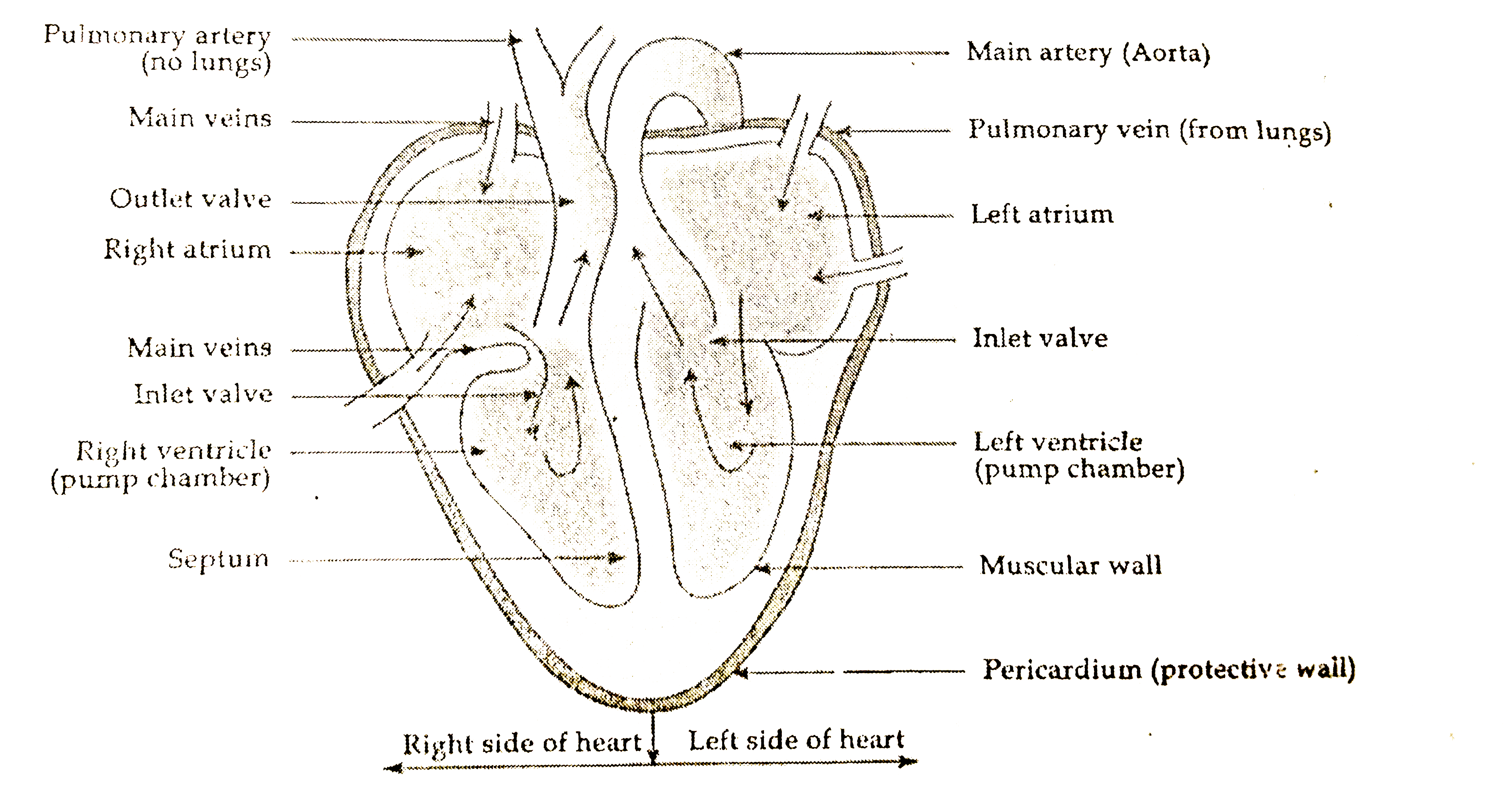
The heart is roughly cone shaped hollow organ. They will be to the lower left of the Aorta.
Draw A Labelled Diagram Of Human Heart
IG 263 describe the structure of the heart and how it functions DP 621 Draw and label a diagram of the heart showing the four chambers associated blood vessels valves and the route of blood through the heart.

Draw a labelled diagram of human heart and explain it. The heart though small in size performs highly significant functions that sustains human life. Science 29012020 1515 jack626259 Draw a diagram of human heart and label its parts. Draw a labelled diagram of Human Heart.
The heart lies in the thoracic cavity in the space between the lungs mediastinum anterior to the vertebral column and posterior to the sternum. In this video our mentor has showed how you can draw a basic anatomical diagram of human heartLike and comment your thoughts and feedback belowSubscribe. The heart is responsible for the circulation of blood in our body.
Find an image that displays the entire heart and click on it to enlarge itStep 2 Find a piece of paper and something to draw with. Heart rate varies depending on the phase of respiration. Draw Label Annotate Guidelines You are drawing labelling and annotating the digestive system.
It is not possible to draw the human heart dont u have in your text book or u can search in net. We will review the anatomy function and order of blood through the human heart using pictures of the atria ventricles tricuspid valve mitral valve pulmonary valve aortic valve superior and inferior vena cava pulmonary arteries and veins and aorta. The wall of the heart has three different layers such as the Myocardium the Epicardium and the Endocardium.
The heart consists of 4 chambers. Draw a labelled diagram of human ear and explain its working Get the answers you need now. Watch 1000 concepts tricky questions explained.
Correct answer to the question. FileDiagram of the human heart croppedsvg. There are two of them.
Drawings must be correctly proportioned and clearly drawn showing connections between structures. The drawing may show the heart without contraction or in any stage of contraction. This occurs even in normal human beings and hence it is quite physiological one.
This is known as sinus arrhythmia. This is a file from the Wikimedia Commons. A heart diagram labeled will provide plenty of information about the structure of your heart including the wall of your heart.
It is approximately the size of owners closed fist and weighs about 250-300 gm in female and 300-350gm in the male. Arrhythmia refers to irregularity in the beating of heart. Apne doubts clear karein ab Whatsapp par bhi.
Loading DoubtNut Solution for you. 611 600 pixels. Markscheme Remember up to TWO quality of construction marks per essay.
Every single part of our body is so well designed that it works continuously throughout our life. A well labeled human heart diagram given in this article will help you to understand its parts and functions. Step 1 To find a good diagram go to Google Images and type in The Internal Structure of the Human Heart.
Draw a labeled diagram of human heart. The heart one of the most significant organs in the human body is nothing but a muscular pump which pumps blood throughout the body. Draw a labelled diagram of human heart.
The human body is the best machine created by God. When the pacemaker is located in the SA node and heartbeats accordingly it is known as sinus rhythm. The anatomy of the heart is made easy in this post using labeled diagrams of the main cardiac structures and vascular system.
Human Heart Diagram Labeled Science Trends. Start with the pulmonary veins. AND 623 Explain the.
Draw a labelled diagram of the human heart showing the attached blood vessels. 26 K views 100 people like this. Draw the top vein slightly smaller than the bottom veinStep 3 Below the pulmonary veins and slightly to the right begin sketching the.
Heres more about these three layers. A Labeled Diagram of the Human Heart You Really Need to See. 10 Draw And Label A Human Heart.
Draw a table to show the functions of any two chambers of Human Heart. Exterior of the Human Heart. The human heart and its functions are truly fascinating.
244 240 pixels 489 480 pixels 782 768 pixels 1043 1024 pixels 2086 2048 pixels 663 651 pixels. Click here to get an answer to your question Draw a labelled diagram of human heart. Size of this PNG preview of this SVG file.