Are overweight have obesity. The most common causes of heel pain are plantar fasciitis bottom of the heel and Achilles tendinitis back of the heel.

What Is Causing Your Heel Pain Robbins Rehabilitation West
Too much pressure overuse and hard impacts over time can overstretch and damage the ligament.
Why do i have pain in my foot heel. Its caused by inflammation overuse. Plantar fasciitis is a problem with the ligament that attaches to the bottom of your heel and runs the length of your sole. Choose which area of your foot hurts most to read about treatments when to get medical help and possible causes of foot pain.
Heel spurs are often caused by strains on foot muscles and ligaments stretching of the plantar fascia and repeated tearing of the membrane that covers the heel bone. This fascia connects your heel bone to your toes. In addition peripheral neuropathies associated with diabetes alcohol abuse or a vitamin deficiency can cause diffuse foot and heel pain.
Hypothyroidism can cause heel pain in the morning. Plantar fasciitis is characterized by pain in the bottom of the feet involving either the arch or the heel or both it is usually felt when taking the first few steps in the morning then subsides or disappears after resting or gently walking however the pain returns when walking or. Plantar fasciitis is a foot condition characterized by sharp stabbing heel pain that typically occurs when a person gets out of bed in the morning or stands up after sitting for a long period of.
Bursitis joint inflammation Haglunds deformity. Its one of the most complex hard-working regions of your body. You may be more likely to develop heel pain if you.
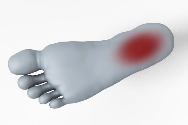
The conditions producing pain in this area are included below common condition Conditions producing pain in this area include common condition. Recently in the past few months I have barely been able to walk at times on my left foot because the heel is so sore. Causes of heel pain also include.
Irritation of a nerve in the lower back called radiculopathy may cause pain of the calf muscle that moves down the leg into the heel. Plantar fasciitis is the most common cause of arch pain and one of the most common orthopedic complaints reported. Sometimes as I am getting out of bed in the morning I can hardly stand to put any weight on that foot at all its too painful.
The way you walk foot mechanics and your foots shape foot structure are also factors. Anything that puts a lot of pressure and strain on your foot can cause heel pain. It has 26 bones and 33 small joints all held together by a network of soft tissue made up of muscles tendons ligaments nerves and blood vessels.
Ankle Impingement Anterior Ankle Arthritis. Most cases of foot or ankle pain are short. Experiencing severe pain in heel of right foot hard to put.
Experiencing severe pain in heel of right foot hard to put wait on it after sitting or laying down for a period of time. Plantar fasciitis is one of the most common causes of foot pain. Sharp stabbing heel pain can be caused by a number of different conditions but the most common one is plantar fasciitis.
Heel pain is often caused by exercising too much or wearing shoes that are too tight. Why do I have pain in my foot or ankle. Your symptoms might also give you an idea of whats causing your heel pain.
Most people experience pain in and around their feet or ankles at some point in their lives. It seems to get better and loosen up a little as I continue walking. After walking awhile lightens up.
2 It results from irritation of a thick band of tissue called the plantar fascia that runs across the bottom of your foot. The disruption of chemicals and hormones in the body can lead to inflammation and swelling in the feet ankles and heels. Im a 52 yr old female with no previous foot problems of any kind.
Common causes of heel pain.
Your feet take a beating every day and can be particularly prone to injury. Hes an athlete so hes not sure whats causing it.
Yellow Feet 6 Potential Causes
A man wrote into The Doctors saying he had orange palms and orange soles of his feet.

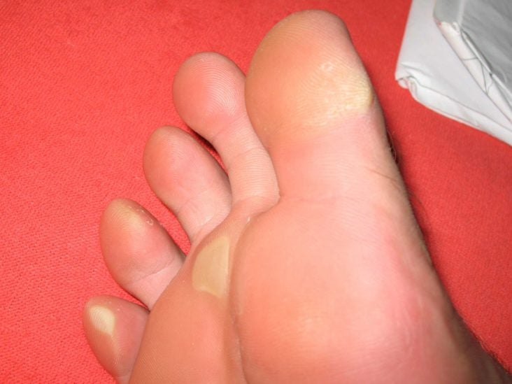
Why do i have orange feet. Ive posted a pic of her feet now compared to what they were before. Bright orange coloring suggests that a male duck also known as a drake is getting all his vitamins particularly carotenoids such as beta-carotene and vitamin A. Hi i was hoping you could tell me why the bottoms of my feet are a kind of orangy colour.
He said theyre so orange people have commented on it. Looking her over the only thing I noticed was that her feet have become bright orange. He wonders whether something in his diet could be causing the orange color.
I dont drink or eat large ammounts of Orange coloured food and am currently awaiting more blood tests for thyroid. I looked at. Our rooster a buff orpingtons had feet like this but I thought it was just breed since thats all hes ever had.
It can also caused from underlying disease states like hypothyroidism diabetes mellitus anorexia nervosa nephrotic syndrome liver. Posthinking01s reply seems plausible because I have always taken cod liver oil which I believe. Because your feet are complicated structures with many moving parts foot redness can also be due to inflammatory environmental and vascular causes.
Because nobody wants to go in public when youre suffering a self tan meltdown most of these tips are at home remedies from products you probably already have. Jennifer Ashton said that this is usually caused by people who take excessive amounts of. Kevin Omland is an evolutionary biologist at the University of Maryland at Baltimore County and he knows as much about mallard-duck coloring patterns as anyone.
Soles of feet with an orange tinge. Visiting doctor should is always a embarrassment freeAs disease spares nonehigh colored or pink feet can be due over blood supply to feet or vaso dilatation so consult doctor get it treated. General Surgeon Dr.
A fungal infection obtained from a public shower may cause orange toenails. However hers are usually much. It can be developed from increase oral intake of carotenoids food like carrot XXXXXXX spinach pumpkin or sweet potato.
Is it hard skin although I do moisturise and buff. Eating carrots and other foods rich in carotene may cause people to have orange skin. Soles of feet with an orange tinge.
Orange colouration of sole of your feet might be due to the condition known as carotenoderma. I have put together a list of tips that others have used to undo what usually takes days to be undone. Walking barefoot in public places may cause toenail fungus that changes the color of the toenails.
Since being placed on his hormone replacement therapy to replace his cortisol he has these orangebrown spots on his palms from time to time. You can also rub your hands with a small slice of lemon to. If yellow feet are the only symptom that a person has the cause is most likely to be a callus or a high intake of carotenoid-containing foods.
Tinted a bluish green or gray. I have had an orange rusty tint to the soles of my feet for many years probably started in my early twenties and it has always bothered me and left me wandering why. Justin writes that he has noticed that the palms of his hands and soles of his feet have an orange tint.
Actually many species of ducks have feet and legs. One of the main causes why the skin may turn yellow or orange is due to the affected individuals dietary intake. It was his graduate thesis.
Theyre more common in areas that experience a lot of friction or. Calluses are thick layers of hardened skin that often develop on the bottom of your feet. Why are they orange looking.
However trauma is only one of the potential causes of foot redness. Orange toenails can be caused by a fungal infection. Consuming a large amount of orange and yellow vegetables such as carrots pumpkins and squash may result in a condition known as carotenemia.
To lighten up the results of a self tanner apply lemon juice to your body. The most common causes of orange skin are carotenemia a usually benign condition where people ingest too much beta carotene and their bodies cannot clear it quickly enough and jaundice a. Improperly cutting toenails can lead to trauma and infection.
But for the ducks that do have orange feet well its all about attracting the ladies. We just re-homed him last week because he got too aggressive with our kids. A podiatrist can treat calluses and other foot.
Than the nest person also the skin there is hard and there is a semi circle of non orange skin. Rajinder Bajoria s Response.
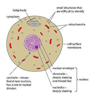
Use cleandry mounted slide while placing it under the lens of the microscope. As you can see in the above labeled plant cell diagram under light microscope there are 13 parts namely Cell membrane.
Animal Cell Structure And Organelles With Their Functions Jotscroll
To use a light microscope to examine animal or plant cells.

Draw the labelled diagram of animal cell which you have observed under microscope. Draw a well labelled diagram of animal cell as observed human cheek experiments on microscopes 4 schools if onion peel cells and cheek are observed through a investigating cells with a light microscope bbc com the inner epidermis of the onion bulbs cataphylls the looking at onion epidermis using a microscope techknow onion root mitosis. A squamous epithelial cell looks flat under a microscope. As you evaluate their diagrams you may choose to note diagrams.
83 an overview of cell you have earlier observed cells in an onion peel andor human cheek cells under the microscope. On this page we will learn about what is a plant cell definition structure model labeled plant cell diagram its cell organelles and the difference between plant cell and animal cell. In this lab we adjusted the resolution on the microscope to have a better look at the specimens that were observed.
The average microscope has a resolving power of up to 02 micrometers. Aims of the experiment. Peel off a thin layer of epidermal tissue from the inner surface 4.
46 Draw The Diagram Of An Animal Cell And Label The Following ImagesIllustrate only a plant cell as seen under electron microscope. Thats the major difference between plant and animal cells under microscope. The vacuole in an an animal cell is smaller.
Its this cell membrane that will contain all of the different parts of the cell. So it is important to note that what we are drawing is definitely not life. Title the drawing with the type of cell magnification and phase of mitosis.
Once slides have been prepared they can be examined under a microscope. For example something that you draw as 3cm long may in fact be 10 000 times smaller in real life. As observed in the labeled animal cell diagram the cell membrane forms the confining factor of the cell that is it envelopes the cell constituents together and gives the cell its shape form and existence.
Onion peel the cells are very clearly visible as compartments with. Use unusednew toothpick for scraping of cheek cells. Human cheek cell under microscope diagram.
Draw a labelled diagram of the section that you observed under the microscope. How to Sketch a Microscope Slide Identifying Cell Structures and Adding Dynamic Elements. Continue with your observations until you have found cells in each phase and both you and your partner can easily identify.
Animal Cells Under Microscope Labelled But at the same time it is interpretive. The Plant Cell is the basic structural and functional unit found in the members of the kingdom Plantae. Generalized Cell is used for structure of Animal Cell and Plant Cell to present the.
At the end of the observations you will be reviewing the diagrams students drew. Students should describe what they see in the microscope with words and by making a labeled diagram. You may want to state some of the expectations you have for their diagrams before they get started.
Cell structure and function class 8 science chapter 8 as per ncert book used in cbse and other schools. Name the longest cell in human body. As you can see in the above labeled plant cell diagram under light microscope there are 13 parts namely Cell membrane.
Use a light microscope to observe and measure. Learning how to sketch a microscope slide requires an open-mind patience and a willingness to learn the basic drawing principles of perspective size shape and negative space. You know Animal cell structure contains only 11 parts out of the 13 parts you saw in the plant cell diagram because Chloroplast and Cell Wall are available only in a plant cell.
Draw and label an animal cell as seen under an electron microscope. Draw and label a typical bacterial cell then provide functions for at least five of the labeled structures. Labelled diagram of a plant cell under microscope posted on march 18 2011 by admin onion cells stained with methylene blue look at the images of onion cells as they would be seen under a microscope draw each magnification label appear high picture plant and animal cell.
Now the first thing to point out when looking at images under an electron microscope is the scale. To use a light microscope to examine animal or plant cells. Label all important structures nucleus chromosome cell plate.
2017 q9 b ii - In relation to an investigation you carried out to prepare and examine with a microscope a transverse section of a dicot stem answer the following. Neha observed a slide of human cheek cells under a microscope in its i low magnifying power ii high magnifying power settings. Animal fat Butter is also an animal fat because it comes from cows milk In this investigation the student will observe record Title April 15th 2019 - Draw and label 3 plant cells Take another strip of cells from your plant.
Obtain a slide of epithelial tissue from the instructor. Humans only have a resolution the ability to separate or distinguish two or more objects that are close together of 01 millimeters. Sketching specimens will provide you with a better understanding as you study the intricacies of the image you see through the.
Animal cells have a basic structure. Draw A Diagram Of Animal Cell And Label Any Three Parts Which Differentiate It From Plant Cell Brainly In - Cells are covered by a cell membrane and come in many different shapes. To make observations and draw scale.
When you have found one make a detailed observational drawing of that cell.