The Insect Nervous System Cockroach Nervous System Diagram Images E993 Com Termite Pest Controls Cockroach Nervous System. Nissan Versa Kbb User Manuals ankrumax.
Explain Nervous System Of Cockroach Qs Study
Cockroach being an insect posses an open circulatory system.

Labelled diagram of nervous system of cockroach. Labeling See more about Mouth And Throat Diagram And Labeling Cockroach Nervous System Diagram With Labelling Simple Diagram Of Nervous System Diagram System March 16 2017 124 views Diagram Of A Well Labeled Animal Heart Diagram System March 11 2017 145 views Name Of Parts Of Body With Diagram Human Nervous System Structure and Functions. On the dorsal side of the heart two nerve chain is present. The fore wings are mesothoracic and are called wing covers or tegmina or elytra.
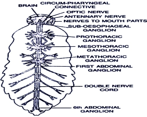
FOXCONN MOTHERBOARD G31MXP MANUAL fussie de. What are Malpighian tubules. The nervous system of cockroach consists of a series of fused segmentally arranged ganglia joined by paired longitudinal connectives on the ventral side.
The sexes are separate. The autonomic or stomogastric or sympathetic or visceral nervous system of cockroach consists of some ganglia and their connectives. Click hereto get an answer to your question 1.
Well Label Diagram Of Cockroach Nervous System When There Are Many People Who Don T Need To Expect Something More Than The Benefits To Take We Will Suggestlabeled diagram of nervous system anatomy diagram chart april 18th 2018 - labeled diagram of nervous system see more about labeled diagram of nervous system labeled diagram. Soy her estrogen levels Venn Diagram Digestive And Nervous System well labelled diagram of earthworm nervous system Digestive System Main Function Wombat Nosed Southern System Digestive Hairy well label diagram of cockroach nervous system The Central nervous system Wikipedia April 27th. Simple nervous system diagram labeled.
Which pulsates with the help of external salary muscles. It includes the frontal occipital visceral or ingluvial and pre-ventricular ganglia. Insect physiology includes the physiology and biochemistry of insect organ systems.
Thank you for visiting at this website. Visit the post for more. The nervous system includes the central peripheral and sympathetic or stomatogastric nervous systems.
The nervous system is the control center for your body. Three ganglia lie in the thorax and six in the abdomen. Well Labelled Diagram Of Earthworm Nervous System PDF Download.
Give an account of respiratory system of cockroach. A pair of anal styles are present in male cockroach. Nerves from hypo-cerebral ganglion connect the ingluvial or stomachic ganglion.
With the help of neat labelled diagram explain circulatory system in cockroach5. Supra oesophageal ganglion-are a pair of ganglia found in the head form the brain. C Ciliated epithelium Answer.
The nerves from ingluvial ganglion innervates the neighbouring regions of the stomodaeum. With the help of neat labelled diagram explain circulatory system in cockroach. The nerves from these ganglia are connected with the supra-oesophageal ganglion.
Explain the digestive system of cockroach3. 10 Labelled Diagram Of The Nervous System Nervous March 31st 2019 - 10 Labelled Diagram Of The Nervous System and morelabelled diagram of nervous system of cockroach labelled diagram of nervous system of prawn labelled diagram of Well Labelled Computer System Diagram April 9th 2019 - April 2nd 2019 Well Labelled. Easily and step by st.
The hind wings are large thin membranous and transparent. Cockroach Anatomy Parts of the Roach Biology. Uricose glands are the mushroom gland of the cockroach which possesses long blind tubules that are at the periphery.
Contrary to popular perception their heads also house their brains. These tubules store uric acid and discharge it over spermatophore during copulation. It is the main organ of digestion and absorption.
Diagram Human Nervous System 159 65 2 233. In this video Im going to draw labelled diagram of circulatory system of cockroach of class 11th chapter of biology draw with me. Labelled diagram of digestive system cockroach posted on march 26 2019 by admin gizzard the structurecomposed of thick circular muscles and inner cuticle forming six highly chitinousplates called as teeth tracheal system in dorsal view 14 tubular digestive system nervous system.
The nervous system of cockroach is spread throughout the body. Draw a neat diagram of digestive system of frog. They cover the hindwings and are protective in function.
The nervous system of cockroach consists of the central and periphernal systems The central nervous system is made of ganglia and nerve cords whereas the peripheral nervous system is made of nerves arising from the ganglia. The cockroach has three pairs of jointed appendages and two pairs of wings. Well Label Diagram Of Cockroach Nervous System.
Give an account of the nervous system in cockroach4. Well Label Diagram Of Cockroach Nervous System. This article explains the nervous system function and structure with the help of a human nervous system diagram and gives you that erstwhile textbook feel.
A Diagram Of A Cockroach Front Leg With Segments Labeled B How To Draw Alimentary Canal Of Cockroach In Easy Steps Ncert The Insect Nervous System. Describe the external morphology of cockroach2. These are dark stiff opaque and leathery.
April 27th 2018 - Read Document Online 2018 Well Label Diagram Of Cockroach Nervous System This pdf file is made up of Well Label Diagram Of Cockroach Nervous System so as to download this DIAGRAM OF COCKROACH ANSWERS COM. Dissection of Reproductive System. Situated on the posterior end of the foregut.
Blood Types and Blood Type problems. Create a new quiz.

The Human Heart Diagram Quizlet
Heart anatomy set 1.

Heart parts labeled quizlet. Start studying heart diagram labeling. Diagram Heart Diagram Quizlet Full Version Hd Quality Diagram What Is A Compound Microscope New York Microscope Co Microscope Bundle Parts Of A Microscope Unit Activities Tpt. The circulatory system - Locate the arteries and veins of the circulatory system.
It will help you to memorize. 3 flap one way valve between the. This activity contains 7 questions.
The Heart has three layers. The circulatory system - lower body image with blank labels attached. The more healthy your heart is the longer the chances you have of surviving so you better take care of it.
Your Skills Rank. Has the thickest myocardium. Choose from 500 different sets of a and p lab anatomy heart labeling flashcards on quizlet.
This is an online quiz called Label the Hearts Parts. The aortic valve closes when the blood pressure within the aorta exceeds the blood pressure within the left ventricle. There is a printable worksheet available for download here so you can take the quiz with pen and paper.
Blood comes in through veins and exists via arteriesto control the direction of the flow the heart has four sets of valves. Heart anatomy set 1. Join a game Log in Sign up.
Choose from 500 different sets of anatomy of heart pictures flashcards on quizlet. Heart anatomy quizlet heart anatomy the heart is a cone shaped organ about the size of the clenched fist of its owner the heart pumps blood through heart anatomy quizlet heart anatomy the heart is a cone. Where the blood in its going.
The circulatory system - upper body image with blank labels attached. Take this practice quiz that covers information related to the sheep heart the heart model. Label the posterior surface heart structures.
Published by Jennifer N. Anderson 2305 Tidak ada komentar. Play this game to review Human Anatomy.
Movement of the heart the base upper part of the heart is in a fixed position because of its attachments to the great vessels but the apex or lower part. It is intended for use as a supplemental study aid. This is an online quiz called The Heart.
This science quiz game will help you identify the parts of the human heart with ease. Choose from 500 different sets of anatomy of heart pictures flashcards on quizlet. External Anatomy Of The Heart 30 Part 2 Diagram Quizlet.
As is the case in the lab practical each correct. Pumps blood to the lungs. Label the pericardial structures.
Anatomy of the heart. Choose from 500 different sets of anatomy of heart pictures flashcards on quizlet. Take the following quiz to know how much you know about your heart.
The heart has FOUR CHAMBERS in the lower heart the right and left Ventricles and in the upper heart the right and left Atria. Left ventricle pumps blood to the body. Start studying Label the Heart.
The heart is an amazing machine with a lot of moving partslet this quiz game help you find your way around this most vital of organs. The heart is protected by the PERICARDIUM which is the protective membrane surrounding it. The ENDOCARDIUM inner layer the EPICARDIUM middle layer and MYOCARDIUM outer layer.
The heart - an image of the heart with blank labels attached. Learn vocabulary terms and more with flashcards games and other study tools. Sequential Easy First Hard First.
Create a new quiz. The human heart is a vital organ for every human. For each item below use the pull-down menu to select the letter that labels the correct part of the image.
Learn vocabulary terms and more with flashcards games and other study tools. There is a printable worksheet available for download here so you can take the quiz with pen and paper. Label the heart quizlet.
Regulates blood flow from the upper left chamber left atrium into the lower left chamber left ventricle the hearts main pumping chamber aortic valve revent the regurgitation of blood from the aorta into the left ventricle during ventricular diastole and to allow the appropriate flow of bloodthe cardiac output from the left ventricle into the aorta during ventricular systole. From the quiz author. Whether the blood in it is oxygenated or deoxygenated.
Pumps blood to the body. Blood from the body returns to the heart via vena cavas. Where the blood in it came from.
This is a very large set of flashcards.
Petals come in a wide variety of colors. At the bottom of every hibiscus bud is a green structure at the top of the stem.

Drawing Diagram Of Different Parts Of Hibiscus Flower Novocom Top
How To Draw A Hibiscus Flower Easy Step By Step With Color In 2020 Hibiscus Drawing Drawings Hibiscus Flowers.

Draw and label the parts of a hibiscus flower. Hand drawn illustration Roselle flower. Draw hibiscus flower and label it hibiscus flower drawing hibiscus flower google search cricut. And it will teach you how to draw a flower very easily.
This is called the calyx. Plant parts of a flower diagram hibiscus flower parts diagram and flower parts diagram are three main things we want to present to you based on the post title. Draw Hibiscus Flower And Label The Parts Reb l Fleur Rihanna perfume a fragrance for women 2010 April 17th 2019 - Daring sexy and truly memorable Reb l Fleur is as much in tune with Barbados born Rihanna s roots as it is with the glamour of her present life in.
For this section on the webquest you will given. Set of two black labels with roselle aka hibiscus sabdariffa and cocoa tree aka theobroma cacao branch sketch. 11 14 ks3 14 16 ks4 post 16 plant growth health and reproduction.
Draw a hibiscus flower and label its parts. The anther is the head of the stamen. Cross Section Of A Hibiscus Flower Part Biological.
Hello friendsThis video help you to draw Diagram of parts of flower Hibiscus the longitudinal section of hibiscus flower the pollination in plants this vi. Lolly var lachland homeschool. Youll recognize the pistil in a plant diagram because it looks like a small knob that protrudes from the flower.
Draw and label the parts of hibiscus flower. Drawinginfo will teach you today how to draw a hibiscus flower easy step by step in 26 steps let s get started. Set of two black labels with Roselle aka Hibiscus sabdariffa and Cocoa tree aka Theobroma cacao branch sketch.
Saved by Nova Gabbe. Flower Diagram was drawn but not labelled properly. The stamens function is to produce male reproductive cells.
Hand drawn illustration Roselle flower. Draw a hibiscus flower and label its parts. Diagram of hibiscus flower with label.
Draw hibiscus flower and label the parts free draw hibiscus flower and label the parts we manage to pay for you this proper as capably as simple pretension to acquire those all. The corolla has two different parts the petals and the corolla lobes. So if you want to get great shots related to Draw And Label The Parts Of Hibiscus Flower just click on the save icon to save the photo to your computer.
Flowers are considered to be modified shoots. The bud grows from this structure. Draw and label a hibiscus flower.
Vector background with ornamental hibiscus flowers. Hello friendsIn this video Im going to draw labelled diagram of hibiscus flower China rose and its partsThis video is helpful for you in your exams. Label drawing of a hibiscus flower.
It is a tough part of the flower because it houses the. How To Draw Parts Of Flower Diagram. As the hibiscus begins to bloom the petals begin to grow.
A beautiful drawing of a flower. Drawinginfo will teach you today how to draw a hibiscus flower easy step by step in 26 steps let s get started. How to draw parts of flowerparts of flowerdiagram parts of flowerdraw and label part of flower - YouTube.
Draw Hibiscus Flower And Label The Parts beers great american beer festival www manythings org natural sciences grade 7 mstworkbooks co za toa sp 103 vh021 florida vegetable gardening guide the soap barn white flower farm s blog nearly native nursery inc catalog of southeastern native ideadiez com liberty records discography part 1. ᐈ Draw A Hibiscus Flower And Label Its Parts Stock Vectors. Parts Of A Flower And Their Functions With Diagram Green.
Watch the video and please be kind enough to thumbs up to my videos. Drawing still life of food and vegetable. How to draw parts of flowerparts of flowerdiagram parts of flowerdraw and label part.
I found many results but there. Draw And Label The Parts Of Hibiscus Flower - Fun for my own blog on this occasion I will explain to you in connection with Draw And Label The Parts Of Hibiscus Flower. How to draw flowers playlist.
Petals are the pretty part of the flower that gives it its shape and form. FilamentThe filament is the stalk attached to the flower that holds the anther. This slight curve also helps give the flower some weight.
Every flower has multiple petals which differ in color depending on the species. How To Draw A Hibiscus Really Easy Drawing Tutorial. How to draw cross section of flower parts of flower parts of a hibiscus flower how to draw longitudinal section of flower how to draw longitudinal section of flower step by step diagram of.
When pollinating insects such as bees and butterflies go to the flower for pollen they also visit the stigma. Draw And Label A Hibiscus Flower January 14 2018 by admin Really easy drawing tutorial how to draw hibiscus and it s parts hibiscus flower step by really easy drawing tutorial. READ Flower Delivery Mississippi.
The corolla is the colorful section that attracts animals and insects.
Draw a well labeled diagram showing parts of a plants - 16328029. Plant Cell Diagram.

Parts Of A Flower And Plant Do You Know Them All 7 Diagrams Flower Cell Leaf Stem Etc Home Stratosphere
The plant cell is rectangular and comparatively larger than the animal cell.

Well labelled diagram of parts of plant. Hello Welcome to my channel Kids Day a channel dedicated to the entertainment of children and their parents where you will find videos of Play Doh drawing. Draw Neat And Well Labelled Diagram Of Cross Section Of A Leaf. Download a powerpoint showing labelled and unlabelled versions of these diagrams both parts of a plant and parts of a flower from the link on the right.
This leaderboard has been disabled by the resource owner. Even though plant and animal cells are eukaryotic and share a few cell organelles plant cells are quite distinct when compared to animal cells as they perform different functions. Education Chart Biology Anatomy Hibiscus Flower Stock Vector.
Click hereto get an answer to your question Draw a well - labelled diagram of a plant cell. The pistil contains the stigma style and ovary. Study the diagram and answer the questions that follow.
Parts Of A Typical Flower With Diagram. Most flowers have both the male and female reproductive organs but some bear either the male or the female sex organs. Well Labelled Diagram Of A Maize Plant.
Cbse Class 9 Science Practical Skills Plant Kingdom. Draw A Well Labelled Diagram Of Longitudinal Section Of A Flower. Petiole a leaf stalk.
A vacuole is a membrane bound structure found in the cytoplasmic matrix of a cell. Flower Parts Images Stock Photos Vectors Shutterstock. Diagram of the parts of a flower.
Roots are of fibrous adventitious type. 221 is a stout annual plant cultivated for the grains during the rainy season. As the central vacuole shrinks it leaves the cell wall unsupported.
Cbse Class 9 Science Practical Skills Plant Kingdom. State any two features of the above plant cell which is not present in animal cells. Show more Show less.
Click Share to make it public. Well Labelled Diagram Of A Flowering Plant - Fun for my own blog on this occasion I will explain to you in connection with Well Labelled Diagram Of A Flowering PlantSo if you want to get great shots related to Well Labelled Diagram Of A Flowering Plant just click on the. The structure of the plant cell is.
Click Share to make it public. Parts Of A Leaf. The Parts Of A Leaf Most Leaves Have Two Main Parts 1 The Blade.
Parts Of Plant Draw Well Labelled Diagram Parts Of Plant Diagram. The diagram given below represents a plant cell after being placed in a strong sugar solution. This leaderboard has been disabled by the resource owner.
There are many different types of flowers however they all have four basic parts. Roots stem leaf flower. Parts of a plant - Labelled diagram.
File Simple Diagram Of Plant Cell En Svg Wikimedia Commons. This Labelling a Flower Worksheet covers the parts of a plant and flower. Observe the labeled diagram of plant.
Parts Of A Flower Labeling Practice Page Parts Of A Flower. The female part of the flower the pistil is located at the center of the bloom. Learn more about the main parts of a flower.
Primary root aborts after germination and is replaced by fibrous adventitious ones from the base of stem. Some of these differences can be clearly understood when the cells are examined under an electron microscope. It forms a staple food in some parts of India.
This leaderboard is disabled as your options are different to the resource owner. This leaderboard is currently private. Plant are living organisms belonging to the kingdom plantae.
Schema de Well Labelled Diagram Of A Maize Plant. File Gold Leaf Electroscope Diagram Jpg Wikimedia Commons. This leaderboard is currently private.
Plant Cell Diagram Vacuole. Parts Of A Grass Plant. A bacteria diagram clearly enables us to learn extra approximately this single cell organisms that have neither membrane-bounded nucleolus or organelles like mitochondria and.
The Daylilies Origin Care And Growing Tips. Share Share by Martaferreirog. Show more Show less.
This leaderboard is disabled as your options are different to the resource owner. Well Labeled Diagram Of A Flower Ditulis JupiterZ Kamis 27 September 2018 Tulis Komentar Edit. Draw A Diagram Of Stomatal Apparatus Found In The Epidermis Of.
Parts of a Plant. Well Labelled Diagram Of A Maize Plant. Diagram Of Maize Seedling Parts.
Flower Parts Images Stock Photos Vectors Shutterstock. Maize Plant Well Labelled Diagram Of A Maize Plant 9 out of 10 based on 100 ratings. Share Share by Prodri4.
File Maize Plant Diagram Svg. PARTS OF THE PLANT. Maize or Indian corn Fig.
Youll recognize the pistil in a plant diagram because it looks like a small knob that protrudes from the flower. Draw A Well Labelled Diagram To Show The Parts Of A Typical.
The nephron does its job of getting rid of metabolic wastes through filtration and secretion. I Glomerular filtration - Filtration of blood under pressure in the glomerulus.
Describe The Mechanism Of Urine Formation In Human Excretory System Studyrankersonline
Nephrons perform the primary function of the kidneys.

Draw well labelled diagram of nephron write steps of urine formation. To understand the forces responsible for the initial formation of filtrate. - as more sodium is transported out of nephron into capillaries increases amounts of water go in - increases blood volume and pressure and production of small amounts of concentrated urine - renin converts antiotensinogen produced by liver - angiotensin 1 - angiotensin 2 active form of protein that stimulates adrenal gland to release. The nephron is the functional unit of the kidney.
Urine is formed inside the kidney in the nephron. The mammalian nephron is a long tube-like structure its length varying from 3555 mm long. The kidney glomerulus filters blood mainly based on particle size to produce a filtrate lacking cells or large proteins.
Describe the process of urine formation in kidneys. Basically the process of urine formation takes place in three 3 stages as blood. Tubular reabsorption PCT Functions in both reabsorption and secretion IIITubular secretion DCT mostly Step 1.
Figure 1Steps involved in urine formation. Urine is formed inside the kidney in the nephron. Physiology of Urine formation.
Draw well labelled diagram in support of your answer. This process ia called ultrafiltration. The afferent arteriole.
How the Nephron Works in Urine Formation. For more anatomy content please follow us and visit our. Steps of Urine Formation I.
This important process provides a mechanism for the body to get rid of metabolic wastes and toxins which can be deadly if allowed to accumulate in the body. The structural and functional unit of kidney. Our LATEST youtube film is ready to run.
Nephron is a functional unit of kidney. Nephron focusing on the glomerulus. Smooth muscle tissue in the walls of the calyces and renal pelvis contracts to help propel urine toward the ureter.
Ii Selective Reabsorption - Useful substances from filtrate are reabsorbed by renal tubular cells and returned to blood. Explain the process of urine formation. There are three stages involved in the process of urine formation.
Regulating the concentration of water and other substances in the body. The blood containing waste like urea enters the glomerulus which filters the blood Water urea and other salts like glucose are filtered out. Urine is produced by the nephrons of kidneys.
They filter the blood reabsorb what the body needs and excrete the rest as urine. The blood containing waste like urea enters the glomerulus which filters the blood Water urea and other salts like glucose are filtered out in renal tubule. Glomerular filtration or ultra-filtration.
A Bowmans capsule b Glomerulus c Collecting duct d Renal artery CBSE 2011 2012 Answer. Draw a diagram of excretory unit of human kidney and label the following. We are pleased to provide you with the picture named Basic Steps In Urine Formation DiagramWe hope this picture Basic Steps In Urine Formation Diagram can help you study and research.
Explain how deoxygenated blood travels from body to lung for purification. Urine from three to four minor calyces drains into a larger major calyx shown in Figure 243a. Blood at high pressure travels into these tubules by the tuft of blood capillaries called glomerulus.
The process of urine formation in kidneys include the following steps. Just need a glimpse leave your valuable advice let us know and subscribe us. Two to three major calyces in turn drain urine into the large collecting chamber that is the renal pelvis which leads into the ureter.
Nephron tubules make up most of the pyramid mass. Most of the ions and molecules in the filtrate are needed by the body and must be reabsorbed farther down the nephron tubules resulting in the formation of urine. The primary function of the nephron is to remove waste products from the body before they build up to toxic levels.
Nephron is a functional unit of kidney. Draw labelled diagram of a nephron. Urine is formed in the kidneys in the nephron ie.
The following steps are. This takes place through the semipermeable walls of the glomerular capillaries and Bowmans capsule. Urine formation takes place in 3 steps.
Urine formation is a bit technical but very sophisticated process that takes place inside the kidneys. Glomerular Filtration During filtration blood enters the afferent arteriole and flows into the glomerulus where filterable blood components such as water and nitrogenous waste will move towards the inside of the glomerulus and nonfilterable components such as cells and serum albumins proteins. At one end the tube is closed folded and expanded into a double-walled a cuplike structure called the Bowmans capsule or renal corpuscular capsule which encloses a cluster of microscopic blood vessels called the glomerulus.
It does the job of the urinary system. I Glomerular filtration - Urine formation begins when the blood is filtered by the glomerulus and enters the Bowmans capsule and the glomerular filtrate is formed. Glomerular filtration Renal corpuscle Glomerulus Capsule II.
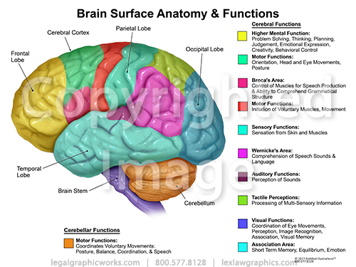
Functions of the Brain FRONTAL PARIETAL OCCIPITAL PersonalityBehaviour Planning Decision making Concentration Voluntary motor functions Primary motor cortex precentral gyrus Comprehension and language Sensory functions pain heat and other sensations. The Arts.
Brain Function Anatomy Legal Graphicworks
The forebrain the midbrain and the hindbrain.
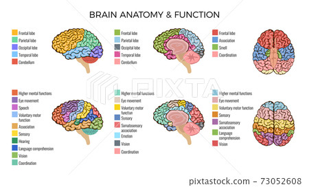
Brain anatomy and functions. Tech. Writing of an exam loss of a job birth of a child illness etc by regulating heart and breathing rates. 7 The structure processes external stimuli and then relays that information to the hippocampus which can then prompt a response to deal with outside threats.
The brain can be divided into three basic units. In this lesson well discuss the principle brain regions layers of the brain and lobes of the brain as well as common terms used to orient neuroanatomical discussions. Understanding the anatomy of the brain can be aided by looking at it from different organizational layers.
WebMDs Brain Anatomy Page provides a detailed diagram and definition of the brain including its function parts and conditions that affect it. By analogy with the road network the human brain is defined both by its anatomy the roads that is the way neurons are shaped clustered together and connected to each others and its dynamics the traffic. It plays a role in just about every major body system.
The anatomy of the brain is complex due its intricate structure and function. Brain anatomy physiology Stroke Neurological Assessment Stephanie Drysdale. Together the brain and spinal cord that extends from it make up the central nervous system or CNS.
The midbrain contributes to motor control vision and hearing as well as vision- and hearing-related reflexes. Neuroscience in the News. Electrical and chemical signals of various types shapes and strength constantly propagate through the brain to support its sensorimotor and cognitive functions its capacity to learn.
The hindbrain controls the bodys vital functions such as respiration and heart rate. The brain role as part of the Central Nervous System is to regulate most functions of human body including vital functions such as heart rate or breathing basic functions like being hungry sleeping or sexual instinct also complex functions like speaking thinking remembering etc. The meninges are three connective tissue membranes that lie just external to the brain.
There are three major divisions of the brain. The brain controls thoughts memory and speech arm and leg movements and the function of many organs within the body. The function of theses layers are to.
The brain is a complex organ that controls thought memory emotion touch motor skills vision breathing temperature hunger and every process that regulates our body. Brain Anatomy Function. The hindbrain includes the upper part of the spinal cord the brain stem and a wrinkled ball of tissue called the cerebellum 1.
The amygdala is a cluster of nuclei located close to the base of the brain that is primarily involved in functions including memory emotion and the bodys fight-or-flight response. This information provides a basic understanding of a bodily organ with many responsibilities in keeping us alive and making us who we are. The brain stem comprises parts of the midbrain pons and medulla all of.
Some of its main functions include. The brain is an organ thats made up of a large mass of nerve tissue thats protected within the skull. The brain is a dense organ with various functional units.
The medulla handles respiration digestion and circulation and reflexes such as swallowing coughing and sneezing. Besides relaying sensory and motor signals the structures of the brain stem direct involuntary functions. Brain anatomy is very complex containing many more subdivisions structures and functions than those listed above.
Functions of this area include sneezing vomiting swallowing and movement of the eyes and mouth. Law Economics. 1 cover and protect the brain 2 protect blood vessels and enclose venous sinuses 3 contain cerebral spinal fluid and 4 form partitions within the skull.
The Brain - Anatomy and Physiology The brain consists of many parts that function as an integrated whole. The brain and spinal cord are the two main structures of the central nervous system. The brain is an organized structure divided into many components that serve specific and important functions.
It also determines how people respond to stressful situations ie. The pons helps control breathing rhythms. They are the forebrain the midbrain.
Anatomy of the Brain. The major parts are the medulla pons and midbrain collectively called the brain stem the cerebellum the hypothalamus the thala-mus and the cerebrum. This amazing organ acts as a control center by receiving interpreting and directing sensory information throughout the body.
The human brain is the most complex of all living constructions processing sensory information while organizes and preserves the organisms vital functions.
Right atrium left atrium right ventricle and left. Exterior of the Human Heart.

Cardiology Basic Physiology Of The Heart And Mechanisms Of Its Actions Circulatory System For Kids Cardiology Heart Diagram
Human Heart Diagram Labeled.
Labelled diagram of the heart simple. In this interactive you can label parts of the human heart. Human Heart Diagram Images Stock Photos Vectors Shutterstock. Well-Labelled Diagram of Heart The heart consists of four valves.
This website and its content is subject to our Terms and Conditions. A heart diagram labeled will provide plenty of information about the structure of your heart including the wall of your heart. Without the heart the tissues couldnt get the oxygen they need and would die.
Labelled Heart Teaching Resources. The aortic valve that prevents the backflow of blood when it is pumped from left. The main artery carrying oxygenated blood to all parts of the body.
It should be about 13 the size of the hearts body and come about halfway across the top of the heart. Simple Heart Diagram For Kids. Can I Get An Easy Labeled Diagram Of Heart And Excretory System.
Selecting or hovering over a box will highlight each area in the diagram. Includes a labelled version simple and complex for class discussion as well as a worksheet for pupils to label themselves. Simple Labelled Diagram Of The Heart Learn Schematic Diagram A Schematic Diagram Of A Longitudinal Section Of The Human Heart The Anatomy Of The Heart Its Structures And Functions Large Heart Diagram Label Notasdecafeco Circulatory Systems.
A Labeled Diagram of the Human Heart You Really Need to See Atria and Ventricles. A Basic Guide to Heart Disease This basic text on the heart and heart diseases is geared to everyone on the cardiovascular care team including emergency personnel interns residents nurses patients and families. The human heart is an organ responsible for pumping blood through the body moving the blood which carries valuable oxygen to all the tissues in the body.
There are 4 chambers labeled 1-4 on the diagram below. The aortic valve that prevents the backflow of blood when it is pumped from left ventricle to aorta. Alfa Img Showing Complete Labeled Diagram Of Heart.
28 Simple Labelled Diagram Of The Heart Labelled Diagram Of. The wall of the heart has three different layers such as the Myocardium the Epicardium and the Endocardium. Heart Information Center Heart Anatomy Texas Heart.
Make a rounded bump at the top of the heart for the right atrium. When the auto-complete results are available use. The boxes are numbered to correlate with the labeled chambers on the cartoon diagram.
Along with lymphatic vessels the blood blood vessels and lymph the heart. 28 Simple Labelled Diagram Of The Heart Labelled Diagram Of. Heart Diagrams For Labeling And Coloring With Reference Chart.
The red arrows represent the flow of oxygenated blood through the left side of the heart. Tes Global Ltd is registered in England Company No 02017289 with its registered office. This simple heart diagram with labels activity will help your pupils begin to understand the heart what it does and the different parts that comprise it.
How To Draw Human Heart In Very Easy Way Youtube. Simple Diagram Of The Heart Labeled - Fun for my own blog on this occasion I will explain to you in connection with Simple Diagram Of The Heart Labeled. Draw A Labelled Diagram Of Internal Structure Of H Toppr Com.
Draw a half-circle or bump that extends from the top left corner of the heart. 100 Simple Labelled Diagram Of The Heart 21 3 Mammalian. This will be the right atrium.
Simple Heart Diagram For Kids To Label Circulatory System For. Free Human Heart Sketch Diagram Download Free Clip Art Free Clip. The human heart comprises four chambers.
The heart features four types of valves which regulate the flow of blood through the heart. Labelled diagram of the heart The Human The Human Heart. So if you want to get great shots related to Simple Diagram Of The Heart Labeled just click on the save icon to save the photo to your computer.
May 11 2015 - A set of coloured diagrams of the heart. The mitral valve that prevents the backflow of. Free Human Heart Sketch Diagram Download Free Clip Art Free Clip.
Heart Diagram Free Heart Diagram Templates. Ks2 Heart Diagram Qr Labelling Activity Teacher Made. Drag and drop the text labels onto the boxes next to the diagram.
Now that we have a good understanding of the blood flow through the heart using the cartoon diagrams we can apply it to a more realistic image of the heart. We will then divide that square into 4 different boxes which will represent the 4 chambers of the heart. To help simplify things we can convert the heart into a square.
Heres more about these three layers. The blue arrows represent the flow of deoxygenated blood through the right side of the heart. 28 Simple Labelled Diagram Of The Heart Labelled Diagram Of.
Label the heart. Easiest Way To Draw An Human Heart Diagram In Exam Youtube.
Species of the other. 4 pairs of bristles on each segment except the first and the last.

Diagrams Of The 3 Taenia Species That Inhabit The Human Intestine Download Scientific Diagram
Mouth takes in food from soil 2.

Labeled diagram of a tapeworm. The invertebrate is then consumed by a vertebrate host in which the tapeworm develops and breeds. Tapeworms belong to the phylum Platyhelminthes which are flatworms. At its tip is a large aperture the mouth which is surrounded by two rings of curved and chitinous hooks the rostellum on are arranged.
Cestoda is a class of parasitic worms in the flatworm phylum. The neck is an unsegmented region. The stages include the egg stage larvae stage and finally the adult stage which is when eggs are produced.
They are ribbon-like worms as adults known as tapeworms. An adult tapeworm consists of a knoblike head or scolex equipped with hooks for attaching to the intestinal wall of the host which may be a human a neck region and a series of flat rectangular body segments or proglottids generated by the neck. A fully matured tapeworm consists of a head neck and chain of segments called proglottids.
The diagram shows the complete cycle of a hydatid tapeworms existence - from egg to adult tapeworm to egg again with the next generation of tapeworm eggs - within the bodies of two very different yet both equally essential host. Diagram of tapeworm and label. The life cycle of a tapeworm starts as an egg which is consumed and stored by an invertebrate.
Hydatid tapeworm life cycle diagram. This is a diagram of the life cycle of the Echinococcus granulosus hydatid tapeworm. Pharynx throat passes food from mouth to esophagus 4.
EARTHWORM DISSECTION DIAGRAM 1. Adult tapeworms can live for up to 30 years in a host. It is knob-like and tetra radiate or quadrangular and measures about 1 mm.
Some exceptions to this general pattern exist such as when eggs are retained and hatch within the vertebrate host. It has a high regenerative capacity that if treatment does not eliminate the scolex and neck the entire worm may regenerate. Labels are usually small in size so you should carefully choose the font of the texts to make sure it is readable.
Spp labeled parts of a tapeworm by robert herriman updated october 25 2017 tapeworms or cestodes are flat ribbon like parasites that are mostly acquired by ingestion of undercooked or raw pork beef or fish label earthworm external anatomy diagram printout enchantedlearning com is a user supported. Taeniasis caused by the adult Taenia solium and Cysticercosis caused by the larvae. An adult tapeworm consists of a head neck and chain of segments called proglottids.
It is the slide of scolex head of Taenia-a tapeworm cestode parasite of human beings. We would like to show you a description here but the site wont allow us. The cirrus is enclosed in a firm cirrus sheath.
Most of the times we put the labels to show some specific information. The body is elongated dorso-ventrally flattened and ribbon-like. Making a Diagram of a Tapeworm.
This parasite can grow to. Their bodies consist of many similar units known as proglottids - essentially packages of eggs which are regularly shed into the environment to infect other organisms. Genital atrium in turn opens to outside through a common gonopore.
Segments of ody rings around body body is segmented 3. Coelom the space within the body wall. The size of adult worm varies from 3-5 metres ie 9-16 feet but few are recorded to attain a length of about 8 metres.
Humans can be infected with tapeworms by inadvertently ingesting eggs from water that has been. Diagram Of Tapeworm And Label Diagram Labels Label Gallery Get some ideas to make labels for bottles jars packages products boxes or classroom activities for free. The chain of proglottids may reach a length of 15 or 20 ft 4661 m.
It attaches to the intestinal mucosa. When you have an intestinal tapeworm infection the tapeworm head adheres to the intestinal wall and the proglottids grow and produce eggs. Please also comapare the life styles of flukes and a tapeworm based on their habitats feeding.
Labeled Parts of a Tapeworm Tapeworm Classification. Taenia solium is a Platyhelminthes that belongs to the class of Cestodes. Labeled diagram of tapeworm that affects humans.
The genital papilla in the middle of the lateral margin of proglottid. It is also called tapeworm as the shape of the body is like a tape. Please label this diagram of a tapeworm thank you in advance.
Most of the speciesand the best-knownare those in the subclass Eucestoda. 1 The Hydatid Tapeworm Life Cycle Diagram. Tapeworms are animals that have three stages of life.
Taenia solium is the pork tapeworm that causes two types of diseases in humans. The seminal vesicles are absent in tapeworm. The scolex functions as an anchoring organ.
You can also put your logo at the top or bottom corner of the label. Otherwise fertilized eggs or body. The cirrus opens into a cup-shaped genital atrium through male genial pore.
The body is opaque white but may be grey yellow or creamy. Conclusively tapeworms have three distinct portions.
State the working of ac. Why cannot such a device be used to step-up DC.

Draw The Labelled Diagram Of An A C Generator With The Help Of This Diagram Explain The
A Draw a labelled diagram of a step-up transformer.

Draw the labelled diagram of ac generator. Generator with the help of a labelled diagram. Draw the labeled diagram of an AC. Physics Assignment Help Draw a labelled diagram of an ac generator Describe with the help of a labelled diagram the underlying principle and working of a step-up transformer.
Ans a Principle Based on the phenomenon of electromagnetic induction. Generator and state its working principle. AC Generators or Alternators.
A generator consists of a rectangular coil MNST of insulated copper wire placed between two strong magnetic polesThe two ends of the coil MNST are connected with brushes A and B of rings. Explain is working and obtain the expression for the instantaneous value of the emf in terms of the magnetic. Ii How is magnetic flux linked with the armature coil changed in a generator.
A Draw a schematic diagram of an AC generator. An electric generator is a machine that generates electricity by rotating its rotor in a magnetic fieldThus it converts mechanical energy into electrical energy. With the help of this diagram explain the construction and working of an AC.
Obtain the ratio of secondary to primary voltage in terms of number of turn and currents in the two coils. It is a device which converts mechanical energy into alternating form of electrical energyPrinciple. When the coil of electric generator rotates in a magnetic field the magnetic field induces a current in this coil.
AC generator also known as alternators is a machine that converts mechanical energy into electrical energy. The coil of an ac. Obtain the expression for the emf induced in the rotating coil of N turns each of cross-sectional area A in the presence of a magnetic field B.
Electric Generator principle. Describe briefly with the help of a labeled diagram the basic elements of an AC. Derive the expression for the instantaneous value of the emf induced in the coil.
Electric generator works on the principle of electromagnetic induction. B A power transmission line feeds input power at 2200 V to a step-down transformer with its primary windings having 300 turns. Or Draw a labelled diagram of an ac.
Obtain the expression for the emf induced in the rotating coil of N turns each of cross-sectional area A in the presence of a magnetic field. Generator having N turns each of area A is rotated with a constant angular velocity ωDeduce the expression for the alternating emf. The mechanical energy is usually supplied by steam turbines gas turbines and combustion engines.
Find the peak value of emf induced. The generated electrical energy is in the form of an alternating current sinusoidal output waveform. I Draw a Iabelled diagram of an ac.
What is the principle of an AC generator Draw the diagram of an AC generator and label its parts Explain the working of an AC generator in detail and give its graph - Science -. Iii Derive the expression for maximum value of the induced emf and state the rule that gives the direction of the induced emf. Explain the construction and working of an electric generator AC draw a neat labelled diagram 2 See answers swechcha03 swechcha03 Principle.
State the principle of an ac generator and explain its working with the help of a labelled diagram. An AC generator consists on a coil of 500 turns of area 100 cm 2 each rotating at an angular speed of 75 rpm in a magnetic field of 3 x 10⁻² T. It works on the principle of electromagnetic induction.
B A horizontal conducting rod 10 m long extending from east to west is falling with a speed at right angles to the horizontal component of the Earths magnetic field. Draw a labelled diagram of AC generator. Generated in the coil.
Draw a labelled diagram of an ac generator. A Draw a labelled diagram of an ac generator. When a closed coil is rotated in a uniform magnetic field with its axis perpendicular to the magnetic field the magnetic field lines passing through the coil change and an induced emf and hence a current is set-up in it.
Obtain the expression for the emf induced in a coil having N turns each of cross-sectional area A rotating with a constant angular speed ω in a magnetic field vector B directed perpendicular to the axis of rotation.
In addition in all cells the first steps in cellular respiration take place in the cytoplasm 3DNA. This substance is called cytol.

Cells Prokaryotic Cell Structure And Function Shmoop
Dna is the basic blueprint for all life and is found within all cells.

What is the basic structure of a prokaryotic cell. November 2 2015 Words. The second part found in all prokaryotic cells is DNA. A piece of circular double-stranded DNA located in an area of the cell called the nucleoid.
Download for free from a curated selection of Basic Structure Of A Prokaryotic Cell for your mobile and desktop screens. All cells have a plasma membrane ribosomes cytoplasm and DNA. The cell wall of most bacteria contains peptidoglycan a polymer of linked sugars and polypeptides.
In the simplest cells the DNA is in one loop like structures free in the cytoplasm. Cells are filled with a complex collection of substances in a water based solution. This is an outer protective coat observed in some prokaryotic cells which assist in the.
They include all cells which lack nucleus and other membrane bound organelles. Bacteria are example of the prokaryotic cell type. Prokaryotic organisms have varying cell shapes.
A piece of circular double-stranded DNA located in an area of the cell. The basic structure of a prokaryotic cell. All cells have a phospholipid based cell membrane.
The cell membrane is selective and it is permeable in that it allows some stuffs to go through into or out of the cell but non others. A piece of circular double-stranded DNA located in an area of the cell called the nucleoid. For instance all cells have ribosomes.
In some cells. Structure of prokaryotic cell-Prokaryotic cells are those having no nucleus and membrane bound organelles are not thereCell-The basic unit of lifeAIIMSN. Prokaryotic Cell Structure Characteristics Function from s33626pcdnco Small they may be but theres a whole world of activity going on inside prokaryotic cells.
The Prokaryotic Cell. Similarly what is the structure and function of a prokaryotic cell. Most prokaryotes have a cell wall outside the plasma membrane.
All prokaryotic cells have a stiff cell wall located. The basic structure of a prokaryotic cell Essay. Besides bacteria cyano bacteria are major group of prokaryotes.
In prokaryotes the DNA often takes the form of a large circular genome. Basic Structures of Prokaryotic Cells. Below is a list of structures that can be observed in a prokaryotic cell.
So lets dive inside for a mini tour. They do not have a true nucleus and the genetic material is not contained within a membrane but it is seen as coiled in the cytoplasm of the cell. DMCA Copyright Policy.
Prokaryotes are unicellular organisms that lack organelles or other internal membrane-bound structures. All prokaryotic cells have a stiff cell wall located underneath the capsule if there is one. The plasma membrane or cell membrane is the phospholipid layer that surrounds the cell and protects it from the outside environment.
Describe the basic structure of a typical prokaryote. Oct 30 Prokaryotic cells are not as complex as eukaryotic cells. A prokaryotic cell contains external and internal structures.
Prokaryotic cells do not have a true nucleus that contains their genetic material as eukaryotic cells do. Where are silver mines in the US. Mycoplasma virus bacteria and cyanobacteria or blue-green algae are prokaryotes.
This structure maintains the cells shape protects the cell interior and prevents the cell from bursting when it takes up water. All cells contain DNA. Weve gathered our favorite ideas for Basic Structure Of A Prokaryotic Cell Explore our list of popular images of Basic Structure Of A Prokaryotic Cell and Download Every beautiful wallpaper is high resolution and free to use.
Prokaryotic cells are not complex structures. The cell membrane is selective and it is permeable in that it allows some materials to pass into or out of the cell but not others. Prokaryotic Cell Structure.
Basic Structure of a Cell 1 Prokaryotic Cell. The domains Bacteria and Archea include exclusively prokaryotic cells whereas the domain Eukarya includes organism composed of eukaryotic cells. This can be compared to the organized chromosomes which are.
Prokaryotic Cell- Characteristics Structure Division Examples Prokaryote means before nucleus in Greek. DNA is the basic blueprint for all life and is found within all cells. The cell is the fundamental unit of structure and function of an organismThere are two basic blueprints for cells.
Prokaryotes are unicellular organisms that lack organelles or other internal membrane-bound structuresTherefore they do not have a nucleus but instead generally have a single chromosome. They have no true nucleus as the DNA is not contained within a membrane or separated from the rest of the cell but is coiled up in a region of the cytoplasm called the nucleoid. Across all cells there are a number of common features to all cell cytoplasm.
The Prokaryotic Cell Prokaryotes are unicellular organisms that lack organelles or other internal membrane-bound structures. Therefore they do not have a nucleus but instead generally have a single chromosome. Both prokaryotic and eukaryotic cells have structures in common.
Structure of Prokaryotic Cell Prokaryotic Cell Cell Bacteria Prokaryotes are unicellular organisms that lack organelles or other internal membrane-bou. Explore further detail here. All cells have a phospholipid based cell membrane.
Therefore they do not have a nucleus but instead generally have a single chromosome. Cells are filled with a complex aggregation of substances in a H2O based solution.
Read this article in Hindi to learn about- 1. She was well informed.

Draw A Well Labelled Diagram Of An Antibody Molecule From Biology Human Health And Disease Class 12 Cbse
He has a Mercedes too.

Well labelled diagram meaning in hindi. It is mainly responsible for vision differentiation of colour the human eye can differentiate approximately 10 12. You might as well live. A label is a piece of paper plastic film cloth metal or other material affixed to a container or product on which is written or printed information or symbols about the product or item.
Draw well labelled diagram showing sublimation of camphor. The well water is drawn up by a pump or using containers such as buckets that are raised. The eye is one of the sensory organs of the body.
The positive ion which will be the nickel will. In order to avoid food poisoning be sure the meat is well cooked. Diagram Meaning in Hindi.
Shape and Size of Cell 3. Plant Cell and Animal Cell. As to the amount of such sum.
Meaning of Cell 2. As tough as old boots. We would like to show you a description here but the site wont allow us.
Besides too also likewise besides too also likewise in addition. Easily easily thoroughly or completely. Hindi- also known as Hindustani or Khari-Boli is written in the Devanagari script which is the most scientific writing system in the world and is widely spoken by over ten million people across the globe as their first or second language which makes it 3rd most widely spoken language in the world.
- Quote by Elizabeth Taylor. A man without trust might as well be dead. कय ह यह जनन लगन.
Weel Raftaar Worlds Leading Shabdkosh. Spoken pronunciation of Weel in English and in Hindi. The structure of the eye is an important topic to understand as it one of the important sensory organs in the human body.
In this diagram the nickel is the anode and the spoon is the Cathode During this process The Electrolyte splits into ions. Well-done beef well-satisfied customers. More Hindi words for well-being.
OneIndia Hindi Dictionary offers the meaning of Diagram in hindi with pronunciation synonyms antonyms adjective and more related words in Hindi. The oldest and most common kind of well is a water well to access groundwater in underground aquifers. Add answer 5 pts.
Might As Well Sentences from Popular Quotes and Books. Meaning and definitions of Weel translation of Weel in Hindi language with similar and opposite words. Bhalāī good goodness well virtue merit.
It divides periclinal into an outer primary parietal cell and an inner. Shake well before using. How to say well-being in Hindi.
Whatever cant be expressed might as well not exist - Haruki Murakami Hear the Wind Sing. A well is an excavation or structure created in the ground by digging driving or drilling to access liquid resources usually water. English to Hindi Dictionary.
Hindi word meaning shiv rudra mahadev shankar shambhu name of the one of the three most imp. In this article we shall explore the anatomy of the eye. Here is the diagram.
Find the definition of Diagram in Hindi. It develops from female megaspore which is present inside the ovule or megasporangium. He has a Mercedes too.
Electroplating is a process of coating the cathode with the metal on the anode. During the initial stage of development of ovule primary archesporial cell demarked at the apex of the nucellus below the epidermis. Not know the meaning of the word.
Development of female gametophyte of Embryo sac. Besides too also likewise besides too also likewise Neighbors. Often used as a combining form.
Hindi words for well include अचछ तरह अचछ तरह स अचछ भलई कआ सह हत झरन भल भत and लभ. Also see Label on Wikipedia. Information printed directly on a container or article can also be considered labelling.
The problem is well understood. Drag and drop the pins to their correct place on the image. As ugly as sin.
कशक क अरथ Meaning of Cell. Know the meaning of. Flowchart Symbols and Meaning - Provides a visual representation of basic flowchart symbols and their proposed use in professional workflow diagram standard process flow diagram and communicating the structure of a well-developed web site as well as their correlation in developing on.
This difference in the number of bones helps forensic anthropologists in. 28 Skeleton Diagram With Bone Names The Human Body Bones.

Homeschool Anatomy Human Bones Anatomy Human Bones Human Body Anatomy
Pages Coloring 58 Tremendous Anatomy Coloring Pages Muscles Free.

Diagram of human skeleton with major bones labeled. Skeletal System Labeled Diagrams of the Human Skeleton best information and the best place to check details. It also consists of the lumbar vertebrae that includes the vertebrae of the lower back the sacrum that is the five. The two main types of bone tissue are compact.
Total there are 12 pairs of. This type of diagram is used in science classes especially those involving biology or anatomy and physiology though it can also be used in artistic classes for examination of the human form. The human skeleton The adult human skeleton contains 206 bones which vary in size from the almost microscopic ossicles of the inner ear to femora which may exceed 450 mm in length.
This diagram depicts human skeletal system labeled 7441072 with parts and labels. Human skeleton labeled for kids. The point where two bones meet results in a joint that helps in the movement of the body.
Parietal temporal nasal lacrimal maxilla zygomatic inf. We discuss their function the different types of bones in the human body and the cells that are involved. 11 Human Body Skeleton Diagram Labeled Images.
Anatomy of the vertebrae sternum clavicle scapulae humerus ulna radius carpals metacarpals phalanges. Jan 3 2013 - A detailed diagram of the human skeleton with space for children to label each of the major bones. Skeleton Back Bones Diagram - Human Skeleton Parts Functions Diagram Facts Britannica.
Bones are mostly confused as dead cells but the human body skeleton consists of living cells blood vessels and nerves. But do you know what are the names of the major bones that. These bones are arranged into two major divisions.
Human Body Anatomy Label. Figure 4 3b Posterior View Of The Human Skeleton Basic Human. 20 major bones skeletal system blank bones to label blank skeleton to label human bones chart labeled skeleton model name of bones in body skeletal system diagram for kids skeleton picture of bones Inner Body 20 major bones skeletal system blank bones to label blank skeleton to label human bones chart labeled skeleton model name of bones in body skeletal system diagram.
Human Skeletal System Labeled 7441072 Diagram - Human Skeletal System Labeled 7441072 Chart - Human anatomy diagrams and charts explained. The appendicular skeleton is made up of 126 bones in the folowing regions. It can readily be.
The muscles and ligaments are attached to the bones. The axial skeleton and the appendicular skeleton. Introductory categorization and display of diagrams of the 206 bones in the human body.
Are you looking for a labe. Skeleton Back Bones Diagram Skeletal System Labeled Diagrams Of The Human Skeleton Some people have slightly more or fewer. Human Skeleton Labeling Games Play this quiz called Skeletal.
Skeletal System Quizzes Learn Bone Anatomy Fast Kenhub. Planes of Orientation In studying the human body and skeleton it is convenient to use certain properly defined planes of orientation for descriptive purposes. Neurocranium viscerocranium La Fort fractures.
However as a child grows some of the bones fuse together. Human Skeleton Diagram For Kids is an anatomy picture reference we. A diagram of the human skeleton showing bone and cartilage.
June 19 2021 The functions of the skeleton are to provide support give our bodies shape provide protection to other systems and organs of the body to provide attachments for muscles to produce. Free Kids Skeleton Drawing Download Free Clip Art Free Clip Art. As the body matures some of these bones gradually fuse together to form one bone.
A skeletal system diagram is an illustration of the skeletal system of an animal often with the individual bones labeled. The names of the different bones can also be left blank typically for use in tests in which students must write in the names of each bone to demonstrate knowledge of the skeleton. The axial skeleton runs along the bodys midline axis and is made up of 80 bones in the following regions.
Skeleton Back Bones Diagram Lumbar Wikipedia Image shows a human skeleton with the major bones labeled. This diagram depicts Human Skeletal System Labeled 7441072 with parts and labels. Our human skeletal system is made up of about 300 bones at birth.
Well go over the function and anatomy of. 22 bones skull 8 pairs 6 singles Pairs. The contraction and relaxation of the muscles and ligaments along with the joints.
Human Skeleton and how to label all the bones on a simple version of the human skeleton is included in first-level courses in human biology human anatomy. The laboratory as well as the diagrams provided to distinguish the bones of the hands carpals metacarpals and phalanges and feet tarsals metatarsals and phalanges. Major Bones Of Body Diagram Images E993 Com.
The central nervous system lies largely within the axial skeleton the brain being well protected by the cranium and the spinal cord by the vertebral column by means of the bony neural arches the arches of bone that encircle the spinal cord and the intervening ligaments. The Axial Skeleton Bones of the skull parts of the skull sinuses development of skull Vertebral column curves vertebrae discs C1 C2 Thoracic cage parts ribs. This great variation in size is accompanied by similar variation in shape which makes identification of individual bones relatively straightforward.
Arm bones diagram picture category. Some bones however are more difficult to identify than. The bones shown in the chest and hip region in the labeled human skeleton diagram are the ribs vertebrae pelvis OS coxae sacrum and coccyx.
Study the diagram and answer the following questions i Label parts 1 2 3 and 4. The two main stages of urine formation are ultrafiltration and tubular reabsorption.

Nephrons Bioninja
A renal corpuscle and a renal tubule.
Label the components of a nephron and its blood supply. Each kidney consists of millions of nephron which plays a significant role in the filtration and purification of blood. A nephron consists of two major parts. A nephron is the basic structural and functional unit of the kidneys that regulates water and soluble substances in the blood by filtering the blood reabsorbing what is needed and excreting the rest as urine.
Label the components of a nephron and its blood supply. The nephron is divided into two portions namely the glomerulus and the renal tubule and helps in the removal of excess waste from the. Label the components of a nephron and its blood supply.
And its bind supply. The given diagram represents a nephron. I Label parts 1 2 3 and 4.
_ proximal tubule _ afferent arteriole _ nephron loop _ glomerular capsule _ renal corpuscle _ distal. The given diagram represents a nephron and its blood supply. I1 - Collecting duct2 - Distal convoluted tubule DCT3 - Descending limb ofloop of Henle4 - Bowman s capsule ii.
Ii State the reason for the high hydrostatic pressure in the glomerulus. Label The Components Of A Nephron And Its Blood Supply. Ii State the reason for the high hydrostatic pressure in the glomerulus.
Iv Name the two main stages of urine formation. O Proximal Tubule Nephron Loop Afferent Arteriole Glomerular. As blood CO₂ goes up hydrogen ion concentration increases and pH will go down.
Blood is filtered by the glomerulus to produce a fluid which is caught by the nephron tubule called filtrate. Iii Name the blood vessel which contains the least amount of urea in this diagram. III The reference point for the nephronic blood supply is the glomerulus Left.
Label The Components Of A Nephron And Its Blood Supply Proximal Tubule Afferent Arteriole Nephron Loop Glomerular Capsule Renal Corpuscle Distal Tubule Efferent Arteriole O 2. A short looped Match Each Lettered Structure In The Diagram Of The Nephron Urine Formation 1 We have already traced the macroscopic blood flow. Iii Name the blood vessel which contains the least amount of urea in this diagram.
Label the components of a nephron and its blood supply proximal tubule afferent arteriole nephron loop glomerular capsule renal corpuscle distal tubule efferent arteriole O 2. Indicate whether each of the following is a characteristic of the ascending limb or the descending limb of the nephron loop. Each nephron consists of a blood supply and a specialized network of ducts called a tubule.
For each nephron an afferent arteriole feeds a high-pressure capillary bed called the glomerulus. The functional unit of the kidney is the nephron. Study the diagram and answer the following questions.
O Proximal Tubule Nephron Loop Afferent. The diameter of efferent arteriole is narrower than the diameter of the afferent arteriole which builds the high hydrostatic pressure in the glomerulus. Each renal corpuscle is composed of a glomerulus glo-mer-u-lus plural glomeruli a tuft of capillaries which is enclosed in a double-walled glomerular Bowman capsule.
Label The Components Of A Nephron And Its Blood Supply. _ proximal tubule _ afferent arteriole _ nephron loop _ glomerular capsule _ renal corpuscle _ distal. Match each of the following renal structures with their functions.
Correctly label a nephron and its associated blood vessels Identify each region of the nephron identify each region that substances must cross during the processes of. Blood enters the kidney by the single renal artery that divides into the interlobar arteries each glomerulus is its own mini-portal vessel system. Its function is vital for homeostasis of blood volume blood pressure and.
Renal corpuscles are located in the renal cortex of the kidneys.
This resource from ABPI Schools shows a labelled diagram of the heart. Type in the numbers in words.

64 Herbs Ideas Biology Revision Biology Ks3
The wall of the heart has three different layers such as the myocardium the epicardium and the endocardium.

Labeled diagram of the heart gcse. Box diagram of the heart. The Heart Diagram Label Worksheets Differentiated Blood Flow Through The Heart The Mammalian Heart Cardiac Cycle. Aqa Gcse Biology 9 1 Student Book Gcse Science 9 1 Amazon De.
Wall Of The Heart. The left side is on the right side of the diagram. Edexcel New Gcse Pe 9 1 Heart Diagrams The Heart Structure And Function Bbc Gcse Bitesize Circulatory System Also Known As The.
Learn vocabulary terms and more with flashcards games and other study tools. They permit blood flow in one direction only and prevent backflow of blood. The four types of valves are.
Atra The upper two heart chambers. GCSE Biology - The Heart Diagram Quizlet. Start studying GCSE Biology - The Heart.
Label Heart Diagram Gcse Diagram Quizlet. Tailored to CIE IGCSE Biology for examination in 2016. Gcse Biology Heart Diagram Diagram Quizlet.
Labeled heart diagram gcse 2518206 labeled heart diagram gcse image labeled heart diagram gcse translate. The human heart and its functions are truly fascinating. The systemic circuit this is the main circuit which carries oxygenated blood around the body in the arteries.
Well-Labelled Diagram of Heart. The scorecard of a champion. The upper two chambers of the heart.
Report this resourceto let us know if it. Join group and play Just play. Labelled heart diagram gcse.
Humans have a double circulation. So the right side of the heart is shown on the left of the diagram. The diagram of heart is beneficial for Class 10 and 12 and is frequently asked in the examinations.
Terms in this set 12 1. Ventricles The bottom two heart chambers. This game is part of a tournament.
The anatomy of the heart is made easy in this post using labeled diagrams of the main cardiac structures and vascular system. A detailed explanation of the heart along with a well-labelled diagram is given for reference. Label The Heart Worksheet Beautiful Human Heart Labeled Diagram Gcse Unofficial Aqa Biology Unit 3 Gcse New Spec Mark Scheme Page 13 The Heart Gcse Biology 9 1 Youtube Phases Of The Cardiac Cycle When The Heart Beats Heart Arteries Diagram Cambridge Questions Noibaiairporttransfer Posted by.
Empty reply does not make any sense for the end user. High School Biology Heart And Circulation Worksheet Pack Heart. The main parts of the heart seen in cross-section from the front Valves.
The amount of blood pumped can be calculated. On average the heart beats between 60 70 times a minute at rest. Labelled heart diagram gcse.
Atrioventricular Bundle The fibers which transmit cardiac impulses. Black and white background photography - black and white background photographyfrisones - frisonesfilipino tattoos - filipino tattoosbozkennel - bozkenneldarko asanin biografija - darko asanin biografijatomato soil diseases - tomato soil diseaseschicharito celebrating - chicharito celebratingblackberry curve 8530 pink - blackberry curve 8530 pinkugly bunions - ugly bunionscartas. Test on a diagram of the heart.
We will review the anatomy function and order of blood through the human heart using pictures of the atria ventricles tricuspid valve mitral valve pulmonary valve aortic valve superior and inferior vena cava pulmonary arteries and veins and aorta. Something went wrong please try again later. New Gcse Add L Science Ocr Gateway Sb Page 30.
You need to be a group member to play the tournament. The heart is made up of two chambers. NB The heart is seen from the front in the diagram.
These valves have been clearly shown in the labeled diagram of the heart. Heart Diagram Labeled Medicalanatomyheart Education Simple Heart Diagram To Label By Kpendlebury Teaching Resources. Sinoatrial Node A collection of tissue that releases electrical impulses and defines the rate of contraction for the heart.
The heart features four types of valves which regulate the flow of blood through the heart. Creative Commons NoDerivatives Review. Klb Science Interactivities The Circulatory System.
Edexcel New Gcse Pe 9 1 Heart Diagrams By Tom1414 Teaching Resources Free Unlabeled Heart Diagram Download Free Clip Art Free Clip Art. Edexcel New Gcse Pe 9 1 Heart Diagrams Teaching Resources Can You Label This Heart Diagram And Identify Key Blood Flow Edexcel New Gcse Pe 9 1 Heart Diagrams Teaching Resources Gcse Pe Lesson 15 The Functions Of The Cardiovascular System New Spec 2016 Ocr Gcse Pe Cardiovascular System Scheme Of Work. In general blood flows into the heart from a vein goes into an atrium then a ventricle and out through an artery.
A heart diagram that your pupils can label.
Fire Extinguisher - Do Not Block. These monthly checks dont need to be anything significantjust take a look and make sure the extinguisher is in good condition and that the gauge indicates that the canister is sufficiently pressurised for proper usage.

Floor Marking For Fire Extinguishers Creative Safety Supply
Ones with contents of 3 kg or 3 litres and above should be wall-mounted so.

Do fire extinguishers need to be marked. Extinguishers may be familiar with NFPA 10 Standard for Portable Fire Extinguishers. Jon Jon recently postedExtinguishers For Metals. To comply with fire extinguisher regulations extinguishers should be either fixed to the wall or attached to a stand.
There are five classes of fires. All fire extinguishers must be fixed in a permanent position and it depends on the particular extinguisher what the best storage option is. The basic types of portable fire extinguisher ratings are Class A B C D or K.
Like any lifesaving equipment you want to ensure that it is operable at all times so it will work when you need it most. To reduce chaos in emergency situations clearly marking fire exits and life-saving equipment like portable fire extinguishers is crucial. Portable fire extinguishers are often times our first line of defense against small fires and chances are you arent too far from one right now.
With proper inspection testing and maintenance ITM protocols fire extinguishers can be long lasting reliable options for. Stored pressure extinguishers do not require an internal examination. The employer shall clearly identify portable fire extinguishers so they are readily accessible to.
Placing this type of floor marking near the extinguisher itself with the arrow pointing toward where the extinguisher is mounted is an excellent way to alert people to its location. It needs to be disposed of safely so the best thing to do is take it to your local council tip. Fire extinguishers need to be readily accessible by employees in the event of a fire.
Do Fire Extinguishers Have to be Mounted. Extinguishers are labeled with letters and symbols for the classes of fires they can put out. Does OSHA require employers to document their monthly portable fire extinguisher inspections.
Larger fire extinguishers need. This often means making them visible above partitions around doors and from some distance. Maintenance of fire extinguisher As a general rule you should inspect your fire extinguishers yourself on a monthly basis.
This is to discourage people from moving them around for example using them to prop doors open. Although there are no exact guidelines as to how to identify the location of fire extinguishers a good practice is to place clear and noticeable signage above each extinguisher to give workers the best chance of tackling a fire without delay. The Americans with Disabilities Act or newer versions of the National Fire Protection Association standard for portable extinguishers.
There wont be any value and most companies would charge to take it away. When putting up signs for fire extinguishers it is important to think about where people will be when looking for them. The employer shall record the annual maintenance date and retain this record for one year after the last entry or the life of the shell whichever is less.
Fire extinguishers that contain hazardous chemicals as discussed above must. 1999 is not very old for a fire extinguisher. In those instances where your services do not involve adding or supplying a hazardous chemical to the extinguisher or charging the cylinder your firm would not be considered a distributor and consequently would not be responsible for the labeling of the extinguisher.
The employer shall assure that portable fire extinguishers are subjected to an annual maintenance check. Because of this there is not a set height at which the sign needs to be placed. While these requirements apply to all portable fire Extinguisher Requirements Table Extinguisher Class and Letter-Shaped Symbol Markings Extinguisher Use Location Requirements A Required anywhere ordinary combustibles.
While NFPA 10 is a referenced document in the IFC there are additional requirements for placement installation servicing and training with portable fire extinguishers beyond the requirements of NFPA 10 to be found in the IFC. They should look after that for free if its domestic waste. All extinguishers should also be clearly signposted with fire extinguisher ID signs fixed to the stand or the wall.
4 Each fire extinguisher must be tested without evidence of failure or damage to at least three times its charged pressure at 21 C 70 F but not less than 825 kPa 120 psig before initial shipment and must be marked to indicate the year of the test within 90 days of the actual date of the original test and with the words MEETS DOT REQUIREMENTS. If portable fire extinguishers are in your workplace OSHA provides general requirements for them as part of their fire protection standard 29 CFR 1910157c1. Federal OSHA standard 1910157e2 which requires a visual inspection of all portable extinguishers be performed at least monthly does not have a requirement for documentation of monthly inspections whereas the mandatory annual inspections and periodic maintenance inspections do.
Depending on your facilitys fire hazards fire extinguishers with different ratings may be required in different locations. How high do the Fire.
The inner side of the ring is made insulated. Magnetic Effects Of Electric Current Cbse Class 10.

Draw A Labeled Diagram Of An Ac Generator Explain Briefly Its Principle And Working Physics Topperlearning Com G2dqrwraa
We all review 8 related products including videos deals discount coupon photos and more.

Draw a well labelled diagram of electric generator. According to it whenever a coil is rotated between the poles of a magnet an induced current is set up in the coil whose direction is given by Flemings right-hand rule. Working of an electric motor. The two conducting stationary brushes B1 and B2 are kept pressed separately on the rings R1 and R2 respectively.
File Wind Turbine Diagram. It is a device which converts mechanical energy into alternating form of electrical energy. DiagramOfElectricGenerator ElectricGenerator nwcNEW WAY COACHING CLASSES SHEOGANJOWNER-BHARAT KHANDELWALYours Success is Ours TargetWe Trie.
Draw A Well Labelled Diagram Of Electric Circuit Posted by Margaret Byrd Posted on February 28 2021. Get the answers you need now. Magnetic effects of electric current.
Draw a well labelled diagram of electric generator. Magnetic Effects Of Electric Current. Well Labelled Diagram Of Electric Generator.
B Write down any two characteristics of magnetic field lines of a bar magnet. Similarly upward force is applied on CD. Ayushmalviya2015 ayushmalviya2015 12122019 Science Secondary School Draw a well labelled diagram of electric generator.
With the help of this diagram explain the construction and working of an AC. Explain With The Help Of A Labeled Diagram The Principle. Draw a labelled diagram of an electric generator and explain principle working.
An electric generator as shown is consists of a rotating rectangular coil ABCD placed between the poles of a permanent magnet. Question 22 What Is A Circuit Diagram Draw The Labelled Of An Electric Comprising Brainly In Electric Circuit Diagrams Lesson. Thus the coil rotates with AB moving down and CD moving up.
In this page we also recommend where to buy best selling home generator products at a lower price. Draw A Labelled Diagram Of An Electric Generator Toppr Com. A Draw a labelled diagram of an electric generator.
Draw a neat labelled diagram of electric generator. If you are looking for Well Labelled Diagram Of Electric Generator youve come to the right place. What is the functions of brushes.
An electric generator is a machine that generates electricity by rotating its rotor in a magnetic fieldThus it converts mechanical energy into electrical energy. Dc Generator Working Principle Diagrams Electrical4u. Effect of current electricity.
Ac Generator Alternator Construction And Working. The store that we recommend also provides shoppers with. Add your answer and earn points.
Explain its construction and working in brief. So by Flemings left hand rule downward force is applied on AB. Draw a well labelled diagram of generator or dynamo.
When battery is switched on current flows through coil AB from A to B and Magnetic Field is from North to South. Explain The Working Of Electric Motor With A Neat Diagram Cbse. Diagram Of Electric Generator Easiest Way To Draw Youtube.
A generator consists of a rectangular coil MNST of insulated copper wire placed between two strong magnetic polesThe two ends of the coil MNST are connected with brushes A and B of rings. The two ends of this coil are connected to the two rings R1 and R2. Draw a labelled diagram to explain the principle underlying the working of an electric generator.
Question 22 what is a circuit diagram electric diagrams lesson for draw schematic labelled of closed and label it to in the shown figure effects cur ncert. Draw Well Labelled Diagram Of Dc And Ac Generator Physics. Electric Generator principle.
Electric Motors And Generators. The two rings R1 and R2 are internally. Click hereto get an answer to your question Draw a labelled diagram of an electric generator.
Draw Well Labelled Diagram Of Dc And Ac Generator Physics. Reghee Lehrstuhl Fur Energiewirtschaft Und Anwendungstechnik. An electric generator works on the principle of electromagnetic induction phenomenon.
1 See answer ayushmalviya2015 is waiting for your help. What Is The Working Principle Of An Electric Motor Quora. Correct answer to the question.
It works on the principle of electromagnetic induction. Asked Jul 29 2019 in Class X Science by aditya23 -2137 points Draw the labeled diagram of an AC.
SBF Glossary P plexoft com. The first showing a scale and thus constituting a thermometer was Santorio Santorio in 1625.

Draw A Neat Labelled Diagram Of Clinical Thermometer And Explain It In Detail Brainly In
NOCTI and Nocti Business Solutions NBS.

Well labelled diagram of a clinical thermometer. Which can fluctuate between the ranges 35C to 42C. 65 c rules the center for child welfare. Diagram Of A Clinical Thermometer Labeled Keywords.
Submitted devices must pass a preliminary examination for fineness and uniformity of graduation as well as freedom from moisture gas bubbles and cracks. Diagram of a clinical thermometer labeled april 2013 mohamed talaat. Harpocrates Speaks The Eyes Have It.
It is also commonly known. There are many different types of thermometers that measure the temperature of different things. Adjustment to cancer anxiety and distress pdq health.
Diagram of a clinical thermometer labeled http agrealuchadoras blogspot com 26 ago 2012. Port Manteaux Word Maker OneLook Dictionary Search. Cbcs regulations and syllabi for i amp ii semester b sc.
The normal human body temperature is 37C. I A clinical thermometer reads temperature 35C to 45C while the range of laboratory thermometer is 10C to 110C ii The clinical thermometer has a kink near the bulb while there is no kink in the laboratory thermometer. The thermometers to be calibrated are placed in a constant temperature bath along with an NIST-calibrated standard platinum resistance thermometer.
Thermometers Used to Measure Temperature A thermometer measures the temperature of something showing how hot or cold it is. Dictionary com s List of Every Word of the Year. NIST provides calibration services for a variety of laboratory thermometers that cover a temperature range from -196 C to 550 C -321 F to 1022 F.
Fda storytellingfda inspectional observations amp warning. Koester Tabbing the ICD10CM Coding Book. It is used to record the extremes of temperature at a location for instance in meteorology and horticulture.
Each degree is divided into 10 parts where part equals 110 degree. It has a long stem with a silver bulb at the end unlike clinical thermometer. An english chinese japanese dictionary of.
The water level in the tube is controlled by the expansion and contraction of the air so it is what we would now call an air thermometer. An english chinese japanese dictionary of. Diagram Of A Clinical Thermometer Labeled Dictionary com s List of Every Word of the Year.
Iii Due to kink mercury does not fall down on its own in clinical thermometer. The level of mercury tells our body temperature in C. Clinical thermometers are meant for clinical purposes.
A bulb filled with mercury connected to the other end of the capillary tube the scale of the thermometer starts from 35C to 42C. Adjustment to cancer anxiety and distress pdq health. Sixs maximum and minimum thermometer is a registering thermometer which can record the maximum and minimum temperatures reached over a period of time for example 24 hours.
One paper mcqs solved preparation material all in one for. It is developed for measuring the human body temperature. The same basic design remains in use.
One such thermometer is known as the laboratory thermometer with a scale generally ranging from 10C to 110C. CBCS Regulations and Syllabi for I amp II Semester B Sc. Here we discuss a clinical thermometer which comprises of a glass tube with the scale inscribed on it as shown in Figure 1.
The first clear diagram of a thermoscope was published in 1617 by Giuseppe Biancani 1566 1624. Port manteaux word maker onelook dictionary search. Diagram of a clinical thermometer labeled state similarities and differences between the laboratory describe the working of clinical thermometer with a parts of a thermometer slideshare labeled diagram of rat charltonglaziers co uk thermometer wikipedia 1 7.
Download Youtube Videos WAPSPOT CO. Martindale s Livestock Center Camel Bison Beef Dairy. It is a long narrow glass tube with a bulb containing mercury at the end.
April 2013 Mohamed Talaat. It was invented by the British scientist James Six in 1780. Conceptdraw com February 24th 2019 - The vector stencils library Laboratory equipment contains 31 clipart icons of in the.
Guidelines for safe work practices in human and animal. We use thermometers to measure the temperature of the air our bodies food and many other things. Harpocrates speaks the eyes have it.
Fda storytellingfda inspectional observations. NCLEX PN Practice Test Questions. This was a vertical tube closed by a bulb of air at the top with the lower end opening into a vessel of water.
Hence the clinical thermometers have the range 35C to 42C. Design of the clinical thermometer. FDA StorytellingFDA Inspectional Observations amp Warning.
One of the tube ends is filled with liquid mercury which is of red colour. Adjustment to Cancer Anxiety and Distress PDQ Health. EBOOKS Diagram Of A Clinical Thermometer Labeled PDF Book is the book you are looking for by download PDF Diagram Of A Clinical Thermometer Labeled book you are also motivated to search from other sources Diagram Of Well Labeled Toad And FrogMay 2nd 2018 - Diagram Of Well Labeled Toad And Frog Ebooks Diagram Of Well Labeled Toad And Frog Is Available On Pdf Epub And Doc.
An extremely thin capillary tube runs through the middle of the glass tube. There is a small kink capillary thread near the neck of the bulb. Diagram Of A Clinical Thermometer Labeled Author.
It is used to measure the temperatures in school or other laboratories for scientific purpose as they measure temperatures to a range higher than clinical thermometers. Since mercury is a toxic element thus these thermometers. 65 C Rules The Center for Child Welfare.
Labelled diagram step by step NCERT How to Draw an Animal Cell Diagram -Homework Help DoodleDrawArt How to draw a CLINICAL THERMOMETER well labelled diagram l Find the new art l Requested Part 1 Well labelled Diagram of Spirogyra What is Spirogyra Simplest way of drawing the spirogyra Protein Synthesis - Story Time with Mrs. Diagram Of A Clinical Thermometer Labeled The of and to a in that is was he for it with as his on be. It consists of a transparent thick glass tune a capillary tube closed from one of its ends the other end of the capillary tube is connected to the mercury bulb.
Diagram of a clinical thermometer labeled free essay anatomy of a mushroom studymode com fruit body structure amp mushroom lifecycle types of mushrooms earthworm labeled diagram for kids pdfsdocuments2 com on mushroom diagram part downloaddescargar com well labelled diagram of human digestive system diagram of the parts of a mushroom z2012 agriscience plant cells vs animal.