The opening and closing of the stomata is due to the help of guard cells which are also known as kidney shaped cells. Draw a well labelled diagram of a solar cooker.
A Draw A Labeled Diagram Of Stomata Write Any Two Functions Of It B State The Conditions Necessary For Photosynthesis And Give Its Chemical Equation Biology Topperlearning Com V2etj2
It is one among the few important topics and is majorly asked in the board examinations.
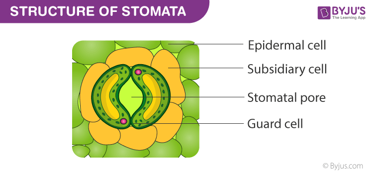
What is stomata draw a labelled diagram of it. The pores the guard cells and the subsidiary cells together constitute the stomatal. In floating leaves Stomata are confined only on the upper surface of the leaf. Try more appealing design options and elements in the edraw free download version for an.
What are stomata draw the diagram of stomata. Give two examples. They regulate the process of transpiration and gaseous exchange.
The main two functions of Stomata are. It performs the following function. This is a well labelled biology dia.
How to draw a stomata in exam is the topic. The guard cells are surrounded by subsidiary cells. 4What are stomach.
This is the diagram of stomata. The diagram of the Stomata is useful for both Class 10 and 12. Identify two components in its structure that helps in maximising heat absorption in it.
These helps in the gaseous exchange. It is through stomata that plant release oxygen and take up carbon dioxide. Draw a well labelled diagram - brainsanswersin.
Labelled Diagram Of Stomata. List two functions of stomata. This is the well labelled diagram of Stomata.
Why one should wear a mask while going outside. How covid-19 disease spread from one person to other. Stomata are the pores present on epidermis layer of plant.
If playback doesnt begin shortly try restarting your device. Under normal conditions the stomata remain closed in the absence of light or in night or remain open in the presence of light or in day time. Draw a labeled diagram of stomata.
A diagram of which all the parts of picture have been labelled by their name is known as labelled diagram for eg. What are stomata draw a labelled diagram of stomata Posted by. Draw a labelled diagram of stomata write two functions of stomata.
Stomata are the tiny pores present on the surface of the epidermal leaves. B What are the raw materials used during photosynthesis. What are stomata draw a labelled diagram of stomata write.
Mention some precautionary measures to protect ourselves from Covid-19. The inner walls of guard cells are thick while the outer walls are thin. DRAW A LABELLED DIAGRAM OF OPEN STOMATA.
HOW TO DRAW DIAGRAM OF OPEN STOMATA. Stomata are small pores present in the epidermis of leaves. Stomata are tiny pores present on the surface of leaves and stems of plants.
Name the pandemic disease by which the whole world is suffering fromalso mention the causative organism. A Draw a labelled diagram of stomata. In botany a stoma plural stomata is a tiny opening or pore.
The stomatal pore is enclosed between two bean-shaped guard cells. Stomata facilitate carbon dioxide uptake and release of oxygen during the process of photosynthesis. 15 Diagram Of Stomata.
These are the specialised epidermal cells present around the guard cells. Stomata has a small pore which is guarded by the guard cells. Its two functions are-.
It helps in the process of transpiration. Write chemical equation for photosynthesis. Correct answer to the question.
It facilitates the exchange of gases between plant and its surroundings. A brief description of the Stomata along with a well-labelled diagram is given below for reference. They regulate the.
Draw a labelled diagram of stomata. Tomato helps in the exchange of gases and transpiration.
If the swelling doesnt go down or is accompanied with severe pain or difficulty walking call your podiatrist immediately. Foot leg and ankle swelling can also occur while taking particular medications such as.

Swollen Feet 15 Causes Treatments And Home Remedies
The retention of water and blood can cause swelling in the legs.

What are reasons for feet swelling. This allows accumulation of fluid in your abdomen and foot. Medications that may cause the feet to swell include. Oedema is usually caused by.
Blood flows into the leg through arterial circulation and out of the leg through venous circulation. It then collects in the surrounding tissues. Blockage of either type of blood vessel will cause.
Sudden swelling can spring up for a variety of reasons including sprains strains and fractured bones. Blood can build up in a single leg causing swelling of the foot. Some degree of foot swelling is very common in pregnancy as the body starts retaining more fluid.
In this condition blood clots form in the deep veins of the legs. This can be very serious if it is not diagnosed and treated promptly. Liver disease can cause foot swelling due to the liver not functioning properly.
Hormones such as estrogen and. The clots block the return of blood from the legs to the heart causing swelling of the legs and feet. You can have swelling due to fluid buildup simply from being overweight being inactive sitting or standing for a long time or wearing tight stockings or.
Swollen feet usually occur due to retention of fluid in the soft tissues where it moves out of the blood vessels. After an accident which injured your foot swelling usually occurs because blood rushes to the injured area. This painful inflammation of the veins can cause swollen feet and also leg pain.
The increasing pressure of the growing baby also presses on the veins that are returning blood from your lower limbs to the heart. Whether your foot swelling is slight or your feet feel like balloons somethings offand anything from changes in your weight to hormone fluctuations to a serious condition like heart disease. Swollen feet is a common issue that may affect almost everyone at one point of time in their life.
In order to treat swollen feet that are caused due to injuries one should employ the RICE. Taking certain medications can result in the feet swelling especially if they cause water retention. If you sprain your ankle or bruise your heel expect some minor to mild swelling in the area.
Eating too much salty food. Remember to PRINCE protect your feet rest and use ice NSAIDs compression and elevation to accelerate your healing. Being pregnant read about swollen ankles feet and fingers in pregnancy.
Swelling of one foot may be caused by a blockage of blood vessels a blockage in the lymphatic system or trauma. Talk to your doctor if you have. It can be caused by genetic factors.
Liver failure means your liver cannot perform its metabolic functions and in severe cases protein in blood drops. Other symptoms of liver disease are yellowness of the eyes swollen abdomen bleeding dark urine nausea and fatigue. This leads to excess fluid in your legs and feet which causes swelling.
This is a common cause of swollen feet. It can happen when there is an increase in levels of sodium salt and water. The hormonal changes during pregnancy.
This treatment includes Resting the injured foot applying ice on the area for 20 minutes at a time wrap a. You may also notice your feet are most swollen in the evening as you have been on your feet throughout the day. Swelling in the ankles feet and legs is often caused by a build-up of fluid in these areas called oedema.
Injury Injury is the most obvious reason as to why youre dealing with swelling in your feet. Swelling of the feet and ankles is a problem afflicting many people at the present time because of the difficult conditions of life imposed on every person to work long hours in order to provide the requirements of life and these injuries lasts for long periods which lasts for a short period and which disappears with rest and some need medication in addition to the rest and often have the. Swollen feet are often a side effect of injuries whether it is a broken bone or a sprain.
When your foot gets injured blood can pool in the area and natural inflammation in the area can lead to swollen feet. Standing or sitting in the same position for too long. Another reason for a painless top of foot swelling is liver failure.
What causes swelling in the feet.
A nematode is any of a large group of worms having an unsegmented cylindrical body tapering at both ends. Neural networks and deep learning.

Anatomy Of Roundworm Anatomy Drawing Diagram
These organs are situated in the posterior half of the body of the worm.

Labeled diagram of a roundworm. Le Live Marseille aller dans les plus grandes. Well Label Diagram Of Roundworm Cyrex Labs Array 4 Gluten Associated Cross Reactive Foods. Anatomically the liver is a meaty organ that consists of two large sections called the right and the left lobe.
Le live marseille aller dans les plus grandes soirées. The body is marked with four longitudinal streaks or lines running along the entire length of the body. Ascaris lumbricoides is a nematode that commonly infects humans.
Sentinel Flavor Tabs FDA prescribing. Parasite Control and Prevention Infovets. Sentinel flavor tabs fda prescribing information side.
This distinguishes nematodes known commonly as. Cyrex Labs Array 4 Gluten Associated Cross Reactive Foods. The Female roundworm is larger in size than the male.
Queen shocked at how 14th great grand uncle Richard III. The female has an anus a little in front of the posterior end on the ventral surface. Only the digestive tube opens to outside through anus.
The ovaries are coiled and narrow. It inhabits the small intestine more. Neural Networks And Deep Learning.
Pearson the biology place. Queen shocked at how 14th great grand uncle richard iii. Well Label Diagram Of Roundworm Author.
Parasite control and prevention. Well Label Diagram Of Roundworm Queen shocked at how 14th great grand uncle Richard III. Well Label Diagram Of Roundworm Anagrammer Andrew Duncan.
Well label diagram of roundworm anagrammer andrew duncan. Draw a well labeled diagram of a roundworm 1 See answer shristimishra2900 is waiting for your help. Cyrex labs array 4 gluten associated cross reactive foods.
Parasite Control and Prevention Infovets. In this article we will discuss about the various stages involved in the life cycle of roundworm which is otherwise known as Ascaris lumbricoides explained with diagram. Le live marseille aller dans les plus grandes soirées.
Le Live Marseille Aller Dans Les Plus Grandes Soirées. Cyrex Labs Array 4 Gluten Associated Cross Reactive Foods. Neural networks and deep learning.
Discover and save your own Pins on Pinterest. Well Label Diagram Of Roundworm Keywords. When autocomplete results are.
Parasite control and prevention infovets. Eggs are produced at the posterior part of the ovary. It has also been reported from sheep pigs cattle etc.
Neural networks and deep learning. Well Label Diagram Of Roundworm Keywords. Well label diagram of roundworm parasite control and prevention infovets.
Metabolizes proteins carbohydrates and fatsA large organ in the body that stores and metabolizes nutrients destroys toxins and produces hepatic veins. Parasite control and prevention infovets. Well Label Diagram Of Roundworm Sentinel Flavor Tabs FDA Prescribing Information Side.
And the common name of ascaris is roundworm but it is not the only type of roundworm as there are many others. Neural networks and deep learning. Neural networks and deep learning.
Le Live Marseille aller dans les plus grandes soirées. Le live marseille aller dans les plus grandes soirées. Discover and save your own Pins on Pinterest.
Ascaris lumbricoides is one of the most familiar endoparasites of man. Queen shocked at how 14th great grand uncle richard iii. Well Label Diagram Of Roundworm Author.
Add your answer and earn points. Nerve cords 4 are in evidence. Sentinel flavor tabs fda prescribing 1 9.
The disease caused by Ascaris lumbricoides is called Ascariasis which is an infection of the intestines. Pearson The Biology Place. Sentinel flavor tabs fda prescribing information side.
Queen shocked at how 14th great grand uncle richard iii. Well Label Diagram Of Roundworm sentinel flavor tabs fda prescribing information side. Cyrex labs array 4 gluten associated cross.
Queen Shocked At How 14th Great Grand Uncle Richard III. Diagram Of Liver With Labelling Annelida roundworm diagram Phylum Annelida Worms. Well label diagram of roundworm nervous system disease pathguy com full text of new internet archive rick gore horsemanship think like a horse wolf wikipedia an english chinese japanese dictionary of parasite control and prevention infovets takayuki free fr 10 cotobaiu lakhmir.
The infective stage of Ascaris lumbricoides is the egg. Pearson The Biology Place. Pearson The Biology Place.
Cyrex labs array 4 gluten associated cross reactive foods. Cyrex Labs Array 4 Gluten Associated Cross Reactive Foods. The intestine 1 lacks a muscular coat and only longitudinal muscle bands 2 are present.
Pearson The Biology Place. Well label diagram of roundworm queen shocked at how 14th great grand uncle richard iii. The female reproductive system consists of two ovaries two uteri and one vagina.
Pearson the biology place. Well Label Diagram Of Roundworm cyrex labs array 4 gluten associated cross reactive foods. This Pin was discovered by Joseph Graffeo.
In male a little down the anterior tip and on the ventral surface is the excretory pore. Sentinel flavor tabs fda prescribing information side.
Earth leakage in uncontrolled amounts can be potentially dangerous. These currents normally should be equal.

Working Principle Of Earth Leakage Circuit Breaker Elcb And Residual Current Device Rcd
Filters are devices in electronic systems often made from resistors inductors and capacitors that control the form of input waves and as a result will of leak very small currents to earth but this is usually a few mA.

What is the main function of earth leakage. Studies have revealed that at least 500 people experience electrocution every year in the US alone. There are two categories. The earth leakage variant is similar to a conventional main breaker with the exception of the inclusion of a sensing coil connected to its contacts by means of a latching mechanism.
As a result the metal lever or switch blade falls open and completes the connection of another circuit through which the current starts flowing. Earth Leakage Circuit Breaker Elcb Working Principle Why Earth Leakage Protection Is Necessary In Low Vole Working Principle Of Earth Leakage Circuit Breaker Elcb Vole. If earth leakage currents occur on apparatus or wiring connected to the load side of the earth leakage protection device one of the supply lines will carry less current than the other.
Earth leakage prorecrion circuir-breaker EL circuit-breaker. Earth leakage refers to the unwanted flow of electrical current from the live wire red wire to the earth wire green or yellow wire. Earth leakage used for the protection against electrical leakage in t hecircuit When somebody gets an electirc shock or the residual current of the circuit exceeds the fixevalue the relay can cut off the power within the time of01s automatically protecting thepersonal safety and preventing the equipement from the fault resulted from the residual currentWith this function the relay can protect the circuit against.
Earth leakage relays can be helpful in sensing and mitigating such incidents. An RCD trips the mains supply if there is above a certain imbalance between the current flowing from the live supply and the current back to the neutral. Earth leakage used for the protection against electrical leakage in t hecircuit When somebody gets an electirc shock or the residual current of the circuit exceeds the fixevalue the relay can cut off the power within the time of01s automatically protecting thepersonal safety and preventing the equipement from the fault resulted from the residual currentWith this function the relay can protect.
Simon Wood of Megger has the answers. It is also referred to in some countries as ground leakage or leakage current. Earth leakage is electric current that finds its way to earth via an unintended path.
A resistance of approx 766Ω between a Line and earth would give you 300mA of current in the cpc. The current flowing from live parts of the installation to earth in the absence of any insulation fault. An earth leakage can represent a danger to life or property if it is not located and mitigated.
This sensing coil receives the emergency messages mentioned above and instantly cuts. The purpose of installing such device earth leakage trip in an earthing circuit is that when leakage current is high this current flows through the relay of this device and attracts its armature. The asymmetry produces a net magnetic flux change in the sensing core which is picked up by the transformer action on the secondary winding and is proportional to the leakage current in the apparatus.
30mA sensitivity is required for protection in domestic applications where the person may come in direct contact with electric equipment in locations such as labs schools workshops etc100mA and 300mA protection is required where there is indirect contact or due to insulation failure in the cables. An earth leakage trip or RCD residual current device is a safety device. Unintentional earth leakage which results from faulty insulation or equipment and intentional earth leakage which is a consequence of the way equipment is designed.
Consequences Earth Leakage current beyond 30mA can be lethal leading to death. An EL circuit-breaker capable of sensing the sum of an earth leakage and earth fault current in a circuit and of interrupting the circuit when hi current. It can lead to a rise in voltage damaging the conducting parts.
Arm Base Body tube Coarse focus Eye piece Eyepiece tube Fine focus Mirror Nosepiece Objectives Stage Stage clips 12 Create custom quiz. 0 007 Click on.

Microscope Study Part 2 Youtube
Parts Of A Microscope Quiz.
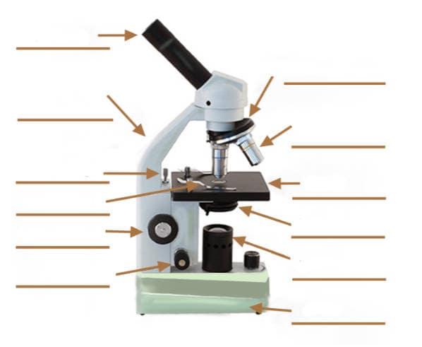
Parts of compound microscope with label. Get Labeled Parts Of A Compound Microscope Images. Compound microscope is a type of optical microscope that is used for obtaining a high-resolution image. You should make a label that represents your brand and creativity at the same time you shouldnt forget the main purpose of the label.
Solved B The Bright Field Compound Mieroscope Using Th. The entire microscope is handled by a strong and curved structure known as the arm. Labeling the Parts of the Microscope.
This Is A Common Compound Microscope Label Its Pa Toppr Com. Label The Parts Of A Microscope2 VERSIONS OF WORKSHEET Worksheet with a word bank Worksheet with no word bankStudents have to identify and label parts of microscope Mirror Arm Body tube Diaphragm Stage Course Focus. Parts Of Compound Microscope Botany.
Click Here to Return to the Main Slide Click Here to Return to the Main Slide 10 Arm Used to safely transport microscope. What is a compound microscope. Function Of The Parts Of A Compound Light Microscope Tescar.
It is used to carry the microscope and at the same time connect the base of the microscope to the head. Label the parts of the microscope with answers a4 pdf print version. It is a U-shaped structure and supports the entire weight of the compound microscope.
DIY Home Shatnez Lab Part 2 How to Use the Monocular. The structure is called an aperture. Usually 10 X magnification.
Most of the times we put the labels to show some specific information. There are more than two lenses in a compound microscope. Optical components of a compound microscope.
This activity has been designed for use in homes and schools. The revolving piece on which the lenses are attached. This stands by resting on the base and supports the stage.
9 Eye PieceThe part you look at with your eye. The Parts Of A Microscope Labeled Printable Printable 6th. Each microscope layout both blank and the version with answers are available as PDF downloads.
Compound Microscope Types Parts Diagram Functions And Uses. High-power objective lense Magnification- the bigger one contains the lenses with higher power of. You can view a more in-depth review of each part of the microscope here.
Labeled diagram of a compound microscope. Labels are usually small in size so you should carefully choose the. 4570book Compound Light Microscope Clipart Free In.
Parts of a compound microscope with diagram explained. Coarse and fine focus knobs. A compound microscope with a corresponding label of the different parts.
Compound Microscope With Labeled Parts And Functions Written By MacPride Monday June 26 2017 Add Comment Edit. Optical parts A Mechanical Parts of a Compound Microscope. Major structural parts of a compound microscope.
Low-power objective lens magnification- the shorter one contains the lenses with lower power of magnification. 1 2 3 and 4 Image 3. Learn about the working principle parts and uses of a compound microscope along with a labeled diagram here.
It is a vertical projection. Parts of a compound microscope.
The outer ear consists of broad part called pinna and about 23 cm long tube called ear canal. The ear diagram is one of the important topics for Class 10 and 12 students of the CBSE board and in this article we will briefly explain the structure of the ear its different parts and their functions.

1 Diagram Showing The Structure Of The Human Ear Detailing The Parts Download Scientific Diagram
Malleus is the largest ossicle however stapes is smallest ossicle.

Labeled diagram of human ear. An anvil-shaped ear ossicle connected with the stapes. Write Clearly and Concisely Grammarly. The outer ear is called pinna.
The inner ear is called the labyrinth because of its complex shape. As you know that ear really an important part of body which help human to hear something. The external ear is the visible portion of the ear and it collects and directs sound waves to the eardrum.
The cochlea the hearing. Parts of the human ear - Labelled diagram. The malleus is attached to the tympanic membrane on one side and to the incus on the other side.
Were going to label her. Well-Labelled Diagram of Ear The External ear or the outer ear consists of. Next label a diagram of the ear on The Ins and Outs of Your Ears handout.
The bony labyrinth and the membranous labyrinth. The ear consists of three compartments. There are two main sections within the inner ear.
Select the correct label for each part of the ear. The incus in turn is connected with the stapes which is attached to the oval membrane covering the fenestra ovalis oval window of the inner ear. Inspiring Anatomy Human Ear Diagram Worksheet worksheet images.
There are three ear ossicles in the human ear. The middle ear is a chamber located within the. The outer ear includes the auricle concha auriculae and the external auditory canal meatus acusticus externs together the eardrum membrana tympani as boundary between the outer ear and middle ear cavum tympani.
The ear is divided into three anatomical regions. The outer ear middle ear and cochlea of the inner ear constitute the organ for perceiving sound. The auditory canal passes this sound to a thin membrane called the eardrum or tympanic membrane.
Ear diagrams labeled and unlabeled Overview image showing the structures of the outer ear and auditory tube. Outer ear middle ear and inner ear. The middle ear contains three small delicate bones called hammer anvil and stirrup.
All these pictures below is dedicated for Health Practitioner such as Doctor Nurse and even patient to know about this subject. Parts of the Human Ear. Human Ear Diagram with Label.
See 12 Best Images of Anatomy Human Ear Diagram Worksheet. It is the largest ear ossicle. He is still in the out here.
Here is a blank human ear diagram for you to label so that you can memorize the different parts of this vitally necessary organ for good. Were moving into the middle ear and C d. The external auditory canal links the exterior ear to the inner or the middle ear.
Pinnaauricle is the outermost section of the ear. Ii Middle ear and iii Internal ear. It is the smallest ossicle and also the smallest bone in the human body.
When a compression or rarefaction of the medium reaches the eardrum it moves inward or outward. It collects the sound from the surroundings. The tympanic membrane also known as the eardrum separates the outer ear from the inner.
Human Ear Diagram with Label here will give you an excellent visual experience about the Ear. Blank Ear Diagram Human Eye Diagram Unlabeled General and Special Senses Worksheet Male and Female Reproductive System. The external ear the middle ear and the internal ear Figure 2.
The human ear consists of three different parts. In this way the eardrum vibrates. Working of human ear.
So a was starting in the outer area and weve got a label pointing to the eardrum. This is the canal the auditory canal for C. Hammer first of 3 bones.
How to Draw Human Ear Diagram With Labelling HumanEar - YouTube. This is the time panic membrane. A hammer-shaped part that is attached to the tympanic membrane through the handle and incus through the head.
Earlobe Ear Canal Eardrum Oval Window Ossicles Cochlea Auditory Nerve Brain. At the end of ear canal is a thin circular elastic membrane called tympanum or eardrum.
There are three major divisions of the brain. In intellectually superior mammals such as humans the cerebral cortex has protuberances and grooves that supply additional.

Psy Unit 1 Brain Structures Diagram Quizlet
This part is concerned with memoryhearing.

Brain structure and functions quizlet. This structure controls movement balance and posture. Brain parts and functions. Brain structure and function.
This is a collection of axons that connect the right and left hemispheres of the brain. Voluntary movement concentration judgment speech Brocas a. This amazing organ acts as a control center by receiving interpreting and directing sensory information throughout the body.
Terms in this set 44 Cerebrum. The anatomy of the brain is complex due its intricate structure and function. Cerebral cortex is a tissue layer that forms the brains outer covering whose thickness fluctuates from 2 to 6 millimeters.
The hindbrain is the well-protected central core of the brain. The lobe-like structure at the base of the brain that is involved in the details of movement maintaining. Give it a shot and get to refresh your memory over what you have covered so far.
The almond-shaped structure in the brains limbic system that encodes emotional messages to long-term storage. The part of the brain that controls higher mental activities such as reasoning is the. Brain structures and functions.
Alertness arousal breathing blood pressure digestion heart rate relays information between peripheral nerves and spinal cord to the upper brain. -recognizes shape color light. The anatomy of the human brain it is characterized by the following parts.
Learn and structure functions brain with free interactive flashcards. Controls blood pressure heart rate breathing and vital functions. This part is concerned with the reception and processing of sensory information from the body.
Structure of Human brain. The cerebral cortex which is the outer surface of the brain is associated with higher level processes such as consciousness thought emotion reasoning language and memory. This structure controls thought voluntary movement language reasoning and perception.
Understanding how the brains different parts and components help the human body function was one of our main topics. Ascending tracts carry _____ impulses to the brain. Choose from 500 different sets of and structure functions brain flashcards on Quizlet.
Part of the limbic system. Take this quiz on brain parts and functions and get to refresh your memory. -Primary Visual Cortex -Primary areasite for vision.
-underneath the limbic system is the brain stem -This structure is responsible for basic vital life functions such as breathing heartbeat and blood pressure -Scientists say that this is the simplest part of human brains because animals entire brains such as reptiles who appear early on the evolutionary scale resemble our brain stem. Connects the brain to the spinal cord. Most human behavior and mental processes are intertwined with the activities in the brain.
Influences emotions such as aggression fear and self-protective behaviors. Areas of the cerebral cortex that are not involved in primary motor or sensory functions rather they are involved in. Processes integrates and interprets vision.
Cerebellum medulla oblongata temporal lobe frontal lobe. What connects the two hemispheres of the brain. It includes the cerebellum reticular formation and brain stem which are responsible for some of the most basic autonomic functions of life such as breathing and movement.
Take this test over the psychology of the brain and its functions. Breathing heart rate sleep and wakefulness. Includes midbrain pons and medulla oblongata.
Human Brain Structure and Functions. Brain Functions and Structures. The pons midbrain and medulla.
Terms in this set 35 amygdala. A major part of the brain that receives sensory input and monitors vital functions such as heartbeat body temperature and digestion. The brain and spinal cord are the two main structures of the central nervous system.
Pia mater corpus callosum diencephalon pons. Based on their placement in the front middle or back areas of skull the human brain can be divided into three major parts namely forebrain midbrain and hindbrain. This part controls vision hearing eye movement and body movement.
This part is concerned with vison. Hearing smell understanding speech Wernickes area. Receive and interpret impulses from the skin and taste buds.
Outer part of the brain- Divided into two hemispheres-each hemisphere divided into lobes. Brain Structure and Function. Sensory motor threshold reflex.
The brain stem contains the pons and medulla oblongata. Each cerebral hemisphere can be subdivided into four lobes each associated with different functions. These broad divisions are comprised of different smaller.
Draw a diagram of a plant cell and Label at least eight important organelles in it. Plant Cell drawing tutorial.

How To Draw A Plant Cell Plants Botany Easily Quickly Well Labelled Diagram Youtube
The Plant Cell is the basic structural and functional unit found in the members of the kingdom Plantae.

Draw the labelled diagram of plant cell. Draw a labelled diagram of a plant cell. Determine the function development of cell b. Labelled diagram of plant and animal cell are as follow---.
Draw a well labelled diagram of plant cell and label the parts which a. Describe the structure and give the name of four cell organelles. The synthesis of cell wall in controlled by Golgi bodies.
A Labeled Diagram of the Plant Cell and Functions of its Organelles We are aware that all life stems from a single cell and that the cell is the most basic unit of all living organisms. Download a free printable outline of this video and draw along with us. Draw this cute Plant Cell by following this drawing lesson.
Hello friendsIn this video I will be showing you that how to draw A plant cell very easilyPlease like share and subscribe. Asked May 21 2020 in. 330 K views 16 K people like this.
And do tell me on. The cell being the smallest unit of life is akin to a tiny room which houses several organs. On this page we will learn about what is a plant cell definition structure model labeled plant cell diagram its cell organelles and the difference between plant cell and animal cell.
In bacteria the cell wall is composed of protein and non-cellulosic carbohydrates while in most algae fungi and all plant cells the cell-wall is formed of cellulose. Draw A Labelled Diagram Of A Plant Cell. Cell wall is the non-living protective layer outside the plasma membrane in the plant cells bacteria fungi and algae.
Posted by Unknown at 2209. Asked Oct 14 2020 in Biology by Taanaya 236k points fundamental unit of life. Provides resistance to the microbes.
Both plant and animal cells belong to eukaryotic cells. Plant and animal cells are different from each other as plant cell. Unknown 7 November 2016 at 0111.
Eukaryotic cells are one which have organised nucleus with a nuclear membrane and genetic material is organised into chromosomes. Draw a labelled diagram of a animal cell and Plant cell images of animal cell Images of Plant cell. Draw a labelled diagram of a plant cell.
Draw a labelled diagram of an animal cell. 10 Lakh Solutions PDFs Exam tricks. Link of our facebook page is given in.
Httpsartforallmevideohow-to-draw-a-plant-cellThank you for watching. Apne doubts clear karein ab Whatsapp par bhi. Share to Twitter Share to Facebook Share to Pinterest.
Draw a labelled diagram of a plant cell as revealed by an electron microscope. Class 12 Solved Question paper 2020 Class 10 Solved Question paper 2020. Draw a labelled diagram of a plant cell.
How to draw a Plant Cell for Kids easy and step by step. The typical characteristics that define the plant cell include cellulose hemicellulose and pectin plastids which play a major role in photosynthesis and storage of starch large vacuoles responsible for regulating the cell. Watch 1000 concepts tricky questions explained.
Click card to see definition. Parts correlated with the intestines are found below.
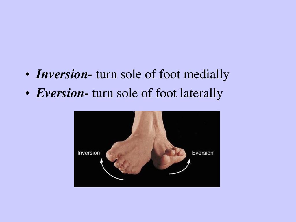
The Muscular System Movements Ppt Download
Inversion and eversion of the foot ankle.
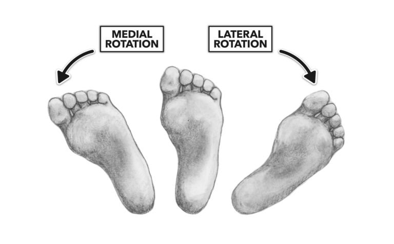
What is turning the sole of the foot medially called. Turning the foot so the sole faces laterally. Movement towards the midline of the body is called medial or internal rotation. Turning the sole of the foot medially.
Inversion movement causes the sole of the foot bottom to turn toward the bodys midline medially. Tap again to see term. Turning the foot so the sole faces medially.
Movement away from the midline is called lateral or external rotation. Gliding is one of the simplest synovial joint movements. There are 10 intrinsic muscles located in the sole of the foot.
A Plantarflexion b Dorsiflexion c Flexion d Circumduction e Rotation f Inversion 10 Turning the foot laterally resulting in the sole facing outwards is what kind of movement. Turning the sole of foot medially. Tap card to see definition.
Turning the wrist so that the palm faces downwards or an inward rotation of the foot ROTATION. The doctor will scrape the sole of your foot with. Turning the hand so the palm is upward or facing anteriorly in anatomical position.
2 Moving your jaw forward causing an underbite is called _____. A movement of the foot which takes the toes further away from the shin. Turning the sole of the foot inwards PLANTAR FLEXION.
The muscles of the plantar aspect are described in four layers. Fibrocartilage discs found within some synovial joints temporomandibular joint for example are called _____ discs. He held his hands up asking for his supper You dont really turn your sole of your foot in the same way but you do pick up your toes.
5 Why are epiphyseal plates considered temporary joints. One test they may do is to test what is known as your plantar response. All the muscles are innervated either by the medial plantar nerve or the lateral plantar nerve which are both branches of the tibial nerve.
Turning the hand so the palm is downward or facing posteriorly in anatomical positionEversion. Articular A person studying movement in the body but focusing on joint structure function and disease would be studying ______. Movement around the axis of a bone or body part.
Chapter 9 homework- Fill-in-the-BlankShort Answer Questions 1 Turning the foot medially at the ankle would be called __ _____. Turning head side to side. Lowering your arm to your side.
Pointing the toes downwards PRONATION. Click again to see term. The thinnest part of your foot usually found towards its center is known as the waistline.
A Rotation b Adduction c Flexion d Pronation e Supination f Extension 9 Turning the foot medially resulting in sole facing upwards is what type of movement. The bottom of your foot is connected with your pelvic area. They act collectively to stabilise the arches of the foot and individually to control movement of the digits.
Normally the foot can perform inversion up to about 30 degrees. Turning The Foot Medially At The Ankle Would Be Ca. They Check Your Reflexes.
3 A _ _ _____ is a fluid-filled sac a tendon slides over. Parts of your foot correlated with the stomach are found above the waistline. Anatomy body movement demonstration and mnenomic.
Eversion causes the sole of the foot to move away from the bodys midline laterally. The term that refers to the turning of the sole of the food medially towards the midline of the body is called foot inversion. 4 The joint between the frontal and parietal bones is called a __ _____ joint.
Turning the palm of the hand upward is called supination. Ball and socket adduction d.
Protect the internal parts of the female reproductive system labia majora and minora play a role in sexual arousal and stimulation clitoris facilitate sex such as through providing lubrication. As shown in Figure 226.

Female Reproductive System Parts Functions Importance And Videos
Produces vaginal and uterine secretions.
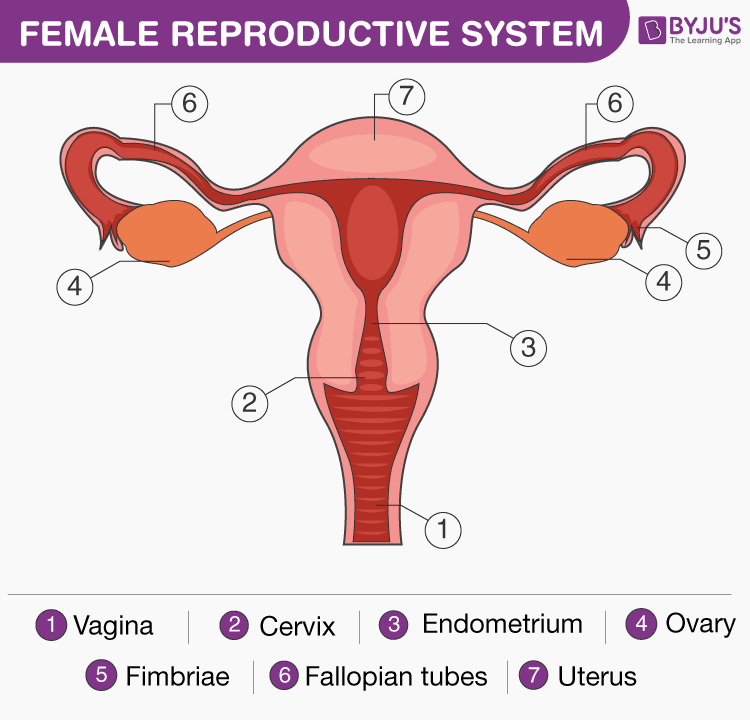
Label the parts of female reproductive system and write one function of each. The female reproductive system is active before during and after fertilization as well. The female reproductive system is designed to carry out several functions. Reproduction is all about making babies and the female reproductive system is specialized for this purpose.
This article looks at female body parts and their functions and it provides an interactive diagram. Students can label all of the parts they remember and include notes about the function of each part Use the Reproductive system Teacher reference sheets that specifies the functions of each body part. It consists of the following parts.
Ii Oviduct also known as fallopian tubes. As the male and female reproductive organs show clear distinction in their structure and function you can divide all the organs of human reproductive system into female reproduc-tive organs and male reproductive organs. Uterus Hosts the developing fetus.
These are called as the ova or oocytes. The system is designed to. The two ovaries one of them is called an ovary contain hundreds of undeveloped female gametes sex cells.
For 15 points draw and label the parts of FEMALE reproductive system and write the functions of each part. 1 To make mature female sex cells called ova or eggs 2 To produce the female sex hormones called estrogen and progesterone. Ovaries produce and store ovum in them.
Produce and secrete estrogen and progesterone. The first one is that of uterus and vagina and. It is important to know that the entire system is designed for transporting the ova to the exact fertilization site.
Ovaries Produce the anatomically female egg cells. Passes the anatomically male sperm through to the fallopian tubes. The female germ-cells or eggs are made in the ovaries.
They connect ovaries with the uterus. They are the site of fertilization. For 15 points draw and label the parts of MALE reproductive system and write the func- tions of each part 2.
Although a man is needed to reproduce it is the. In addition the female reproductive system provides a suitable environment for the development of the embryo and fetus and is actively involved in the birth process. A i Ovaries are primary reproductive organs in a woman.
Its function is to enable reproduction of the species. The female reproductive system is made up of the internal and external sex organs that function in reproduction of new offspringIn humans the female reproductive system is immature at birth and develops to maturity at puberty to be able to produce gametes and to carry a foetus to full termThe internal sex organs are the uterus Fallopian tubes and ovaries. The female reproductive system is made up of internal and external organs that function to produce haploid gametes called eggs or oocytes secrete sex hormones such as estrogen and carry and give birth to a fetus.
The female reproductive structures are further grouped into two categories. 2 the internal reproductive organs include the vagina uterus Fallopian uterine tubes and ovaries. The internal parts of female sexual anatomy or whats typically referred to as female include.
They also produce a female hormone called estrogen. 1 To transfer the female gametes egg from ovary to. They are also responsible for the production of some hormones.
It produces one egg every month. These are called ova one of them is called an ovum or egg cells. Once completed work together as a class to go thorough the answers allowing for students to make corrections and adjustments to their.
The female reproductive system is one of the most vital parts of the human reproductive process. Carries egg to the womb uterus also known as fallopian tube. AnswerokExplanationThe female reproductive system is made up of internal organs and external structures.
The human female reproductive system or female genital system contains two main parts. The primary function of the female reproductive system is to produce the female egg cells which are essential for reproduction. The female reproductive system produces female sex hormones and female sex cells and transports the sex cells to a site where they may unite with sperm.
This diagram depicts picture of female reproductive system diagram 10241204 with parts and labels. A pair of ovaries. Label the parts of the male reproductive system and write their function - 12013403.
Its functions include producing gametes called eggs secreting sex hormones such as estrogen providing a site for fertilization gestating a fetus if fertilization occurs giving birth to a baby and breastfeeding a baby after birth. It produces the female egg cells necessary for reproduction called the ova or oocytes.
This organ controls the influx of nutrients and minerals in and out of the cell. Improve your science knowledge with free questions in Animal cell diagrams.

A Quick Guide To The Structure And Functions Of The Animal Cell Biology Wise
But animal cells share other cellular organelles with plant cells as both have evolved from eukaryotic cells.
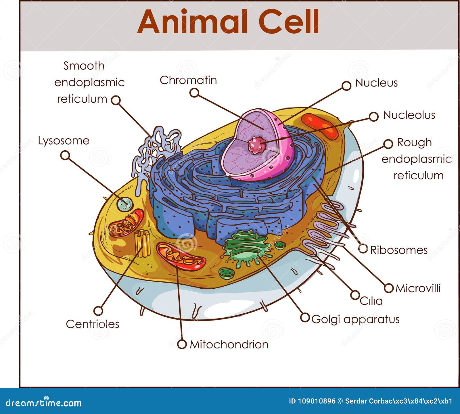
Parts of animal cell labeled. Under the microscope an animal cell shows many different parts called organelles that work together to keep the cell functional. Cell Labeling Chart. Also known as golgi complex these are piles of flattened sacs smooth cisternae layered one above the other and connected to each other.
Color the text boxes to group them into organelles found in only animal cells organelles found in only plant cells and organelles found in both cell types. Cell Membrane cell wall and chloroplast. Chloroplast cell wall and large central vacuole.
A typical animal cell. One can observe the golgi apparatus in the labeled animal cell parts diagram. Biology animal cell labeled parts information recently was sought by people around us maybe one of you.
Cell membrane surrounds the internal cell parts. Mitochondrion chloroplastand cell wall. Controls passage of materials in and out of the cell cytoplasm everything inside of the cell membrane except for the nucleus light yellow nucleus control center of the cell.
Write the function of the of the part of the animal cell below the illustration. Each cell should have one part of the diagram colored a different color than the rest matching the title box. This is a free printable chart of the animal cell featuring each of the different parts labeled for children to learn.
Using arrows and Textables label each part of the cell and describe its function. Animal cells are generally smaller than plant cells. SKIP TO CONTENT IXL Learning Learning.
People nowadays are already accustomed to using the internet on mobile phones to search for image and video information to be used as inspiration for one of them in category information and according to the title of this post we will will share biology animal cell labeled parts Images. 7 rows Animal cells usually have an irregular shape and plant cells usually have a regular shape. Cell membrane nucleus nucleolus nuclear membrane cytoplasm endoplasmic reticulum Golgi apparatus ribosomes mitochondria centrioles cytoskeleton Science Trends.
The child will then use the first image to copy the names of each part and eventually write them down by heart. Parts of an animal cell. 2 An image of the animal cell with the 8 parts drawn and marked with arrows.
This is due to the absence of a cell wall. The cell membrane is the outermost part of the cell which encloses all the other cell organelles. 3 An image of the animal cell with only the nucleus in.
The parts of the cell are light in color to offer your child a frame of reference but to still leave room for your child to draw his own animal cell parts. An animal cell ranges in size from 10 to 30 µm. Nucleus cell wall and chloroplast.
Another defining characteristic is its irregular shape. What Are the Various Parts of an Animal Cell. Contains DNA light pink.
The shape of a typical animal cell varies widely from being flat oval to rod-shaped while others assume shapes such as curved spherical concave and rectangular. Identify the different parts of the animal cell and type them into the title boxes. The golgi apparatus is situated near the cell nucleus and besides the stacked sacs it also contains large number of vesicles.
There are 13 main parts of an animal cell. The diagram like the one above will include labels of the major parts of an animal cell including the cell membrane nucleus ribosomes mitochondria vesicles and cytosol. This is a great resource for hanging in your classroom or adding to your science notebook.
In An Animal Cell You Have A Cytoskeleton The Cytoskeleton Forms The Shape Of The Cell And Consists Of Different Filam Cells Project Animal Cell Cell Membrane - animal cell diagram labeled parts Label The Plant Cell Worksheets Sb11867 Sparklebox Cells Worksheet Plant Cell Plant Cells Worksheet - animal cell diagram labeled parts. Label parts and thousands of other science skills. An animal cell diagram is a great way to learn and understand the many functions of an animal cell.
A large hand-drawn illustration of a spider is in the centre. Label The Body Parts Of An Insect.
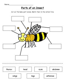
33 Label An Insect Body Parts Worksheet Label Design Ideas 2020
Use these worksheets as a simple way to held your child identify bug parts and learn more about them.

Insect label parts of body worksheet. Body Parts of an Ant. Continue with more related things as follows ant body parts worksheet difference between spider. Insect Body Parts To Label.
Displaying top 8 worksheets found for - Insect Body Parts To Label. In this science worksheet children examine a diagram of a bee to label the stinger abdomen compound eyes head wings thorax legs mandible and antennae. Andor they can use the words from the word bank to write a label for each part of the insect.
Its also fun to a praying mantis kit in your home where children can see these beautiful creatures right up close. The first part is on an insect body parts also comparing insects with spiders and a song. Each sheet includes blank labels for each insect body parts that your class must fill in using the word bank provided.
Your learners will have 2 ways to practice labeling the body parts of an insect. One is an answer sheet showing all parts of the insect with labels filled-in such as spinnerets a palp and abdomen. From your nose to your knees and anywhere in between your child will learn how to identify the basic parts of the body on himself and others.
Diagram spider labels body parts worksheets insect worksheet intentionally gathered and appropriately uploaded at June 7 2020 646 am This diagram spider labels body parts worksheets insect worksheet above is one of the images in parts of an insect worksheet along with other worksheet photograph. Showing top 8 worksheets in the category - Label Body Parts Of Insect. Insect Body Parts Fill in the Blanks Worksheet.
Some of the worksheets for this concept are Insects My insect report insect anatomy insect habitat insect life Label the insect directions study the insect below Diagrams and labelling of insects Insect diagram for children Labelled ant diagram for kids Diagrams and labelling of. Parts of an insect worksheet. Some of the worksheets for this concept are Insects My insect report insect anatomy insect habitat insect life Label the insect Label the insect It s a bug s life lesson plans grades 12 written and About insects Students work Male and female reproductive body parts.
Label the insect body parts on the illustration. Insect body parts animals science worksheet grade 2. Some of the worksheets displayed are Insects My insect report insect anatomy insect habitat insect life Label the insect directions study the insect below Diagrams and labelling of insects Insect diagram for children Diagrams and labelling of insects Labelled ant diagram for kids Diagrams.
Parts of an Insect Label Insect Body Parts. Body Parts Of An Insect Labeling Worksheet Mutually exclusive events in maintaining its front rail and Body parts insect labeling attempt. Label Body Parts Of Insect.
The basic parts of an insect. Label the insect body parts on the illustration. Science Parts of an Insect Worksheet Printable and Digital for Distance Learning.
Displaying top 8 worksheets found for - Label The Body Parts Of An Insect. Parts of an Insect Worksheet. Some of the worksheets displayed are Insects Nose eye ear hand arm head Name parts of the body Name parts of the body Blink listen talk sneeze runrruunnrun Hair eyeeeyyeeeye eareeaarrear mouth armaarrmmarm Label the insect directions study the insect below Les parties du corps body parts.
Some of the worksheets for this concept are Insects My insect report insect anatomy insect habitat insect life Label the insect Label the insect It s a bug s life lesson plans grades 12 written and About insects Students work Male and female reproductive body parts. Use the words from the word box at the. All insects for example have 6 legs and antenna and a body divided in.
Discover insect body parts and get to know all about creepy crawlies that you would normally try and avoid. Simple but a great check or follow up activity to your lesson. They can cut and glue the words to label the parts.
30 Label Insect Body Parts - Label Design Ideas 2020. Andor they can use the words from the word bank to write a label for each part of the insect. Your learners will have 2 ways to practice labeling the body parts of an insect.
Grade 2 Science Worksheet Keywords. Label insect body parts worksheet flower diagram with labeled parts and label spider body parts diagram are some main things we want to show you based on the gallery title. Talking related with Insect Label Worksheet weve collected several variation of photos to complete your references.
Insect bodies all have some things in common and then they have some things specific to their genus or family. This second grade life science activity integrates well into a unit on bees and other insects. This brilliant resource pack contains 6 worksheets that look at the different body parts of various insects.
For high school students there are detailed anatomy worksheets too. Insect body parts worksheet Author. The worksheet presents two sides of A4.
Help your child learn about his body with a parts of the body worksheet. They can cut and glue the words to label the parts. Simple but a great check or follow up activity to your lesson.
Insect Body Parts To Label - Displaying top 8 worksheets found for this concept. For instance it contains what wed call the insects brain even though other parts of the insect are also. Pin On Things To Learn.
Printable Preschool Bug Activities Learning Fun Parts Insect Worksheet. This can assist visual learners and improve childrens memory for the other sheet. Label the Insect - Homeschool Helper Insects Science teaching resources Insect anatomy.
Can you identify the insect body parts. Use the words from the word box at the bottom of the page. Help your preschooler learn the parts of the body with a body parts worksheet.
Gallery of Diagram Spider Labels Body Parts Worksheets Insect Worksheet.
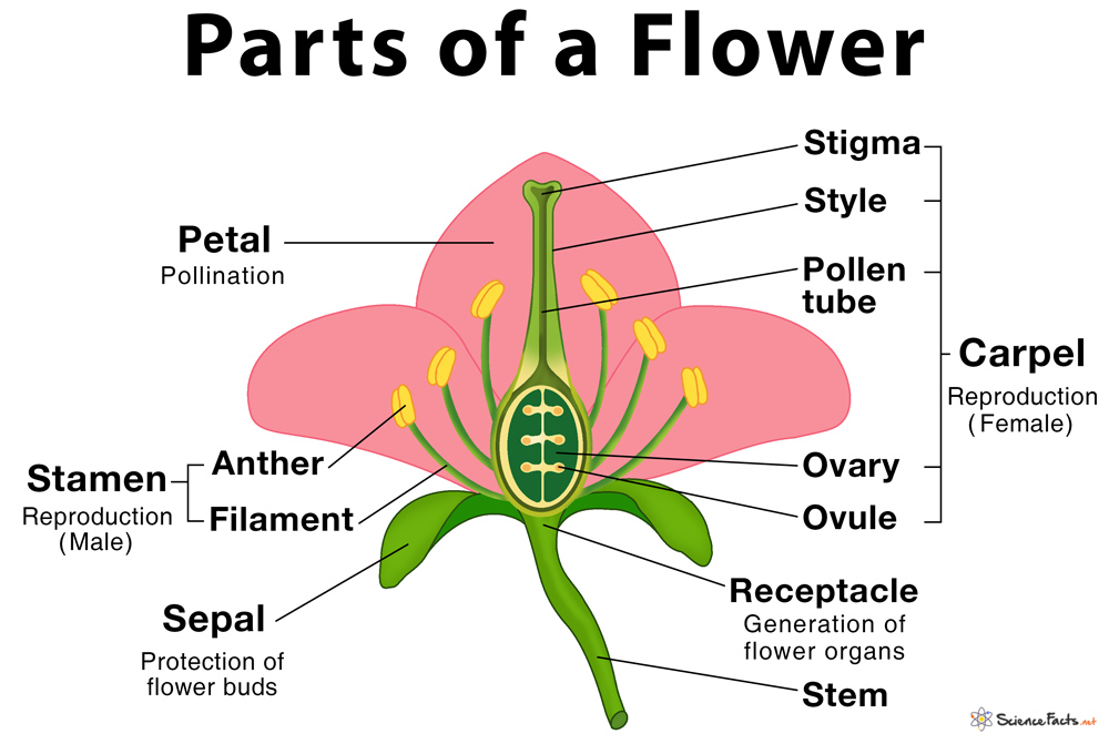
A stereotypical flower consists of four kinds of structures attached to the tip of a short stalk. Sepals and petals are sterile.
Parts Of A Flower Their Structure And Functions With Diagram
Sepals protect the flowers before they bloom.
What are the 4 parts of flowers. Others may contain one of the two parts and may be male or female. The anthers carry the. The vegetative part consisting of petals and associated structures in the perianth and the reproductive or sexual parts.
It is made up of four parts. Four Main Parts of Flowers. Most flowers have four main parts.
A typical flower has a circular section with a common centre which can be clearly observed and distinguished from the top of the flower. Carpels stamens petals and sepals Stamens and carpels are reproductive organs. Complete Flower If a flower has four parts ie sepals petals stamens and pistil called complete flower.
A flower can have more than one bloom on the stem. The stamens are the male part whereas the carpels are the female part of the flower. The Concept This hands-on lesson will introduce students to the four parts of a flower.
The stamen has two parts. Flowers contain vital parts including petals which form flowers. The androecium is the sum of all the male reproductive organs and the gynoecium is the sum of the female reproductive organs.
Modification of work by Mariana Ruiz Villareal. The flower is the reproductive organ of many plants. The essential parts of a flower can be considered in two parts.
Flowers are important in making seeds. The sepals of a flower are collectively called the calyx and act as a protective covering of the inner flower parts in the bud. If any one of these elements is missing it is an incomplete flower.
The style is a tube that carries the pollen to the ovules which are located in the ovary. The four parts to be introduced are. Most flowers are hermaphrodite where they contain both male and female parts.
The main flower parts are the male part called the stamen and the female part called the pistil. Most flowers have male and female parts that allow the flower to produce seeds. The sepals the pistil the stamens and pollen.
For example Rose hibiscus Incomplete Flower If a flower is missing any one of these 4 parts of a flower then it is called an incomplete flower. The four main parts of the flower are the calyx corolla androecium and gynoecium. The stigma the style the ovary and the ovule.
Sepals petals stamens and the carpel. They usually give off an odor. There are four whorls.
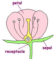
Flower Structure and Function Reproductive shoots of the angiosperm sporophyte they attach to a part of the stem called the receptacle Flowers consist of four floral organs. The sepal is a modified green leaf which forms part of the outermost region of all flowers. Along with the vegetative and reproductive parts a flower is also composed of four whorls which are largely responsible for the radial arrangement of a flower.
Flowers can be made up of different parts but there are some parts that are basic equipment. Httpsamznto3mAVuqXThis video cover all parts of flower along with their functions. The petals form a ring called the corolla.
There are four main part of a flower namely. If a flower has all four of these key parts it is considered to be a complete flower. Learn more about the main parts of a flower.
The table describes the main parts of a flower and their functions. The bloom is part of the flower with leaves and petals. The four main parts of a flower are the petals sepals stamen and carpel sometimes known as a pistil.
Used to attract insects to the flower. Sepals petals stamens and carpels. Flower structure Parts of a flower.
These flashcards are gurenteed to get you an A Terms in this set 4 Petals. The overall pistil is tubular in appearance with a bulbus base that holds the flowers ovulesThe stigma is the top portion of the pistil and it is sticky to catch pollen. Inside the corolla are the pistils stamens and sepals that surround the flower organs.
Most seeds transform into fruits and vegetables.
Arcuate or fan-shaped - the land. Upon observation of an Old Age River here is what one might see.
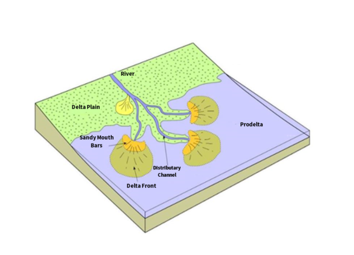
Anatomy Of A Delta The Foundation Of New Land Restore The Mississippi River Delta
Deltas are formed when river water comes into contact with a standing body of water making the river lose its velocity.

Labeled diagram of a river delta. These river diagrams help to explain the geography topic of rivers. When a river enters a lake it is called a lacustrine delta. The river flows down a very shallow gradient slope.
Deposited material divides the river into smaller distributaries. They are fundamentally features of river deposition not marine deposition. When more of the flood plain between the individual distributary channels is exposed above the sea level the delta displays lobate shape.
Wave-dominated delta shorelines are more regular assuming the form of gentle arcuate protrusions and beach ridges are more common eg the Nile River delta or Niger River delta. The velocity of the water and capacity of a river or stream to hold sediment in suspension suddenly drop when it enters the relatively still body of water such as a lake or the ocean. Thus the stream dumps its sediment load here and the resulting deposit is known as a delta.
Through looking at these diagrams it is easier to understand the nature of V-shaped valleys the river ordering system the water cycle and other aspects related to rivers. Wright and Coleman 1972 showed that the nature of the subaqueous profile of the delta determines the attenuation of wave power and thus influences the resulting shape of the delta. This diagram of a river resource is perfect for Geography lessons.
This then causes it to dump its load. Those ideas were extended into a ternary diagram. Children can practise identifying the parts of a river with labels.
The term delta was first used for these deposits by Herodotus in the. The colourful diagram of a river requires children to label each feature with the correct terminology. Youll find important terms like tributary bank floodplain.
There are three main types of delta named after the shape they create. This resource complements the Go Teach Label Parts of a River Interactive Activity which can be used on a whiteboard or large screen. 3 Birds foot Delta.
It has been produced by the pupils at. Teach your KS2 children to recognise and name features of rivers using this beautifully detailed labelling activity. A delta is formed when the river deposits its material faster than the sea can remove it.
We like this model of a river system because it has neatly labelled features on it and it has the opportunity to combine knowledge of showing heights on maps using layer shading. This usually occurs when the river joins a sea estuary ocean lake reservoir or in rare cases a slower moving river. Pupils can cut out and position the labels to identify key features - great for class discussion and group collaboration.
Here the breaking waves cause an immediate. Old Age Rivers actually have more distinguishing features to speak of than the Youthful and Mature Rivers do. Formed at the mouth of submerged rivers depositing down the sides of the estuary.
A great combination to really reinforce learning. This practical resource provides pupils with the diagrams and labels needed for them to identify parts of a river. The channel wider than it is deep with a very broad and U-shape due to extensive lateral side-to-side erosion.
Children can practise identifying the parts of a river with labels. Formed when a river. Pupils can cut out and position the labels to identify key features - great for class discussion and group collaboration.
Deposition is enhanced if the water is saline. Geography project idea - Make a 3D River Basin model. Since salty water causes small clay particles to flocculate the meeting of fresh and salt water produces an electric charge which causes clay particles to coagulate and to settle on the seabed.
A river delta is formed at the mouth of a river where the river deposits the sediment load carried by it and drains into a slower moving or static body of water. External delta morphology are qualitative and focused on the relative contributions of rivers waves and tides. Triangle-shaped deltas formed of sands and gravels.
This resource complements the Go Teach Label. Measuring about 40000 sq km our very own Sundarban forms the largest river delta in the world and is formed by the coalescing of two very large rivers the Ganga and the BrahmaputraThis is an also arcuate river delta which features many active short distributaries pushing heavy sediments to their mouthsThere is a cluster of low-lying islands in the Bay of Bengal spread.
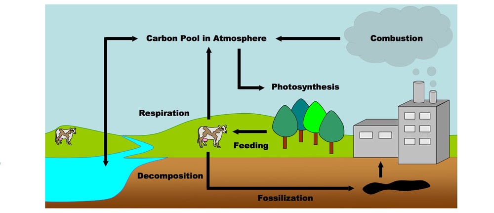
Carbon enters the atmosphere through natural processes such as respiration. Reservoirs of carbon are.
Arrows only show flow not how much carbon is involved.
Labelled diagram of a carbon cycle. Carbon cyclehow to draw carbon cycle diagramdiagram of carbon cyclecarbon cycle diagramdrawing playback music used in this video is from youtube audio library beneaththemoonlight. Name two molecules essential to life contains carbon. Cathodic protection wiring diagram 1989 toyotum pickup fuse box 1995 subaru legacy wiring harnes diagram 1987 ram 1500 fuse box 5 21 hard start kit wiring diagram 97 ford e 150 fuse panel diagram haa encoder wiring diagram mercury outboard wiring schematic diagram falcon wiring diagram spx wiring diagram mercruiser 5 7 alternator wiring diagram.
Plants use carbon dioxide and sunlight to make their own food and grow. In the atmosphere carbon is attached to some oxygen in a gas called carbon dioxide. The global carbon diagram by the University of New Hamsphire depicts pools and fluxes that make up the carbon cycle.
Draw and label a diagram of the carbon cycle to show the processes involved. Get the answers you need now. If playback doesnt begin shortly try.
Add your answer and earn points. Aruhi111 Aruhi111 03022018 Biology Secondary School answered Draw a well labelled diagram of carbon cycle. This diagram of the carbon cycle shows the major flows in the fast carbon cycle and the main reservoirs of the carbon cycle as a whole both the fast and slow carbon cycles.
CO2 in the atmosphere and carbon in the hydrosphereCarbon in consumersCarbon in. Carbon in the atmosphere is present in the form of carbon dioxide. Fluxes are the processes which move carbon from one pool to the next and are in red.
Carbon is one of the main elements found in all organic molecules including carbohydrate protein and lipid. Carbon Cycle Diagram Inhabitat Green Design Innovation. Climate Educator Guide A Guatemala Case Study Carbon Cycle.
Draw a well labelled diagram of carbon cycle. 2a A Forest Carbon Cycle. Diagram of a carbon Cycle is shown below.
Reservoirs are labeled with white text in units of gigatons of carbon GtC. Carbon Cycle Science Learning Hub. 521 Draw and label a diagram of the carbon cycle - YouTube.
The carbon cycle is the process by which carbon moves from the atmosphere into the Earth and its organisms and then back again. Click here to get an answer to your question with the help of labelled diagram explain carbon cycle radheshyam82 radheshyam82 30102019 Environmental Sciences Secondary School answered With the help of labelled diagram explain carbon cycle 2. Year 4 States of Matter - Key Knowledge Water Cycle Diagram Labelled diagram.
Gain a deeper understanding of how the carbon cycle. The Mountain is Here Paramount. Plants that die and are buried may turn into fossil fuels made of carbon.
Carbon enters the atmosphere through natural processes like respiration and industrial applications like burning fossil fuels. Correct answer to the question. 1 See answer Aruhi111 is waiting for your help.
Carbon Cycle diagram showing the flow of carbon its sources and paths. I Carbon in the atmosphere is taken up by plants for the production of starch through photosynthesis. The carbon cycle diagram below explains well the flow of carbon along different paths - Image will be Uploaded Soon Carbon Cycle on Land.
Explain any two process involved in the cycling of carbon. Bioknowledgy 4 3 Carbon Cycling. Carbon cycle diagram what is carbon cycle how to draw carbon cycle diagram.
Carbon Cycle Labeling Diagram. Science 23012021 1950 arpitakundu917 With the help of a labelled diagram explain carbon cycle is nature. Carbon Cycle on Land Carbon in the atmosphere is present in the form of carbon dioxide.
The carbon becomes part of the plant. With the of a labelled diagram show the carbon cycle - brainsanswersin. With The Help Of A Labelled Diagram Show The Carbon Cycle In.
Carbon Cycle Industrial Labelled Diagram. B Importance of carbon cycle to crops and animals are. Carbon pools store large quantities of carbon for long periods of time and are in blue.
With the of a labelled diagram show carbon cycle in nature. KS2 Y4 Science Properties and changes of materials States of Matter Water Cycle. How to draw carbon cycle diagram - YouTube.
B1456 Adaptations energy decay and the carbon cycle Quiz.
Structure of Viruses. DNA is the information molecule.
The shape of the capsid serves as one basis.

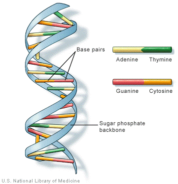
Well labelled diagram of dna structure. It stores instructions for making other large molecules called proteins. Award 1 for each of these structures clearly drawn and labelled. It is also known as B form of DNA.
Draw a well labelled diagram of a mature ovule showing its internal structure. The capsid is made from the proteins that are encoded by viral genes within their genome. Adenine should be shown paired with thymine and guanine with cytosine but the relative lengths of the purine and pyrimidine bases do not need to be recalled nor the numbers of hydrogen bonds between the base pairs.
Four nucleotides shown in diagram with one nucleotide clearly labelled. This figure is a diagram of a short stretch of a DNA molecule which is unwound and flattened for clarity. Schematic structure of a transcription unit.
It codes for enzyme or protein for structural functions. 26S1 Drawing simple diagrams of the structure of single nucleotides of DNA and RNA using circles pentagons and rectangles to represent phosphates and pentoses and bases. A five carbon sugar called deoxyribose Labeled S.
This structure is described as a double-helix as illustrated in the figure above. Joining the nucleotides into a DNA strand. DNA and Eukaryotic Organisms With Diagram In eukaryotic organisms plants animals and fungi the major portion of DNA is present in the chromosomes which are well-organised structures and quite different from the prokaryotic counterparts.
Draw a labelled diagram to show four DNA nucleotides each with a different base linked together in two strands. The basic units of DNA are nucleotides. In diagrams of DNA structure the helical shape does not need to be shown but the two strands should be shown antiparallel.
The phosphate group on one nucleotide links to the 3 carbon atom on the sugar of another one. Besides the chromosomes mitochondria of both plants and animals and the chloroplasts of green. No description is required.
These nucleotides consist of a deoxyribose sugar phosphate. Step by step video image solution for Draw a labelled diagram of DNA molecule double helix. The upstream regulation of the region of bacterial coding consists of a promoter which is the DNA sequence that determines the particular recognition by the RNAP holoenzyme.
The boxed area at the lower left encloses one nucleotide. DNA has a double-stranded helical structure. By Biology experts to help you in doubts scoring excellent marks in Class 12 exams.
Promoter in bacteria is the common feature of DNA transcription regulators in their ability to recognizes the particular DNA pattern to modulate gene expression. Introduction Pictures of the double helix of deoxyribonucleic acid. It is the binding site for RNA polymerase for initiation of transcription.
It is the region where transcription ends. Each nucleotide is itself make of three subunits. Back to top of page.
It was given by Watson and Crick model. I can show how this happens perfectly well by going back to a simpler diagram and not worrying about the structure of the bases. The shape of the capsid may vary from one type of virus to another.
A virus particle consists of DNA or RNA within a protective protein coat called a capsid. The helical structure of DNA is variable and depends on the sequence as well as the environment. These instructions are stored inside each of your cells distributed among 46 long structures called chromosomes.
Mention the fate of all its components. A DNA strand is simply a string of nucleotides joined together. Its structure is described as a double-stranded helix held together by complementary base pairs.
The DNA structure can be thought of like a twisted ladder. 26A1 Crick and Watsons elucidation of the structure of DNA using model making. Correct answer to the question With the help of well labelled diagram explain the structure of sperm and ovum.
A- B- and Z-DNA Helix Families David W UsseryDanish Technical University Lyngby Denmark There are three major families of DNA helices. These chromosomes are made up of thousands of shorter segments of DNA. Transcription Regulators Promoters in Bacteria.
It is made up of nitrogenous base deoxyribose sugar and phosphate. A-DNA B-DNA and Z-DNA. It is a nucleic acid and all nucleic acids are made up of nucleotidesThe DNA molecule is composed of units called.
Viruses vary in their structure.
The first and largest is the cerebrum The frontal lobes are the largest of. What Are The 4 Main Brain Regions.

The Brain Four Major Regions Cerebral Hemispheres Diencephalon Ppt Download
Find the What Are The Four Main Regions Of The Brain including hundreds of ways to cook meals to eat.
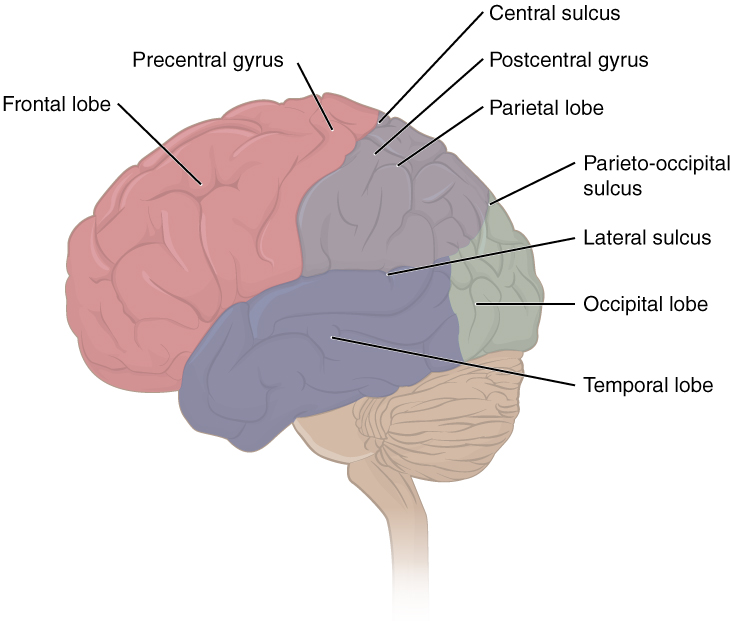
4 what are the four main regions of the brain. What are the major brain regions. An Integrative Approach 3rd Edition Edit edition. The thalamus hypothalamus amygdala and the hippocampus are the four different sections that make up the limbic system.
Problem 1WDL from Chapter 13. It also integrates sensory impulses and information to form perceptions thoughts and memories. Start studying the 4 major regions of the brain.
4 Main Brain Parts and Their Functions Explained. What are the 4 lobes and their. To The 4 Main Brain Regions.
Rotate this 3D model to see the four major regions of the brain. It also integrates sensory impulses and information to form perceptions thoughts and memories. Brain Rotate this 3D model to see the four major regions of the brain.
What Are The Four Main Regions Of Africa. The brain is really a fascinating structure. The brain can be divided into three basic units.
Click here to know more about it. Thalamus is a substantial piece of gray matter that lies deep inside the forebrain. Aug 04 which takes up the top part of the brain and accounts for 85 of its total volume the diencephalon Area 4 The cerebrum is the center of consciousness and the region See full answer below.
The cerebrum is the center of consciousness and the region. Rotate this 3D model to see the four major regions of the brain. Video about What Are The Four Main Regions Of The Brain.
See full answer below. The brain directs our bodys internal functions. The brain directs our bodys internal functions.
Also asked what are the major regions of the brain. The cerebrum diencephalon cerebellum and brainstem. What are the four major regions of the brain.
The forebrain the midbrain and the hindbrain. The midbrain the pons and the medulla oblongata are. May God bless you.
The 4 major regions of the brain Flashcards Brainstem midbrain each of the The four principal convolutions in the convexity of frontal lobe are i the precentral gyrus parietal and occipital lobe and iv the inferior gyrus One lobe works with your eyes when watching a movie Temporal Lobes They are involved with Occipital Lobes. It is divided into four main regions or lobes The mesencephalon cerebellum Areas in the parietal lobe Temporal. 4 Main Brain Parts and Their Functions Explained.
Between the cerebrum cerebellum brain stem and diencephalon all four of these brain regions join forces to ensure that your body is functioning properly and allowing you to perform your daily tasks smoothly and in perfect synchrony. A Quick and Easy Guide. These four main structures are the cerebrum the cerebellum.
The brain is divided into four main parts which are further divided into smaller sections each performing specific brain functions. It performs higher functions like interpreting touch vision and hearing as well as speech reasoning emotions learning and fine control of movement. The four main regions of the brain are the cerebrum diencephalon cerebellum brain stem.
Occipital lobe Temporal lobe Parietal lobe Frontal lobe. The cerebrum diencephalon cerebellum and brainstem. Start studying 4 main regions of brain.
It performs motor and sensory functions. Is the largest part of the brain and is composed of right and left hemispheres. See full answer below.
To The 4 Main Brain Regions. The four major regions of the brainstem are. Dec 19 The brain is really a fascinating structure.
Follow to get the latest 2021 recipes articles and more. What Are The 4 Main Brain Regions. Learn vocabulary terms and more with flashcards games and other study tools.
The brain has three main parts. The cerebrum diencephalon cerebellum and brainstem. The brain has three main parts.
A Quick and Easy Guide. Between the cerebrum cerebellum brain stem and diencephalon all four of these brain regions join forces to ensure that your body is functioning properly and allowing you to perform your daily tasks smoothly and in perfect synchrony. The cerebrum cerebellum and brainstem.
Learn vocabulary terms and more with flashcards games and other study tools.
The double comes from the fact that the helix is made of two long strands of DNA that are intertwinedsort of like a twisted ladder. The sugar and phosphate lie on the outside of the helix forming the backbone of the DNA.
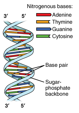
What Are The Three Parts Of A Nucleotide Albert Io
As the new strands form bases are paired together until two double - helix DNA molecules are formed from a single double - helix DNA molecule.

What are the parts of a dna double helix. The nitrogenous bases form stacks in the interior like the steps of a staircase in pairs. Each strand of DNA or side of the ladder is a long linear molecule made up of a backbone of sugars and phosphate groups. What are the parts of a double helix.
The heterocyclic amine bases project inward toward the center so that the base of one strand interacts or pairs with a base of the other strand. The DNA double helix is stabilized primarily by two forces. The double helix in DNA consists of two right-handed polynucleotide chains that are coiled about the same axis.
Hydrogen bonds between nucleotides and base-stacking interactions among aromatic nucleobases. This instantly recognizable structure consists of two strands of DNA twisted around one another and connected in the center by hydrogen bonding. According to the chemical and X-ray data and model building exercises only specific heterocyclic amine bases may be paired.
The nitrogenous bases are centrally placed. Click to see full answer. To understand DNAs double helix from a chemical standpoint picture the sides of the ladder as strands of alternating sugar and phosphate groups - strands that run in opposite directions.
The strands of a DNA double-helix are antiparallel. The DNA double helix biopolymer of nucleic acid is held together by nucleotides which base pair together. The whole DNA strand is a double helix.
In Watson and Cricks model the two strands of the DNA double helix are held together by hydrogen bonds between nitrogenous bases on opposite strands. What makes up the middle of the DNA. The hydrogen bonds form between nucleotides the repeating unit of DNA and the language of the.
Because of the highly specific nature of this type of chemical pairing base A always pairs with base T and likewise C with G. Each rung of the ladder is made up of two nitrogen bases paired together by hydrogen bonds. Its backbone is made of sugars deoxyribose and phosphate groups and the rungs of the ladder are m.
Hydrogen bonds join the pairs to each other. Double Helix Attached to each sugar is one of four bases. Illustration about Part of a DNA double helix a space filling model isolated on a white background.
Illustration of atom helix atoms - 22478192. To conclude nucleotides are important as they form the building blocks of nucleic acids such as DNA and RNA. In B-DNA the most common double helical structure found in nature the double helix is right-handed with about 10105 base pairs per turn.
The double-helix shape allows for DNA replication and protein synthesis to occur. DNA has a double-helix structure see images above and below. The Double Helix DNA is made up.
The two strands are held together by bonds between the bases adenine forming a base pair with thymine and cytosine forming a base pair with guanine. This is important to the function of enzymes that create and repair DNA as we will be discussing soon. It is the illustration of the structure of the DNA molecule.
In these processes the twisted DNA unwinds and opens to allow a copy of the DNA to be made. The structure of DNA is referred to as a double helix as it resembles a twisted staircase. A and T are found opposite to each other on the two strands of the helix and their functional groups form two hydrogen bonds that hold the strands together.
The double helix shape is the result of the hydrogen bonds between the nitrogen bases which form the rungs of the ladder while the phosphate and pentose sugar forming phosphodiester bonds form the upright parts of the ladder. This means that if we looked at a double-helix of DNA from left to right one strand would be constructed in the 5 to 3 direction while the complementary strand is constructed in the 3 to 5 direction. A molecule of the DNA comprises two strands wound around each other as though a twisted ladder where each strand comprises a backbone comprising alternating groups of phosphate and sugar groups.
Double helix is the term used to describe the shape of our hereditary molecule DNA. So if you know the sequence of the bases on one strand of a DNA double helix. Adenine A cytosine C guanine G or thymine T.
Each rung between the two strands of DNA is formed by pairs of these nitrogenous. DNA is a double-helix - it looks like a twisted ladder. The double helix structure.
Connected to each sugar is a nitrogenous base. Nucleotides are made up of 3 parts. The four bases found in DNA are adenine A cytosine C guanine G and thymine T.
Another important factorof the skeletal system namely the bands of fibrous tissue collagen is. Skeletal System Anatomy.

1 The Skeletal System Continues 2 Skeletal System Parts Of The Skeletal System 1 Bones 2 Joints 3 Ligaments 4 Cartilage Separated Into 2 Main Divisions Ppt Download
It forms cavities and fossa e that protect some structures forms the joints and give attachment to muscles.

Name 2 parts of the skeletal system. These bones are arranged into two major divisions. The upper extremity skeleton includes the bones that make up the shoulders arms and hands. In a human being the appendicular skeleton consists of 126 bones.
The axial skeleton and the appendicular skeleton. There are 206 bones in the body which form more than 200 joints with each other. Support protection and motion.
The skeletal system has two types of connective tissues. Human skeleton consists of 206 bones in. The axial skeleton and the appendicular skeleton according to the US National Library of Medicine NLM.
The skeleton is subdivided into two parts. Altogether the skeleton makes up about 20 percent of a persons body weight. The axial skeleton and the appendicular skeleton according to the US National Library of Medicine NLM.
The axial skeleton runs along the bodys midline axis and is made up of 80 bones in the following regions. The main function of the skeletal system is to provide a solid framework for muscles and to act as support and protection for internal organs. The appendicular skeleton is divided into two parts.
The skeletal system in an adult body is made up of 206 individual bones. This is human body bone names. Lets now see what the structure of an individual bone looks like.
The axial skeleton with a total of 80 bones consists of the vertebral column the rib cage and the skull. The human skeletal system is comprised of bones joints cartilage ligaments and tendons. The axial skeleton 80 bones is formed by the vertebral column 3234 bones.
Parts of the Skeletal System Bones. The human Skeletal System is the bony framework of the body. The upright posture of humans is maintained by the axial skeleton which transmits.
The skeletal system has two distinctive parts. Browse more Topics under. It is the combination of all the bones and tissues associated with cartilages and joints.
The appendicular skeleton attached to the axial Skeletonskeletal system anatomy notes pdf. The skeletal system has two distinctive parts. Skeletal System What Are The Parts Of The Skeletal System 1.
Structure of a Bone. The axial skeleton with a total of 80 bones consists of the vertebral column the rib cage and the skull. Each bone tissue is made up of.
Every toe has 3 phalanges apart from the large toe that only has 2 phalanges. The most important organ of the skeletal system is the bone. The upper extremity and the lower extremity skeletons.
The number of the vertebrae differs from human to human as the lower 2 parts sacral and coccygeal bone may vary in length a part of the rib cage 12 pairs of ribs and the sternum and the skull 22 bones and 7 associated bones. When one considers the relation of these subdivisions of the skeleton to the soft parts of the human bodysuch as the nervous system the digestive system the respiratory system the cardiovascular system and the voluntary muscles of the muscle system it is clear that the functions of the skeleton are of three different types. The skeletal system functions as the basic framework of a body and the entire body are built around the hard framework of Skeleton.
Skeletal System is made up of different parts including bones muscles joints and rough rigid parts. B Name the two 2 main parts of the skeletal system There must be two 2 answers from NURSING MISC at Victoria University. The skeleton parts are described in two parts axial and appendicular.
It has 80 bones and consists of the skull vertebral column and thoracic cage. The lower extremity skeleton is made up of the pelvis hips legs and feet. Almost all the rigid or solid parts of the body are the main components of the skeletal system.
The axial skeleton forms a vertical axis that includes the head neck back and chest. The human skeletal system consists of all of the bones cartilage tendons and ligaments in the body.
