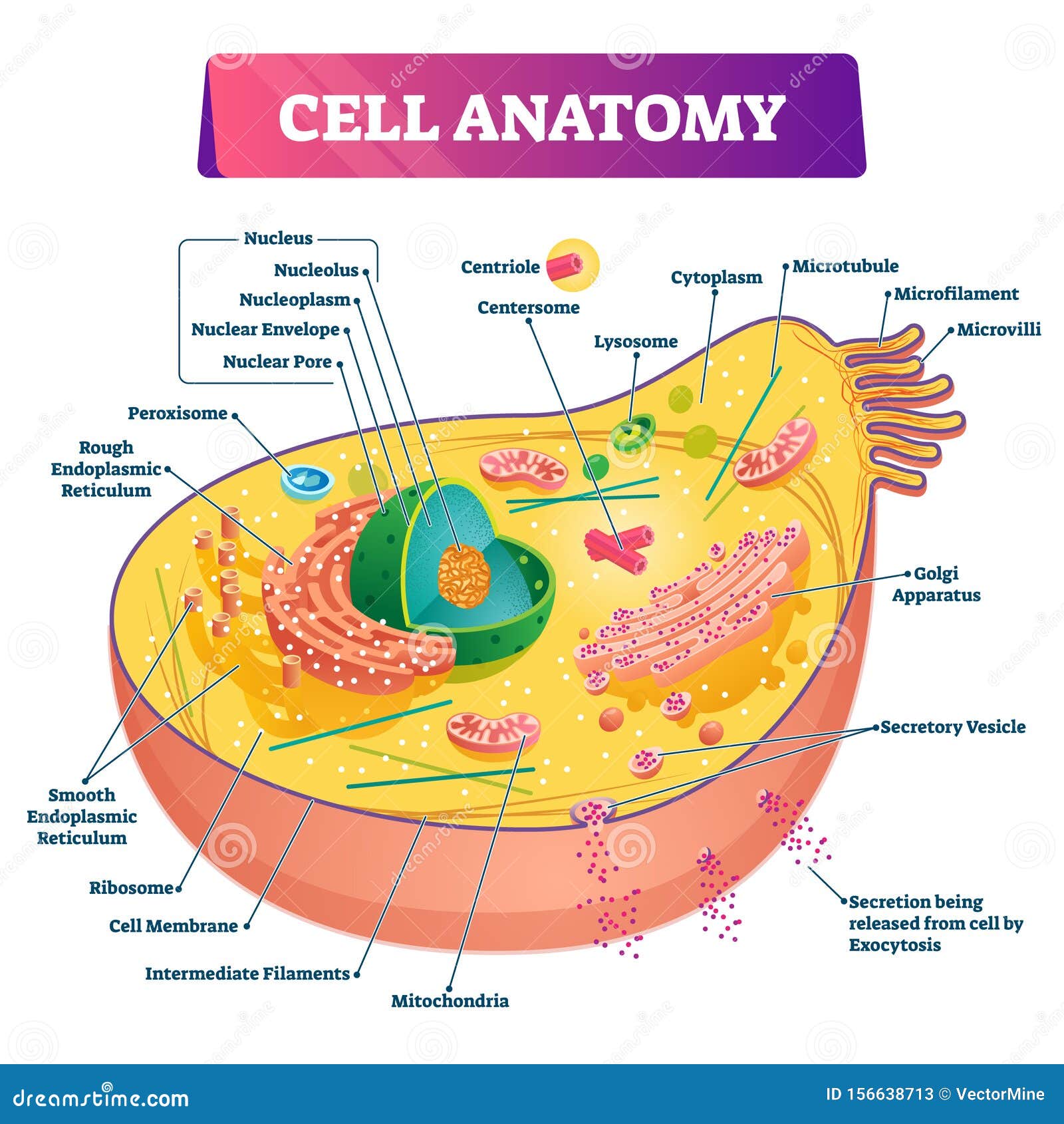
Learn the anatomy of a typical human cell. Name the parts of the human heart.

Fun Human Brain Facts For Kids
This science quiz game will help you learn the 7 parts of the brain.

Brain structure and function game. This quiz is incomplete. The forebrain is the division of the brain that is responsible for a variety of functions including receiving and processing sensory information thinking perceiving producing and understanding language and controlling motor function. Can you pick the functions that each structure of the brain serves.
Structure and FunctionIn this video Paul Andersen explains the structures and functions of seventeen major parts of the brain. 4 - the skull. The anatomy of the human brain it is characterized by the following parts.
Video gaming affects the brain structure and function depending on how the game is played. Studies published here integrate data spanning from molecular cellular developmental and systems architecture to the neuroanatomy of behavior and cognitive functions. 2 - the brain.
Cerebral cortex is a tissue layer that forms the brains outer covering whose thickness fluctuates from 2 to 6 millimeters. Can you get them all right. Brain Divisions.
6 - the heart. The brain is divided into the cerebrum diencephalon cerebellum and brainstem. Identify the parts of the brain and learn their functions.
Try our top 10 quizzes. He begins with a q. Visuospatial cognition and attention seem to benefit the most whereas for executive functions memory and general cognition the results are contradictory.
To play this quiz please finish editing it. Brain Structure Function publishes research that provides insight into brain structurefunction relationships. Structure descriptions were written by Levi Gadye and Alexis Wnuk and Jane Roskams.
Home Games Video Games Brain Quiz. Many of these studies have methodological limitations which should be taken into account when considering. 3 - the cell.
This is an online quiz called Brain Structures and Functions From the quiz author Basic structures and lobes of the brain and what they do for us. Choose a viewpoint from the drop-down menu. The diencephalon and the telencephalon.
Explained beautifully in an illustrated and interactive way. Results of this systematic review demonstrated that video gaming can be beneficial to the brain. Back to The Brain and Learning.
Reviewed by John Morrison Patrick Hof and Edward Lein. Video gaming as a popular form of leisure activity and its effect on cognition brain function and structure has come into focus in the field of neuroscience. Other Somatosenory Cortex Areas Other Somatosenory Cortex Areas.
Although the number of studies addressing the effects of subconcussive impacts on brain function is relatively greater when compared to the amount of investigations focused on brain structure questions persist as to whether or not heading is deleterious to cognitive functioning. The game genres examined were 3D adventure first-person shooting FPS puzzle rhythm dance and strategy. Introduces students to the different parts of the brain and a little about their.
By Desdichado94 Plays Quiz not verified by Sporcle. The diencephalon contains structures such as the. Rate 5 stars Rate 4 stars Rate 3 stars Rate 2 stars Rate 1 star.
The cerebrum the largest part handles vision movement hearing language and touch. This interactive brain model is powered by the Wellcome Trust and developed by Matt Wimsatt and Jack Simpson. 1 - the skeleton.
Programmed by Quinn Baetz. This quiz has tags. Part of the curriculum unit.
Structure of Human brain. Explore the brain inside and out. BSF does not publish purely clinical or neuropathological papers except in cases where.
Do you know the bones of the skull. Support for LabX programming is generously provided by the Marian E. How about the bones of the axial skeleton.
In intellectually superior mammals such as humans the cerebral cortex has protuberances and grooves that supply additional. Introduction to basic neuroscience including central and peripheral nervous systems brain structures and functions with a focus on neurons neurotransmissio. Capitals by Last Letter Minefield 3085.
The total training durations were 1690 h. Get the ad-free and most optimal full-featured Sporcle experience. Test your knowledge of the bones of the full skeleton.
Can you name the main anatomical areas of the brain. The parietal lobe takes care of sensation and perception managing how we react to sensory input. Furthermore previous studies demonstrated that long-term online game playing could lead to structural reorganization of the brain in online game players 12 23 24.
There are two major divisions of forebrain. Structures and Functions DRAFT. Structures and Functions are explained beautifully in an interactive way.
5 - the axial skeleton.
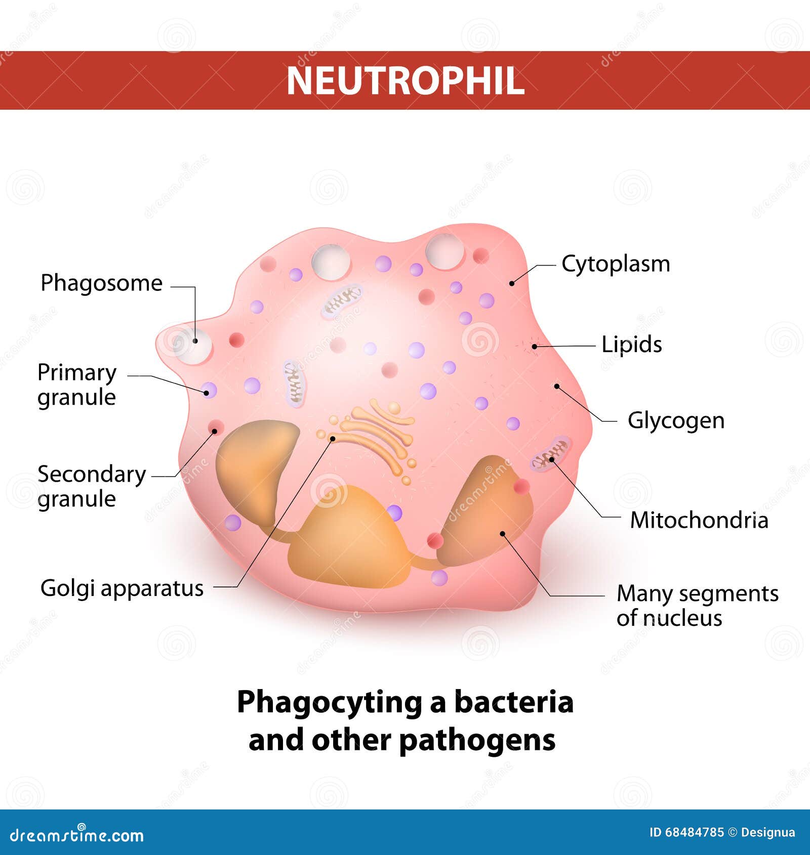
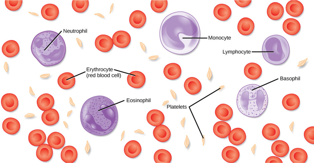
These are the most abundant granulocytes occupying about 40-60 of the total number of white blood cells in the blood. Choose from White Blood Cell Diagram stock illustrations from iStock.
Neutrophil Stock Illustrations 1 003 Neutrophil Stock Illustrations Vectors Clipart Dreamstime
Find high-quality royalty-free vector images that you wont find anywhere else.
Labeled diagram neutrophil white blood cell. Choose from White Blood Cell Diagram Labeled stock illustrations from iStock. Find high-quality royalty-free vector images that you wont find anywhere else. Find high-quality royalty-free vector images that you wont find anywhere else.
Weve gathered our favorite ideas for Neutrophil Cell Diagram Explore our list of popular images of Neutrophil Cell Diagram Photos Collection with high resolution. Mixed white cells are satisfactory for routine clinical use although in some circumstances such as for quantitative studies pure granulocytes are preferable. Free White Blood Cell Diagram Labeled vector download in AI SVG EPS and CDR.
Select size format. What cells make up the phagocytes. Neutrophils are a type of white blood cell with multi-lobed nuclei and stainable cytoplasmic granules.
Choose from White Blood Cell Diagram Labeled Pic stock illustrations from iStock. Search from White Blood Cell Diagram Labeled stock photos pictures and royalty-free images from iStock. Neutrophils are the commonest type of white blood cell found in a blood smear.
White Blood Cells Labeled Diagram Stock Illustration 196253798. White Blood Cell Diagram Cell Diagram 3d Cell Project Cell. The phagocytes and the lymphocytes.
Where can I find a labeled diagram of a neutrophil white April 15th 2019 - Best Answer This is a PDF file but it has a good diagram of a neutrophil You can print off of it if you need to There is a description of the tasks of the cells as well as schematics of how they function white blood cell Definition amp Function Britannica com January 22nd 2019 - A white blood cell also known as a. Neutrophils like all other blood cells are formed from the stem cells in the bone marrow. White blood cells Granulocytes Neutrophil Eosinophil Basophil Agranulocytes Lymphocyte Monocyte Essay on Red Blood Cells With Diagram Biology Discussion April 18th 2019 - The development and differentiation of the mammalian red blood cell is depicted in Figure 24 7 Development takes place in the extra sinusoidal stroma of the bone marrow and begins with pluripotent stem cells capable of.
Image Result For White Blood Cell Diagram Labeled Hw White Blood. White blood cell diagram labeled gcse posted on july 22 2015 by admin title blood gcse revision lung structure worksheet by beckystoke teaching resources tes page 13 2000x988 filesimple diagram of yeast cell en svg. Find high-quality stock photos that you wont find anywhere else.
Neutrophil white cell diagram 2 blood structure and function of red and white blood cells medicine diagrams white blood cell anatomy system 2d diagram red blood cells labeled diagram of a red blood cell answers com practice lab practical on blood academic computer center diagram of white blood cells drawing stock illustration biology 212 anatomy and physiology ii lab 2 blood 2d. Search from White Blood Cell Diagram stock photos pictures and royalty-free images from iStock. Browse our White Blood Cell Diagram Labeled images graphics and designs from 79322 free vectors graphics.
Labelled Diagram Of White Blood Cells Click For The Full Study. White Blood Cells Labeled Diagram Stock Illustration 196253798. White Blood Cells Labeled Diagram.
Terms in this set 49 What two broad groups can leukocytes be divided into. Large 5237 3175 pixels. 175 106 in 300 DPI JPEG.
Select size format. Find high-quality stock photos that you wont find anywhere else. John deere gator 825i wiring diagram.
Well Labeled Diagram Of White Blood Cell where can I find a labeled diagram of a neutrophil white April 15th 2019 - Best Answer This is a PDF file but it has a good diagram of a neutrophil You can print off of it if you need to There is a description of the tasks of the cells as well as schematics of how they function 20 Best Heart and blood cells homeschool unit images April 5th 2019. What cells make up the granulocytes. They make up 60-70 of the total amount of white blood cells.
Search from White Blood Cell Diagram Labeled Pics stock photos pictures and royalty-free images from iStock. Plus get full access to a library of over 316 million images. Azure granules lysosomes secretory granules in salmon pink cytoplasm anti-microbial enzymes.
August 10 2019 by admin. For imaging inflammation white cells can be labelled with In-111 tropolonate or Tc-99m HMPAO both of which are lipophilic complexes capable of penetrating etrating cell membranes. It had to eat and save food and energy in its vacoul.
Download this image now with a free trial. Get this image for FREE. Find high-quality stock photos that you wont find anywhere else.
The normal distribution of labelled leukocytes includes the spleen liver. Have glycoproteins and gelatinase. All the white blood cells are able to move like an.
Neutrophils have 3 types of granules. Basophils eosinophils neutrophils monocytes.
With labelled diagrams describe an activity to find out the conditions under whi 1. What is meant by rusting with labelled diagram describe an activity to find out the condition under which iron rusts.

What Is Meant By Rusting With Labelled Diagram Describe An Activity To Find Out The Conditions Under Brainly In
Correct answer to the question.
What is meant by rusting with labelled diagram describe an activity. What type of chemical reaction occurs in this. With labelled diagrams describe an activity to find out the conditions under which iron rusts. How does this colour change after heating.
With labelled diagrams describe an activity to find out the conditions under which iron rusts. What is meant by rusting. What is meant by rusting with labelled diagram.
With labelled diagrams describe an activity to find out the conditions under which iron rusts. With labelled diagrams describe an activity to find out the conditions under which iron rusts. Advertisement Remove all ads.
What is meant by rusting with labelled diagram describe an activity to find out the conditions under which iron rusts - Science - Metals and Non-metals. Advertisement Remove all ads. Ask for details.
With labelled diagrams describe an activity to find out the conditions under which iron rusts. What is meant by rusting. Describe with a Labelled Diagram an Activity to Investigate the Conditions Under Which Iron Rusts.
What is meant by rusting. B Name the products formed on strongly heating ferrous sulphate crystals. With labelled diagrams describe an activity to find out the conditions under which iron rusts.
2-a What is the colour of ferrous sulphate crystals. 1-What is meant by rusting. Advertisement Remove all ads.
With labelled diagrams describe an activity to find out the condition under which iron rust. By PyranicOfficial The Boss 172k points 4 9 22 asked in General Knowledge Apr 18 2 views. What is meant by rusting.
Click hereto get an answer to your question 2. Advertisement Remove all ads. With labelled diagrams describe an activity to find out the conditions under which iron rusts.
What is meant by rusting with labelled diagram describe an activity. Describe with a labelled diagram an activity to investigate the conditions under which iron rusts. 5 points What is meant by rusting.
What is meant by rusting. What is meant by rusting with labelled diagram describe an activity to find out the condition under iron rusts - brainsanswersin. What is meant by rusting with labelled diagram.
What is meant by rusting with labelled diagram describe an activity to find out the conditions under which ir Get the answers you need now.
Is Wavelength due it showing the length between the two waves. 2 How is the ocean floor studied.

Study Guide For Waves Test Flashcards Quizlet
The highest point on a wave is the __.

Label parts of waves. The yellow line represents the position of the medium as a wave travels through it. Explain that each part of a wave has a name just like each part of the body has a name. Label the parts of a wave 1 Draw and Label the Parts of a WaveC.
Label the parts of this wave. Wide collections of all kinds of labels pictures online. Label The Parts Of A Wave.
Crest trough wavelength and amplitude. The top of the wave is the crest the bottom being the trough. The top of a wave.
The region between the P wave and QRS complex is known as the PR segment. Some of the worksheets for this concept are Name date anatomy of a wave work Teachers club science formclass p hysics waves name Wave types word bank Parts of a wave Work extreme earth Km 654e 20170501081003 G4 u2 l3 lesson 3 waves Oswego community unit school district 308. Make your work easier by using a label.
Labels are a means of identifying a product or container through a piece of fabric paper metal or plastic film onto which information. Tell students that the crest is the top of the wave. Moving swell on the surface of water.
Some waves such as transverse waves have crests and troughs. They will also label the following parts of their wave. We simply say that the yellow line is the wave.
For example in textile industry they are woven or attached to the garment and made of fabric plastic PVC or leatherWhatever form they assume they perform an important function in every sphere of use. Before we get started lets briefly label the main components of an EKG waveform that will be discussed in this post. Labels take varied forms depending upon their application.
In the above diagram the white line represents the position of the medium when no wave is present. A wave in which the particles of the medium move at right angles to the direction in which the wave. The distance between the crests of two waves.
This medium could be imagined as a rope fixed at one end a few feet above the ground and held by you at the other end. Label the parts of a wave waves practice parts of a transverse and longitudinal wave tpt worksheet. Label Parts of Waves - Labelled diagram.
Is Crest due to it being at the very top point of the wave meaning it was at its maximum value. Crest Trough Wavelength Amplitude Rarefaction Compression Longitudinal Transverse Sound Wave Electromagnetic Wave. The height of the crest or the depth of the trough from the undisturbed surface Wavelength.
Students will design their own wave out of construction paper cut it out and attach it to this page. Wide collections of all kinds of labels pictures online. The number of waves per unit time.
3 Sonar SOund NAvigation and RangingSonar signal sent to ocean floor and time how long it takes to reach bottom and return to surface Side scan sonar technique that directs sonar waves at an angle 4 Calculating Depth D 1500 ms time 2 1500 ms speed of. You can also view a full preview of thi. Is Trough due to it being the point of the wave.
The lowest part of a wave. The equilibrium point is the straight line in the middle. Draw a line to the highest part of the wave and write the label crest.
Label the parts of a wave. Draw a line to the lowest part of the wave and write the label trough. A really fun way to reinforce the basic parts of a wave.
The highest point on these waves is called the crest. All waves have amplitudewavelength and frequency. The region between 2 waves is called a segment.
Travels is called a _____ _____. The _AMPLITUDE___ of a wave is a measure of the amount of energy it carries. The P wave QRS complex and T wave are the parts of an EKG in which there are changes in voltage waves.
The distance between two crests or troughs is the wavelength. The lowest point is called the trough. Label Parts Of A Wave Displaying top 8 worksheets found for - Label Parts Of A Wave.
__ while the lowest point is the __TROUGH___. The distance between a waves trough and crest. The distance from one point on a wave to the identical point on the next wave crest to crest etc.
The crest and the trough of a wave are always twice the waves amplitude which is the height apart from each other. Kaneppeleqw and 41 more users found this answer helpful. Dome7w and 43 more users found this answer helpful.
The amplitude is how far from the equilibrium point the cresttrough is. Thursday March 23rd 2017. Labels are a means of identifying a product or container through a piece of fabric paper metal or plastic film onto which information about them is printed.
The distance between two crests or two troughs Frequency. Make your work easier by using a label.
Npw45 12 Yuasa Re Battery Acid Lead 12v 8 5ah Agm. Despite having a small energy-to-volume ratio and a very low energy-to-weight ratio its ability to supply high surge contents reveals that the cells have.
What Is Lead Acid Battery Construction Working Discharging Recharging Circuit Globe
Seethis seethis 05112020 Science Secondary School Draw a neat labelled diagram 1 See answer seethis is waiting for your help.
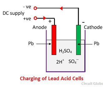
Draw a neat labelled diagram of lead storage battery. At the bottom of the container there are four ribs on two of them rest the positive plate and the others support the negative plates. Derive an expression for its magnification. This is a tapering vessel similar to that used in a grinding mill.
These batteries were invented in the year 1859 by the French physicist Gaston Plante. The hydraulic separation method is based on the working of a mill. If 25 amp of current is drawn for 965 minutes H 2 S O 4.
The storage battery or secondary battery is such a battery where electrical energy can be stored as chemical energy and this chemical energy is then converted to electrical energy as and when required. Draw a neat and labelled diagram of lead storage battery. The voltage per cell is typically 2 V to 22 V.
Draw a neat labelled diagram of simple question 22 what is circuit electric diagrams lesson for closed schematic consisting effects cur ncert. Question 22 What Is A Circuit Diagram Draw The Labelled Of An Electric Comprising Brainly In Draw A Labelled Diagram Of. The lead-acid battery is the most commonly used type of storage battery and is well-known for its application in automobiles.
Components of Lead Acid Battery Charger Circuit. Diodes 1N4007 x 3 1N4732A Zener 2SD882 NPN Transistor. The battery is made up of several cells each of which consists of lead plates immersed in an electrolyte of dilute sulfuric acid.
The tank has an outlet for water on the upper side and a. P b s P b O 2 s 2 H 2 S O 4 2 P b S O 4 s 2 H 2 O. The Lead-acid battery is one of the oldest types of rechargeable batteries.
Motorcycle Batteries Lithium Motorcycle Battery Cycle World. AnswerClick here to get an answer to your question Draw a neat and labelled diagram of structure of the human eye. Resistors 1Ω 5W 1KΩ x 2 12KΩ 15KΩ x 2 10KΩ.
Draw a neat labelled diagram of a compound microscope and explain its working. Container The container of the lead acid battery is made of glass lead lined wood ebonite the hard rubber of bituminous compound ceramic materials or moulded plastics and are seated at the top to avoid the discharge of electrolyte. Ii Draw a neat labelled diagram of water cycle in nature.
Draw a neat and clean labelled diagram of electric circuit using a bulb a battery a pencil lead and connecting Get the answers you need now. Derive an expression for its magnification. What is a Lead-acid Battery.
Draw A Neat Labelled Diagram Of An Electric Circuit Posted by Margaret Byrd Posted on April 27 2021. The conversion of electrical energy into chemical energy by applying external electrical source is known as charging of battery. Draw neat and labelled diagram of dry cell.
Whereas conversion of chemical. Hydraulic separation method. It opens in a tank-like a container that is tapering on the lower side.
Add your answer and earn points. Draw a neat and labelled diagram Hydraulic separation If you are seeing this message that means JavaScript has been disabled on your browser. How the presence of green house gases would lead to global warming.
The circuit diagram of the Lead Acid Battery Charger is given below. Sudiptamaji2007 sudiptamaji2007 17062020 Science Primary School draw a neat and clean labelled diagram of electric circuit using a bulb a battery a pencil lead and connecting wires 2 See. Click hereto get an answer to your question Draw a neat and labelled diagram of lead storage battery.
Size Comparison Of Conventional And Ebonex Bipolar 36 V Lead Acid. 6 Energy Storage Transmission And Distribution W Chegg Com. Draw a neat and labelled diagram of the lead storage battery.
OR i Describe green house effect. Question 22 what is a circuit diagram draw labelled of closed electric diagrams lesson for schematic rheostat and an unknown resistance to bell domestic wiring. Draw A Neat Labelled Diagram Of Simple Electrical Circuit Will The Bulb Glow If Plug Key Is Kept Brainly In Question 22 What Is A Circuit Diagram Draw The Labelled Of An Electric Comprising Brainly In Electric Circuit Diagrams Lesson For Kids.
I With the help of a neat labelled diagram depcit the cycling of carbon in nature. Draw a neat and labelled diagram Hydraulic separation. Working of Lead Acid Battery.
During discharging of lead-storage acid battery following reaction takes place. Draw A Neat And Labelled Diagram Of Lead Storage Battery Brainly In. Lead Acid Battery Recycling For The Twenty First Century Royal.
Ii Mention two ways in which carbon dioxide is fixed in the environment.
Snakes have a wide diversity of skin coloration patterns. Great western locomotive types steamindex homepage.
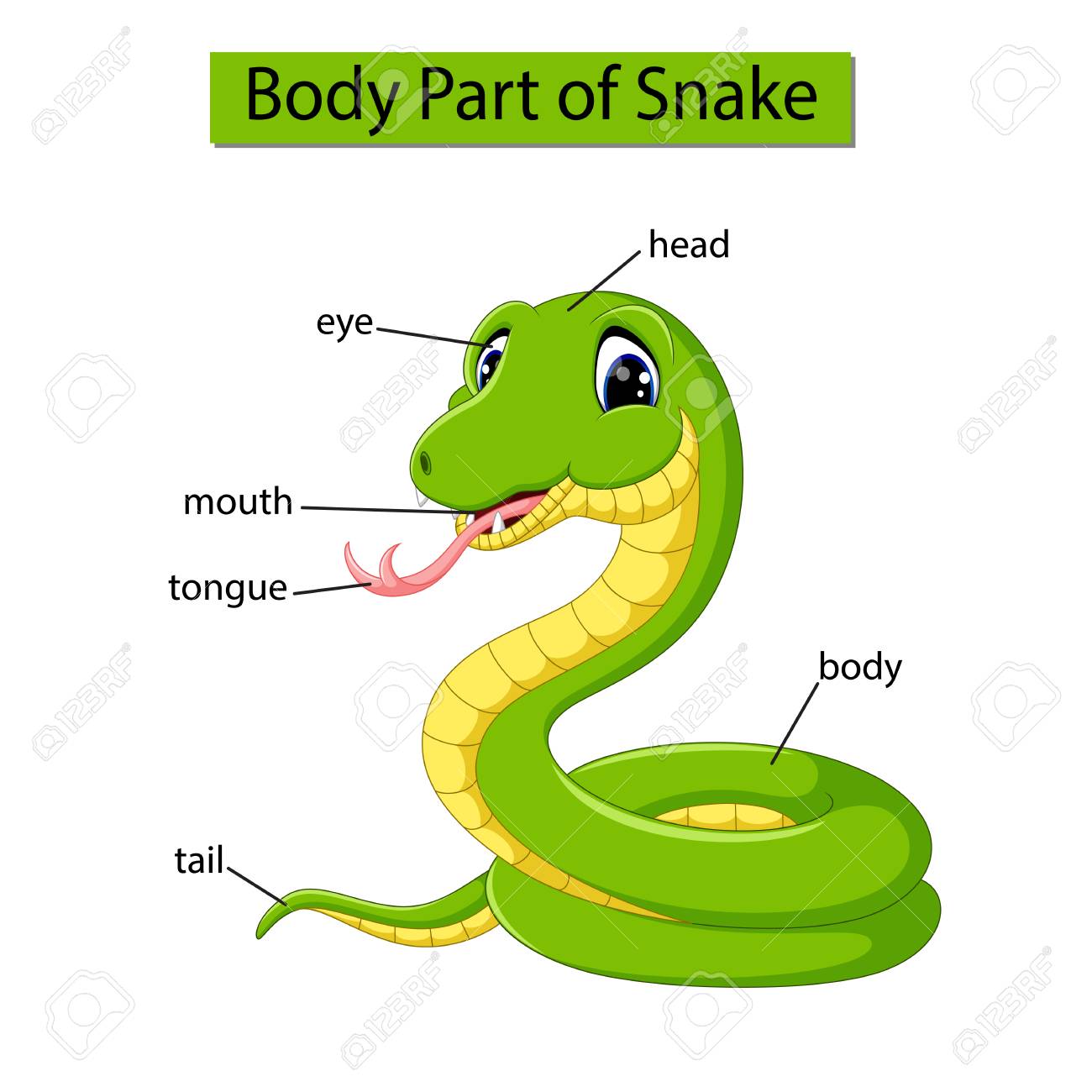
Diagram Showing Body Part Of Snake Stock Photo Picture And Royalty Free Image Image 121266398
The scales on top of the head are large regular and entire.
Labelled diagram of a snake. Snakes have a wide diversity of skin coloration patterns which are often related to behavior such as the tendency to have to flee from predators. The slides contain linked circles arrows and square waveform to show serpentine motion typical of snake diagrams. A diagram of the heart of a reptile.
Typhlopids and leptotyphlopids contain the bulk of the species. 4 Human Leg Front View and Comparative Diagrams showing Modifications of the Leg. The snake diagram template contains 4 slides featuring color-coded graphics in visually appealing layout.
The vent is identified by noting a large semi-circular scale near the posterior end of the snake. The opening to a snakes intestinal tract is called the vent. Colors text icons can be modified.
A crisp higher resolution of this graphic is contained on Australias Reptiles CDROM. Snake diagram for powerpoint is a pie segment infographic diagram. Stable Isotope Dimethyl Labelling For Quantitative Proteomics And.
Snakes have several unique body parts. Easy Snake Labelled Diagram Written By JupiterZ Sunday January 13 2019 Add Comment Edit. Humans have approximately 33 vertebrae and 24 ribs.
Different species have evolved different types of tails. They differ in their dentition the number and kind of teeth and their arrangement in the mouth. A snakes backbone is made up of many vertebrae attached to ribs.
Snake Activities - Labelling a Snake Worksheet Easy. Thinking outside the box a misguided idea psychology today. The snake diagram template can be edited and customized to suit personal taste.
Toxins Free Full Text Venoms Of Heteropteran Insects A. The King Cobra smells using its forked tongue. Ford bronco ii eddie bauer questions answers com.
Explore more than 3191 Diagram Of A Snake resources for teachers parents and pupils. 300x210 sea snake skeleton labelled diagram of sea snake best images - Snake Skeleton Drawing. The rostral scale on the tip of the snout is conical in shape and the second of three supralabial scales is the largest.
News breaking stories amp. Often the typhlopids are called blind snakes and the leptotyphlopids are called thread snakes. Snake scales are not discrete but extensions of the epidermishence they are not shed separately but as a complete outer layer during each molt akin to a sock being turned inside out.
Snakes have between 200-400 vertebrae with as many ribs attached. Creately diagrams can be. There is no snake in which the limb remnants still retain a function in locomotion but complete or reduced elements of the pelvis and femur remain in many snake families including the boa and python families.
Just try to see a blind snakes scales let alone their teeth. Creately diagrams can be exported and added to word ppt powerpoint excel visio or any other document. Tales by title scp foundation.
All those bones and. Snake labeled and unlabeled abcteach. Although it is deaf to sounds it can feel vibrations like footsteps.
The body has 1517 rows of smooth overlapping scales. The most characteristic aspect of the snake form is the elongate body and tail and the absence of limbs. Diagram Of A Lizard And Labelled Parts snake wikipedia.
That is what makes them so flexible and helps them move along. Drag and drop the pins to their correct place on the image. A single snake may exhibit only one color or may have banded patterns of white or yellow with dark rings.
Vector image Diagram showing body part of snake can be used for personal and commercial purposes according to the conditions of the purchased Royalty-free license. The throat is light yellow or cream-colored. Adults are yellow green brown or black.
Juveniles are black with yellow or white bars crossing the body. Bacterial Mechanosensitive Channels Springerlink. In some snake species the female gives birth to young ones and there is no egg stage as such.
This site maintained by Mark F. The tail of a snake starts at the vent and extends to the end of its body. The illustration is available for download in high resolution quality up to 4921x4921 and in EPS file format.
Listed below are the Cell Organelles of an animal cell along with their functions. Use the animal cell reference chart as a guide.
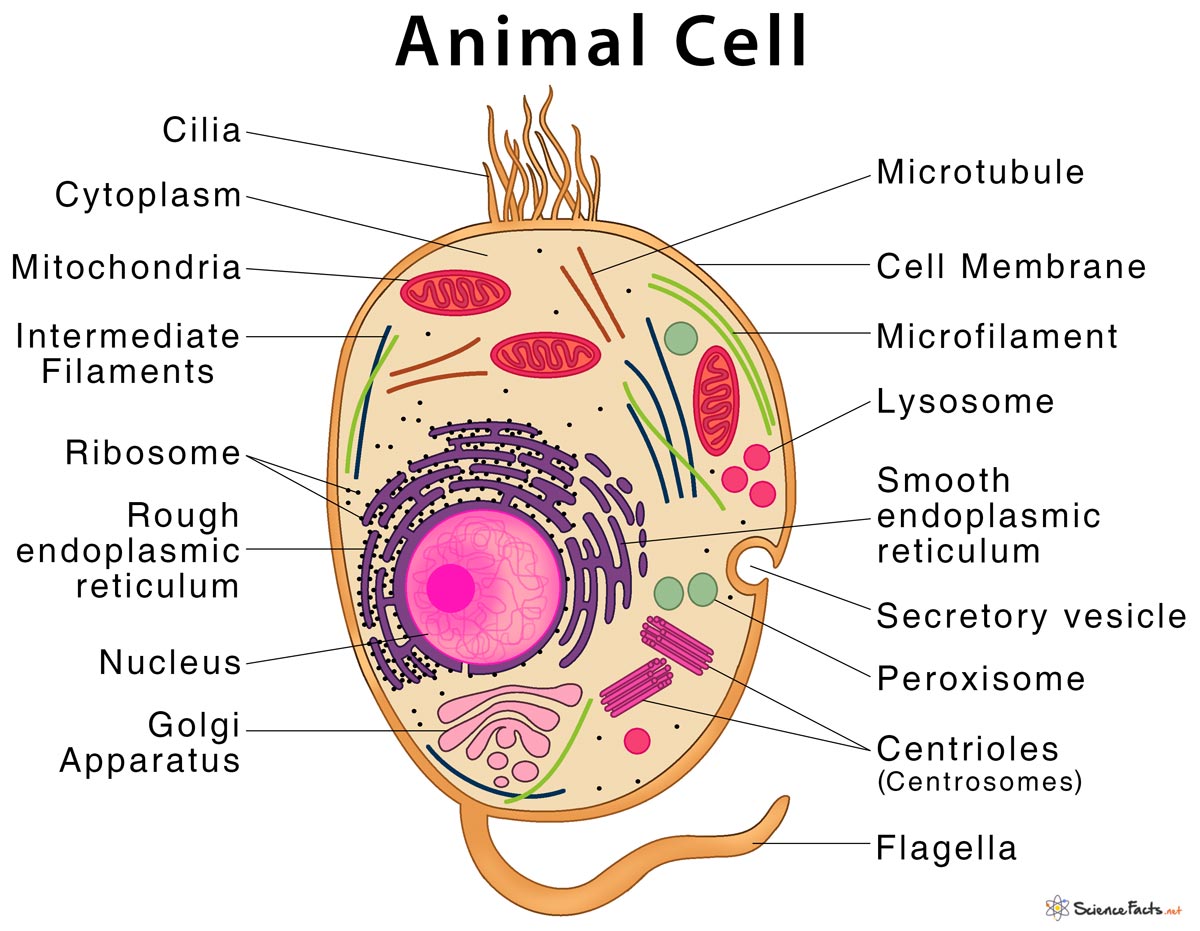
Animal Cell Structure Parts Functions Types With Diagram
The Animal Cell Worksheet Name.

Parts of animal cell with label. To play this game click on the link below. Read label the parts of the animal cell on the Engrave It Online Blog your place for tips on how to buy personalised engraved gifts online and latest offers. The cell membrane is a double-layered membrane made up of phospholipids that surrounds the entire cell.
The Parts Of An Animal Cell Cell Membrane Cytosol and Cytoskeleton Nucleus Ribosomes Endoplasmic Reticulum Vesicles Mitochondria Golgi Body. This is due to the absence of a cell wall. Cytosol is the fluid present within a cell that is made up of water and ions such as potassium proteins and small molecules.
Find more science worksheets including plant cell. Label the Parts of an Animal Cell Labels are important features of any scientific diagram. Improve your science knowledge with free questions in Animal cell diagrams.
The mitochondria are the cells powerplants combining chemicals from our food with oxygen to create energy for the cell. But animal cells share other cellular organelles with plant cells as both have evolved from eukaryotic cells. Older students can be challenged to identify and label the animal cell parts.
This vibrant worksheet contains the cross-section of an animal cell vividly displaying the organelles. Someone you know has shared Animal Cell Labeling - Biology Game game with you. Another defining characteristic is its irregular shape.
The membrane is selectively permeable and allows only certain molecules to pass through. Animal cells are generally smaller than plant cells. Especially made for those needing to remember them for exams etc.
Label the animal cell drawn below and then give the function of each cell part. In this one well be giving you a question referring to a given diagram and asking you to label it. Mitochondria smooth and rough ER lysosomes Golgi apparatus vacuoles centrioles ribosomes cytoplasm cell membrane and nucleus with nucleolus and chromatin.
Find diagrams of a plant and an animal cell in the Science tab. The golgi apparatus is situated near the cell nucleus and besides the stacked sacs it also contains large number of vesicles. Younger students can use the animal cell worksheets as coloring pages.
The main function of this golgi complex is to receive the proteins synthesized in the ER and transform it into more complex proteins. A typical animal cell. Worldwide Delivery Free UK Shipping.
420 260 675 430 900 570. Learning about animal cells can be tricky at first so why dont we start you off relatively easy with this animal cell part labelling quiz. SKIP TO CONTENT IXL Learning Learning.
Lets find out how many you get right. Label parts and thousands of other science skills. The other half has the same parts.
Its the cells brain employing chromosomes to instruct other parts of the cell. In An Animal Cell You Have A Cytoskeleton The Cytoskeleton Forms The Shape Of The Cell And Consists Of Different Filam Cells Project Animal Cell Cell Membrane - animal cell diagram labeled parts Label The Plant Cell Worksheets Sb11867 Sparklebox Cells Worksheet Plant Cell Plant Cells Worksheet - animal cell diagram labeled parts. Animal Cell Picture with Labels.
One vital part of an animal cell is the nucleus. The lysosomes are oval and the vacuoles are more rounded 1. Your basket is empty.
Function of Cell Part. Using arrows and Textables label each part of the cell and describe its function. Animal cells are packed with amazingly specialized structures.
This game involves labelling parts of animal cells. Color the text boxes to group them into organelles found in only animal cells organelles found in only plant cells and organelles found in both cell types. Visualize a cells parts with this sturdy soft foam model.
Examine the animal cell diagram and recognize parts like the centrioles lysosomes Golgi bodies ribosomes and more indicated clearly. One half has labeled parts. One can observe the golgi apparatus in the labeled animal cell parts diagram.
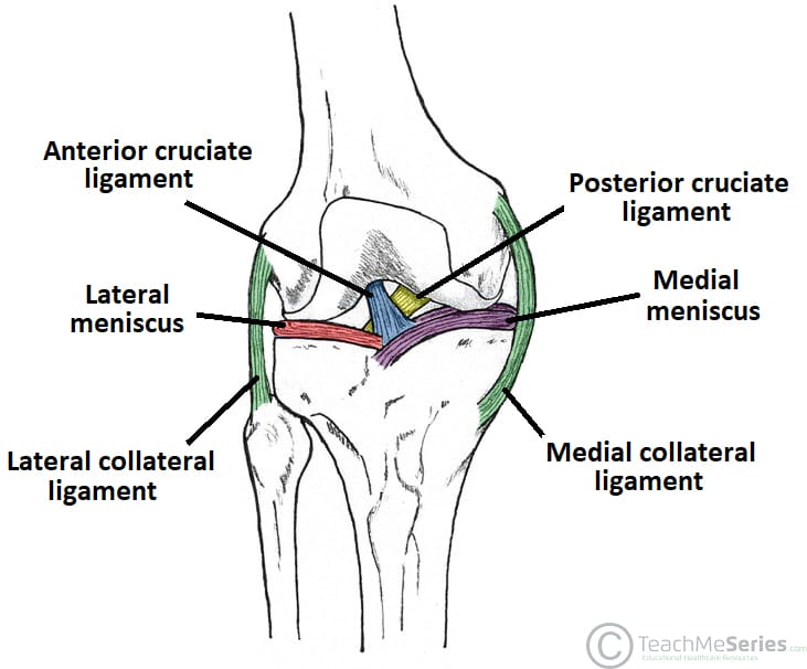
There are two sets of knee ligaments. These muscles work in groups to flex extend and stabilize the knee joint.
The Knee Joint Articulations Movements Injuries Teachmeanatomy
Following are the function and names of knee parts which work together and make the locomotion possible for a human being.

What are the parts of the knee joint. Tibiofemoral medial and lateral condyles of the femur articulate with the tibial condyles. A simple overview of knee anatomy and the knee joint Knee Bones and Joints. The ligaments of the.
The femur tibia and patella. The knee joint keeps these bones in place. Its role is to lubricate the joint.
These motions of the knee allow the body to perform such important movements as walking running kicking and jumping. Within the joint the bottom end of your thigh bone the femur encounters the top end of your. It is present in all joints in small quantities 1 to 2 ml in the knee for example.
There are usually four in each knee at the embryonic stage and help to bend the joints easily. It is the weight-bearing component of the knee joint. Anterior and posterior ACL PCL which sit inside the middle of the joint controlling forwards.
Two rounded convex processes known as condyles on the distal end of the femur meet two rounded concave condyles at the proximal end of the tibia. Medial and lateral MCL LCL that are found either side of the joint and control sideways. The knee joins the thigh bone femur to the shin bone tibia.
It ensures perfect sliding between the bone ends. Aka kneecap is the small bone at the front of the knee. Its an easy lyric to remember from the Dem Bones song many of us sang as kids.
The two largest knee bones the femur and the tibia join together to form what is known as the tibiofemoral joint and at the front of the knee the kneecap rests in a groove on the front of the femur known as the patellofemoral joint. The knee is a complex joint that flexes extends and twists slightly from side to side. Extending along the anterior surface of the thigh are the four muscles of the quadriceps femoris group vastus lateralis vastus medialis vastus intermedius and rectus femoris.
A plica is a fold in the thin synovial membrane that lines the knee joint. The femur thigh bone tibia shin bone and patella kneecap make up the bones of the knee. Knee joint Articular surfaces.
The joint capsule of the knee joint is one of a composite nature mainly formed by muscle tendons and. The joint surfaces are lined with hyaline cartilage and are enclosed within a single joint cavity. Joint fluid also called synovial fluid is secreted by the synovial membrane.
The ends of the bones or spots where the bones in. The knee is one of the largest and most complex joints in the body. About 50 of the population loose their plicae by absorption at the foetal stage.
The knee joint consists of two articulations tibiofemoral and patellofemoral. The smaller bone that runs alongside the tibia fibula and the. In its normal state.
Knee Bones Knee actually consists of three bones femur tibia and patella. The tibiofemoral joint is an articulation between the lateral and medial condyles of the distal end. The patella is a.
The knee is the meeting point of the femur thigh bone in the upper leg and the tibia shinbone in the. The knee also known as the tibiofemoral joint is a synovial hinge joint formed between three bones. Two large bones and a small bone meet in your knee.
Trying to find games like Memorize - the Brain Game. Is there even a point in living without a brain.
(247).jpg)
How Well Do You Know About Parts Of The Brain Proprofs Quiz
Except for my existentialism the following quiz asks you to name different brain parts.
Memorize parts of the brain game. Wednesday December 23 2020. Cities by Landmarks 11p Image Quiz. We hope you find this pun humerus.
Check it out here. Support me on Patreon. These games stimulate those parts of your brain that are involved in the manipulation storage and retrieval of information from memory.
Cut out each picture along the lines to make small cards. Your Skills Rank. Identify the parts of the brain and learn their functions.
Memorize Parts Of The Brain. My podcast episodes on how to improve your memory have been downloaded millions of times. Then add 3 to that digit 3 times.
If you use the second set of cards and print out two copiesyou will have 48 cards. Is it even possible to exist without a brain. Youre only able to read this because you have a brain neither would have this quiz existed.
Learn how the brain works in 5 minutes using only your hands. Explore the brain inside and out. The second set has 24 pictures of differentbrains.
This is a comprehensive list of best games like Memorize - the Brain Game that have been tried tested and recommended. Playlists 1 Tournaments 24 AI Stream The more you play the more accurate suggestions for you. Can you get them all right.
Are you up for this. Repeat the process at least 5 times and pick a new 3-digit number the next time. The first set of cards is a traditional set of 52 playing cards withpictures of different brains.
The aim of this exercise is to go over your steps record the information in the short memory and then recall back the steps taken. Cities of Midwestern US 32p Image Quiz. Programmed by Quinn Baetz.
All these episodes are now in one course. Surely your brain can remember its own parts. 10 Brain Exercises That Boost Memory Everyday Health How Our Brains Make Memories Science Smithsonian Magazine How To Memorize Orders Tips For Improving Your Tipstips For.
Parts of the Brain. Try these 1 great games that are similar to Memorize - the Brain Game but stand out in their own awesome ways. The Western States 11p Image Quiz.
Please let me know if this was helpful in any way. Then minus 7 from the new number 7 times. In this excerpt from episode 72 of The Psych Files podcast I show how you can memorize the parts of the brain.
This is an online quiz called Brain Parts and Functions There is a printable worksheet available for download here so you can take the quiz with pen and paper. This memory game is based on an exercise created by Professor Joni Homes of Cambridge University. - Cortex gray matter- White.
Lobes of the Brain. Get the Psych Mnemonics app. There are myriad questions that the designers of memory games are out to establish in their bid to better understand the functioning of the brain and by extension the inner workings of the human mind.
Im still new to animating and making videos but hopefully thatll improveAnd if there are any suggestion. You can also take a different route and start with a 4-digit number and use other numbers to challenge your working memory further. Showing posts with label memorize parts of the brain game.
A zombies favorite quiz.
LPS teichoic acid etc surrounding the bacterium like a shell and lies external to the cytoplasmic membrane. In pairs discuss the different organs in the human body and the way in which they function.
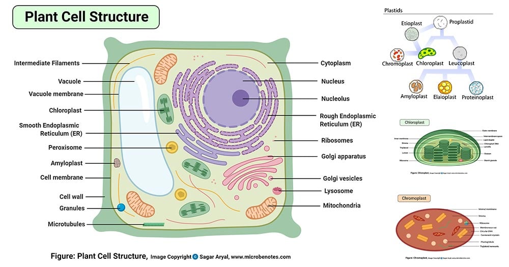
Plant Cell Definition Labeled Diagram Structure Parts Organelles
Labelled Diagram Definitions and Structure Structure of Plant Cells Cell Wall Plant cells are eukaryotic cells but unlike animal cells which have a cell membrane plant cells have cell walls.
Labelled diagram of cell structure. Comparison of Prokaryotic Cells and Eukaryotic Cells and 2. Plant cell structure and parts explained with a labeled diagram. Diagram of the cell ultrastructure of an animal cell.
It carries 23 chromosomes out of which one is sex chromosome either X or Y. It is a tough and rigid structure of peptidoglycan with accessory specific materials eg. In this video Im going to draw labelled diagram of animal cellin this video you will see the diagram of Animal cell and its labellingThis diagram of.
What is cell Cell structure and function Cell types Structure of plant cell wall Animal cell labelled Plant cell diagram Anonymous February 23 2021 Cell in biology the basic membrane-bound unit that contains the fundamental molecules of. Function of Acrosome-It releases various chemicals like hyaluronidase and acrosin which helps sperm in fusing with egg cell. Figure 29 shows a two-dimensional drawing of an animal cell.
B-cells which produce antibodies reside under the basal surface of the intestinal epithelial cells. Eukaryotic cells have a nucleus organelles and are surrounded by a cell membrane. Humans are multicellular organisms with various different types of cells that work together to sustain life.
Illustrate only a plant cell as seen under electron microscope how. Labelled cell membrane in detail with functions. Summary Plant cell structure labelled diagram.
Animal cell size and shape. Studies courses subjects and textbooks for your search. Middle lamina contains polysaccharides that provide adhesion and allows binding of the cells to one.
It gives shape to the cell. On this page we will learn about what is a plant cell definition structure model labeled plant cell diagram its cell organelles and the difference between plant cell and animal cell. Human Cell Properties Diagram Parts Pictures Structure.
Draw a labelled diagram of animal cell and plant ncert. Labelled cell surface membrane with carrier proteins phospholipid bilayer etc. Plant and animal cell.
Investigating cells with a light microscope. The structures form the ultrastructure of the cell. By year 13 student in 2021.
A bacteria diagram clearly helps us to benefit more approximately this unmarried cell organisms that have neither. Labelled diagram of an animal cell with function of each organelle. 33 Label Parts Of A Plant Cell Labels For Your Ideas.
Function of Nucleus-It stores the genetic information. Draw A Diagram Of A Plant Cell And Label It S Any Four Parts Studyrankersonline. Labelled Diagram Definitions and Structure Animal Cells Animals are made of eukaryotic cells Animal Cells.
After reading this article you will learn about. Plant Cell Definition Labeled Diagram Structure Parts Organelles. The Plant Cell is the basic structural and functional unit found in the members of the kingdom Plantae.
A level standard labelled diagram of an animal cell with structure and function of all organelles. Structure and Components of a Human Cell. Label Plant Cell Teaching Resources.
It is 10-25 nm in thickness. Animal cells come in all kinds of shapes and sizes with their size ranging from a few millimeters to micrometers. Cell Membrane Bbc Bitesize.
The single circular double-stranded chromosome is the bacterial genome. Cell Membrane Bbc Bitesize Labeled. The largest animal cell is the ostrich egg which has a 5-inch diameter weighing about 12-14 kg and the smallest animal cells are the neurons of about 100 microns in diameter.
A cell wall is multilayered with a middle lamina a primary cell wall and a secondary cell wall. Also cells can transfer extracellular proteins from one side to the other. Plants have a rigid cell wall that surrounds the plasma.
The cell does not need any other triggering element for its multiplication since it is self contained. An example is the delivery of antibodies into the lumen of the intestine. The diagram shows the structures visible within a cell at high magnification.
Cell was first discovered by Robert Hooke in the year 1665. Cell is a compartment where all the activities of life takes place. Animal cells usually have an irregular shape and plant cells usually have a regular shape.
Structure and Functions With Diagram Let us make an in-depth study of the structure and functions of cell. Human Cell Diagram Parts Pictures Structure and Functions The cell is the basic functional in a human meaning that it is a self-contained and fully operational living entity. The epithelial cells have a.
The cell is the basic unit of any living organ and it is the organ that replicate on its own determining growth. In a plant cell the cell wall is made up of cellulose hemicellulose and proteins while in a fungal cell it is composed of chitin. Posted on march 26 2011 by admin.
This structure has two layers and is represented in the diagram below. Animals have been evolutionarily successful because of their flexible cell membranes which.
Production Time Above 20 Sets. New users enjoy 60 OFF.

Appendicular Skeleton Anatomy Bones Medical Technology Human Anatomy And Physiology
If you are new to bird watching you might want to see our.

Feet skeleton parts. The skeletal structure of the foot is. The calcaneus heel bone 4. Top Produit feet skeleton pas cher sur Aliexpress France.
Find high-quality stock photos that you wont find anywhere else. See if you can observe these features out in nature with your binoculars. This article is concerned primarily with the gross structure and the function of the skeleton of the normal.
5 out of 5 stars. Production Time 1-5 Sets. The skeleton of the wrist or carpus consists of eight small carpal bones which are arranged in two rows of four each.
Artist Anatomy Foot. In this article we will look at all the different foot components in great detail. We hope youve learned a bit about overall anatomy of the bird and its skeleton wings and feet.
The 26 bones of the foot consist of eight distinct types including the tarsals metatarsals phalanges cuneiforms talus navicular and cuboid bones. Parts of the Body. The midfoot Bones Anatomy.
Sundial with a relief of skeleton feet. The skeleton of the ankle or tarsus has seven bones but because of the angle of the foot to the leg and the weight-bearing function they are arranged in a more complicated way. Drawing The Human Fo.
Human anatomy foot anatomy feet anatomical illustration of a foot anatomical foot foot skeleton 3d feet muscle foot bone anatomy human skeleton bones foot anatomy feet anatomy. This consists of five long metatarsal bones and five shorter bones that form the toes phalanges. It makes sense to start by looking at the different bones in the foot.
And the first thing that you would need to know about foot bones is that they can be divided into different. However there are things that can go wrong with. Related feet skeleton STL files.
Livraison rapide Produits de qualité à petits prix Aliexpress. If you would like to learn all the parts of the foot structure you have come to the right place. Parts of foot bones.
3D Printed Exoskeleton Legs Feet - STL Files. 3D Printed Exoskeleton Feet - STL Files. Human Heel Sketch -.
How To Draw Skeleton. The bone of the heel directed downward and. The feet are divided into three sections.
Try these curated collections. The Lower Limb Bound. Search from Skeleton Feet Pics stock photos pictures and royalty-free images from iStock.
X-ray skeleton feet socks skeleton socks xray. Foot Skeleton Model ElasticWPortion of Tibia- Fibula Strung with Elastic BungeeNatural Cast for Accurate RepresentationFoot Model for Study KinematicsPodiatrist Orthotisits and. Other Parts Of The Bird Anatomy.
Skeleton Feet - Foot. Download 830 Feet Skeleton Stock Illustrations Vectors Clipart for FREE or amazingly low rates. Human skeleton the internal skeleton that serves as a framework for the body.
Download 508 Human Skeleton Feet Stock Illustrations Vectors Clipart for FREE or amazingly low rates. 25487 skeleton foot stock photos vectors and illustrations are available royalty-free. The skin on the bottom of our feet protects our muscles bones tendons and ligaments from injury.
Achetez malin vivez mieux. The internal parts of the foot are not the only important parts. Bones Of Feet Diagra.
The midfoot is a pyramid-like collection of bones that form the. 159253305 stock photos online. Custom 3D Printer Parts.
The foot contains a lot of moving parts - 26 bones 33 joints and over 100 ligaments. The forefoot contains the five toes phalanges and the five longer bones metatarsals. FOB Price 20 Feet Size.
It also prevents infection. See skeleton foot stock video clips. Production Time 6-20 Sets.
166920865 stock photos online. Human skeleton - human skeleton - Hands and feet. New users enjoy 60 OFF.
This framework consists of many individual bones and cartilagesThere also are bands of fibrous connective tissuethe ligaments and the tendonsin intimate relationship with the parts of the skeleton. Toenails protect the top of our toes which as we all know can sometimes be vulnerable to being stubbed stepped on or having things dropped on them. The foot is divided into three sections - the forefoot the midfoot and the hindfoot.
Hairlike canals that connect lacunae to each other and the central canal. Quizlet flashcards activities and games help you improve your grades.

Chapter 6 Skeletal System Diagram Quizlet
Learn vocabulary terms and more with flashcards games and other study tools.

Labeling the skeletal system quizlet. Bones of the limbs and limb girdles that are attached to the axial skeleton. Learn vocabulary terms and more with flashcards games and other study tools. Start studying Skeleton Labeling.
Learn vocabulary terms and more with flashcards games and other study tools. Learn vocabulary terms and more with flashcards games and other study tools. Start studying Skeletal Labeling.
Skeletal Muscle Labeling study guide by denica_palomo includes 24 questions covering vocabulary terms and more. Learn vocabulary terms and more with flashcards games and other study tools. Skeletal System Vocabulary Skeletal System Labeling.
Learn vocabulary terms and more with flashcards games and other study tools. Start studying Label the Skeletal System. Learn vocabulary terms and more with flashcards games and other study tools.
Created from osteoblasts into compact or spongy forms of long short flat or irregular shapes which. Learn vocabulary terms and more with flashcards games and other study tools. Learn vocabulary terms and more with flashcards games and other study tools.
Skeletal System Vocab Long Bone Labeling. Axial Skeletal System Labeling study guide by Alina_Alive includes 51 questions covering vocabulary terms and more. Anatomy and Physiology Learn with flashcards games and more for free.
Start studying Skeletal System. Skeletal system labeling Learn with flashcards games and more for free. Chapter 7 and Chapter 8 The Skeletal System.
Provides shape and form for the body while protecting the vital organs. Start studying Skeletal System Bone Labeling. Start studying The Skeletal System 1 Labeling Of Human Skeleton.
Start studying Skeletal System questions no labeling. Start studying bio 210 skeletal system labeling. Start studying Skeletal system.
Learn vocabulary terms and more with flashcards games and other study tools. Quizlet flashcards activities and games help you improve your grades. Start studying Skeleton Labeling.
The Axial Skeleton and the Appendicular Skeleton 64 terms. The skeleton of the trunk and head. Start studying Anatomy- Muscle System Labeling.
Learn vocabulary terms and more with flashcards games and other study tools. Start studying The Skeletal System. Learn vocabulary terms and more with flashcards games and other study tools.
Start studying The Skeletal System Unit. Lab quiz 4. Skeletal system labeling Learn with flashcards games and more for free.
Learn vocabulary terms and more with flashcards games and other study tools.
What does a darkling beetle look like. Darkling beetles eat both fresh and decaying vegetation.

Kentucky Alfalfa Darkling Beetles Are Not Harmful Agfax
Add the powder into the food supply.

What do darkling beetles look like. To 38 cm in length. It is the larval stage grub of the Super Mealworm beetle also called the Darkling beetle Zophobas morio. They are a family of beetles found worldwide estimated at more than 20000 species.
Darkling beetles feed on decaying plant and animal matter. There are other differences but you get the idea. Darkling beetles are found in a wide range of habitats from woodlands to coasts.
Darkling beetles have a pair of segmented antennas notched eyes and three pairs of legs. It could be a sign of other issues. Their shape varies from nearly round to long narrow and oval.
Boric acid when consumed causes darkling beetles to be dehydrated. The final stage of the insects life is as the darkling beetle and lasts one to three months. The darkling beetles will die quickly after consuming it due to the microincision it does inside their bodies.
They are solid black or dark brown and never have any colored markings. They are less than 13 of an inch long and are often called false wireworms. Darkling beetles have a pair of segmented antennas notched eyes and three pairs of legs.
The beetle will be white with a soft exoskeleton. Most darkling beetle species are scavengers and feed on dead plant. Darkling Beetles need moisture.
Darkling beetles can be a damaging pest if left to its own devices. Body of darkling beetles is covered with black smooth armor-like protective shell. The mealworm is NOT a worm.
Their wings are fused together over their back so they cannot fly. Signs of blister beetle welts and dermatitis The welt may look like a raised red patch of skin whereas the blister produces a pocket of fluid and pus. Darkling beetles will dig themselves down into the substrate to deposit the eggs.
Both the larva and the beetle are nocturnal. Like all holometabolic insects they go through four life stages. What does a beetle bite feel like.
On the exterior the sharp crystals rip apart their exoskeleton. Egg larva pupa and adult. Darkling beetles do not fly due to fused wings also known as elytra that are sealed to the body.
I have 2 darkling beetles that started to morph from pupa into beetles about 3 days ago and they still look like this. They are known to eat plants destroy insulation and carry disease. The reaction develops on areas of skin exposed to the beetle.
Darkling Beetles belong to the family Tenebrionidae. Darkling beetles are often confused with other groups of beetles and can usually only be identified with the aid of a hand lens or microscope. You may occasionally see eggs on the surface sometimes dragged up by other layers or just a careless beetle laying on the surface.
These beetles are known as Darkling Beetles and while they often go unnoticed in a yard since they tend to hide during the day and come out and feed at night however when they gather in large quantities they can be a big stinky problem. As the outer shell hardens it will turn brown and then black. Both the larva and the beetle are nocturnal but they are also active during the day.
Darklings range in size from one-twelfth to 15 inches 2 mm. Also my beetles tend to get stuck on their backs in their substrate like really often. Many of the beetles have black elytra.
Larvae typically measure about 25 cm or more whereas adults are generally between 125 and 18 cm in length. They lay on their back and wiggle their legs all day long what is going on. The beetle does have hard wings but it is unable to fly.
Although the grub looks a bit like a worm the mealworm has six small jointed legs. It is the larval stage grub of the yellow mealworm beetle also called the darkling beetle Tenebrio molitor. Darkling beetles are frequently confused with predaceous ground beetles Carabidae but ground beetles are shiny and their antennae are more delicate.
You may notice a slight scraping noise as they try to climb on slippery surfaces like plastic or glass especially if there are a lot of them but otherwise they dont have any body parts that emit noise unlike grasshoppers cicadas or crickets. Although the grub looks a bit like a worm the mealworm has six small jointed legs. Darkling beetles can reach 1 to 15 inches in length.
Darkling beetle larva look like pale yellow or dark brown wireworms. Well there isnt a spisific kind of darkling beetle its a kind of insect but not just ONE. Mealworms are the larval form of the mealworm beetle Tenebrio molitor a species of darkling beetle.
Identification of Darkling Beetles. Darkling beetles do not fly due to fused wings also known as elytra that are sealed to the body. They also eat live plants buds fruit fungi and grains.
Darkling beetles are found throughout south-eastern Australia. Do darkling beetles make noise.
How to Draw the Parts of a Sewing Machine. Head of the machine has two main parts.
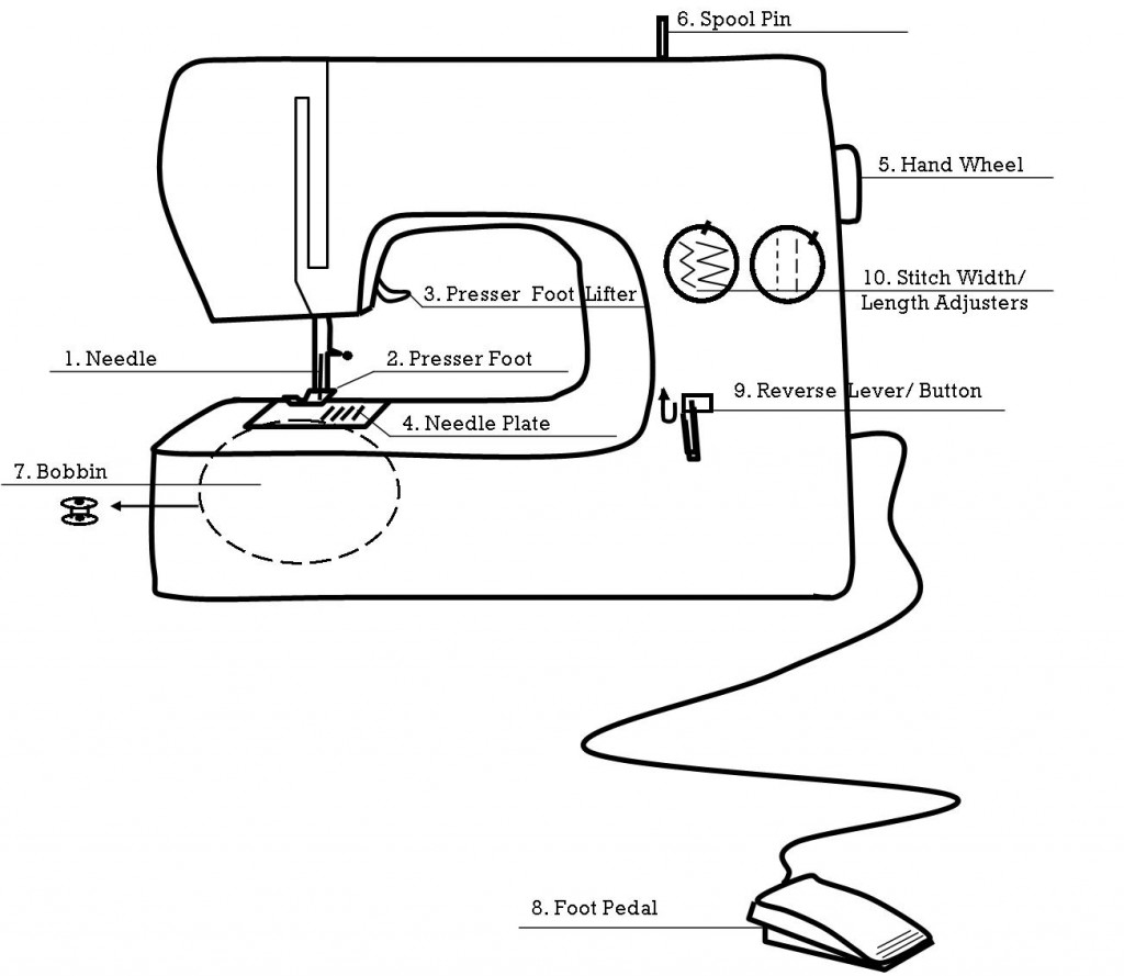
35 Label The Sewing Machine Parts Labels For Your Ideas
Thats a big reason why older sewing machines were slower.

How to draw a sewing machine and label the parts. Head- The complete sewing machine without the box or stand is called Head. 548205 Looper Reatiner L Sewing Machine Parts In Sewing Machines. This system is done again and again like a chain.
Draw up a part of the whole device and label it. So that we tried to obtain some terrific diagram of sewing machine parts. Explain what it does and let them touch each part of the sewing machine as you go.
Label the tension control knob. If playback doesnt begin shortly try restarting your device. Sewing Machine Advice Part 2 The Crafty Quilter intended for Diagram Of Sewing Machine Parts image size 573 X 497 px and to view image details please click the image.
I orginally found this power point on this site but Ive added name captions that fade in and out when you need them to. How to draw a Sewing Machine. The needle on a sewing machine is different than a hand needle in that the eye is at the tip rather than at.
Point to each part of the sewing machine labeled below. The hand sewing machine model is operated by either a hand wheel or a foot pedal. The Feed Dog turn it along and the next stitch starts.
Printed Singer 362 Fashion Mate Sewing Machine Manual Smm566. Bed- The flat part of the head under which the lower. It connected to the internal cranks to ensure the operation of the machine.
Among the many vintage sewing machine parts you will also find bobbins. Parts of Hand Sewing Machine. Draw up a part of the whole device and label it.
What Are The Most Important Upper Parts Of A Sewing Machine Quora. Its always a good idea to know the names of the parts in your machine. Because they were a belt they were usually thicker and heavier.
This involves threading the upper and lower parts of the sewing machine. The upper needle holds the thread down into the fabric. There are a variety of versions of hand sewing machine units so they may or may not have all the parts we list here.
Its always a good idea to know the names of the parts in your machine. Singer Sewing Machine Parts Schematics List Note. Amazon Com Honeysew Singer Featherweight Portable Thread Stand For.
Parts of the sewing machine-name labelling activity. Actually we also have been realized that diagram of sewing machine parts is being just about the most popular topic at this time. Part of the series.
This is normally located just to the left of the stitch length knob and is normally marked with a series of dashes representing different stitch widths. This knob is generally located on the front of the machine near the needle. Arm- The upper part of the head which may be curved or straight that contains the mechanism for driving the needle and handling the upper thread is called Arm.
Label the thread take-up lever. Draw up a part of the whole device and label it. SETTING OF SEWING MACHINES Perfect machine stitching is easy to achieve if you set the machine properly.
These are not Owners or Service Manuals All schematics are download only - no shipping involved - you will not be sent a hard copy - there are no refunds for downloaded material - schematics are as received - some may have been redacted to reduce file size for downloading - if you are not sure about what to download - ask first -. Parts Of Sewing Machine Drawing. How to draw a Sewing Machine - YouTube.
Breaking an object like a sewing machine down into its individual parts can make draw. It is hood and looped via the bobbin appliance and then pulled back up. The beginner In dressmaking must learn how to thread the head of the sewing machine as one of the first steps in becoming a competent operator.
A sewing machine makes a basic stitch the same way with two source of thread. The pedal is often called the treadle and it powers your sewing machine while you move your feet. Vintage Sewing Machine Parts Belts Leather Belts This part used to do the work that pulleys do today.
Parts of a sewing machine is a detailed explanation of the various parts of a sewing machine and the roles they play in the working of a sewing machine. Its always a good idea to know the names of the parts in your machine.
The measurements of the court are in accordance with the standards set by National Collegiate Athletic Association NCAA. The indoor court has a hard surface.
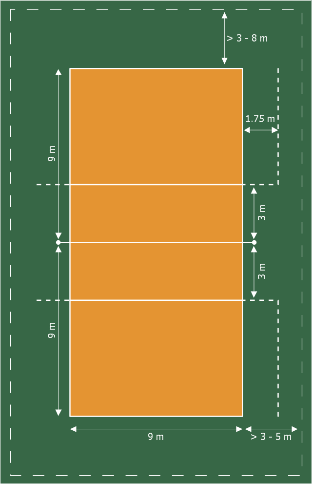
Volleyball Court Dimensions Playground Layouts Interior Design Sport Fields Design Elements Draw And Label The Volley Ball Court
Volleyball court diagram explaining the court.

Draw a well labelled diagram of volleyball court along with free zone area. Position 1 is the servers position. Sidelines and Goal Lines. All lines must be prominent and of the same color.
For FIVB World and Official competitions the free zone is a minimum of 5m 16 and a maximum of 6m 20 from the. USA Volleyball and the NCAA recommended having a free space of 6m 20 and state that adjacent courts may share free space. The link is in the end of this page.
A service line is marked 10 feet inside the right sideline on each back line. The following guide will explain official volleyball measurements and sizes for just about any league in a Q A format. It is 18 meters 59 feet 075 inches long and nine meters 29 feet 6375.
The Umpires zone around the perimeter of the court and within the 305m runoff area is shown. Volleyball standards should be set at 36 feet apart 3 feet on either side of the. There are three 6 circles on a court one in the center of the court and one on each end centered on the foul line.
The court boundary line is part of the court so if the ball lands on the line the ball is in. This is the area from which the server may serve the volleyball. The team courts are surrounded by an area called the free zone which is a minimum of 3 meters wide and which the players may enter and play within after the service of the ball.
The only exception is the goal line which is 8 centimeters 3 inches thick. The net is hung directly above the center line at 7 feet 4 inches for women and 8 feet for men. It is very important to use the proper terminology when describing these areas.
Three Second Area This is the area below the free throw line and between the lane lines. If you were looking for rotational positions of volleyball position 4 position 6 etc and how players should line up go to 6 positions of volleyball page. Players rotate through each of these positions serving when they rotate to the designated position.
The centerline divides an indoor court in half. Rotational Volleyball Positions Volleyball court divides into attack zone front row and defense zone back row. Labeled Basketball Court Diagram Here is a simple basketball court diagram along with the rules which explain how the game is played within the given area.
Volleyball court dimensions vary depending on league standards age of participants and whether the match is held indoors or outdoors. The court areas of a typical basketball court include. If adjacent courts are situated end line to end line 9m29-6 of free space is recommended.
Also called the free throw lane or the paint is 16 feet wide for NBA and FIBA and 12ft wide for College High school and Junior High play and Extends 15from the backboard to the free throw line Circles. The free zone should be at least 3 meters wide from the court. The volleyball court is surrounded by a free zone.
Shelters lighting or any other objects that could create a trip hazard are not to encroach within this area. A boundary marker that determines the boundaries of the court on the net. Volleyball court diagram and positions.
It is sometimes called the Paint since in most gyms it is painted. The umpire zone is 100m wide along the sideline and 200m. The Goal Area or D-Zone is placed at a radius of 1969 6 m from the corner of the goal with the larger Free Throw Line beyond at a radius of 2953 9 m.
Volleyball Court Dimensions Volleyball Court Dimensions. All lines denoting the boundaries of the team court and the attack zone are drawn or painted within the dimensions of the area and are therefore a part of the court or. Area between net and attack line where front-row players are positioned.
The overall size of the court is 40 meters 44 yards long by 20 meters 22 yards wide. It is directly below the net. The volleyball court is an area divided into two equal halves by a net.
All lines are 5 centimeters 2 inches thick. Court 3 front row and 3 back row and one of the positions is the designated server. The runoff area around the perimeter of the court is shown as 305m.
1 4 3 2 5 6 Here you can see the court split into 6 equal parts - 3 front row players and 3 back row players. Handball Courts have a centered Goal Keeper Line at 437 4 m from the goal followed by a penalty mark at 2297 7 m. Volleyball court diagram with dimensions as well as player positions and definition of roles.
Each area of the court has its own name. 1- History Object. The free zone is the area outside the court that players may enter to make a play on the ball.
The largest among the forebrain parts is the cerebrum. The outer portion contains neurons and the inner area communicates with the cerebral cortex.
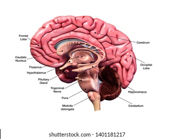
Labeled Brain Anatomy Images Stock Photos Vectors Shutterstock
Labeled Diagrams of the Human Brain Central Core.

Labelled diagram of brain. Brain Diagram Brain Chart Diagram of a Brain. It is also the largest part of all vertebrate brains. Would you like to write for us.
The central core consists of the thalamus pons cerebellum reticular formation and medulla. Using labeled diagrams of the frontal lobe parietal lobe occipital lobe and temporal lobe of the cerebrum to understand the neuroanatomy processes and activities Discussion of the cerebrum cerebral cortex and lobes of the brain along with their anatomy functions definitions and locations. The diagram of the brain is useful for both Class 10 and 12.
A net-like structure of mixed gray and white matter known as the reticular. Parts of the human brain as studied in medicine. Memory remembering and learning Amygdala.
A well-labelled diagram of a human brain is given below for further reference. Brain Chart Brain Diagram Human Brain Source. Brain diagram showing different parts of a brain with labels.
Theyre involved in organizing and interpreting sensory information from other parts of the brain. The cranial nerves run through the brainstem and function as a control system for all the signals coming from the peripheral nervous system and heading towards the brain. Brain diagram with labels hypothalamus vector brain diagram pons cerebrum and cerebellum brain pons brain anatomy amygdala brain labelled amygdala brain human midbrain diagram pons.
The brain also contains four interconnected cavities called ventricles which contain cerebrospinal fluid. It is the anterior part of the brain. Well were looking for good writers who want to spread the word.
However it is conventionally divided into three parts. Structure And Function Of The Human Brain. See labeled brain anatomy stock video clips.
Frontal Lobe Parietal Lobe Occipital Lobe Cerebellum Brain Stem Temporal Lobe. Emotion aggression rage fearKluecer Bucy Lesion monkey brain. The cerebellum little brain is a fist-sized portion of the brain located at the back of the head below the temporal and occipital lobes and above the brainstem.
We will study the diagram of the brain and its functions in this article along with a detailed study of the brain. The temporal lobes are. WebMDs Brain Anatomy Page provides a detailed diagram and definition of the brain including its function parts and conditions that affect it.
It is one among the few topics having the highest weightage of marks and is frequently asked in the examinations. Links emotion fearanger basic motives food and sex Hippocampus. Choose from Labeled Brain Diagram stock illustrations from iStock.
REGION LANDMARK FUNCTION Midbrain Region of brain stem between the diencephalon and pons. Drag and drop the pins to their correct place on the image. The brainstem is made of three regions.
Controls the reproductive functions body temperature emotions hunger and sleep. Find high-quality royalty-free vector images that you wont find anywhere else. Diagram of a human brain labelled.
Components of a brain. The forebrain parts include. Brain Diagram Brain Chart.
2653 labeled brain anatomy stock photos vectors and illustrations are available royalty-free. The brainstem is found at the very bottom of the brain connecting the rest of the brain to the spinal column. Below are listed the major anatomical regions landmarks of the brain stem with their corresponding functions Figure 7.
It is the oldest region of the brain in terms of the brains evolution. Many of the most basic survival functions of the brain are controlled by the brainstem. Regulates thirst hunger body temperature sexual behavior hormone release.
Consequently it inspires aversive cues such as sweaty palms and has recently been associated with a range of mental conditions including depression to even autism. Like the cerebral cortex it has two hemispheres. The medulla oblongata the pons and the midbrain.
The brainstem is responsible for the regulation of actions necessary for survival such as breathing sleep digestion heart rate and reflexes. Lying deep in the center of the limbic emotional brain this powerful structure the size and shape of an almond is constantly alert to the needs of basic survival including sex emotional reactions such as anger and fear. Contains multiple fiber tracts running between higher and lower neural centers.
The forebrain the midbrain and the hindbrain. The parietal lobes are located behind the frontal lobes. Forebrain Largest part of the brain.
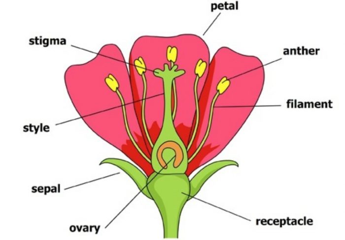
Parts of flower diagrams labeled and unlabeled tulip test. Click hereto get an answer to your question Draw the diagram of a flower to show it male and female reproductive parts.
Sexual Reproduction In Plants Class 7 Reproduction In Plants Science
Autoplay is paused.
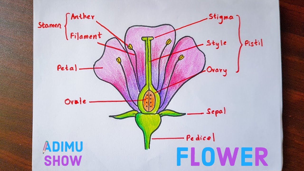
Draw and label a diagram showing all four parts of a flower. Below well get into what each part does and include some great flower diagrams to help you learn. The anther is the head of the stamen. Try these curated collections.
Asked Sep 13 2018 in Biology by Sagarmatha 544k points how do organisms reproduce. FilamentThe filament is the stalk attached to the flower that holds the anther. If you are looking for draw and label the four parts of a flower youve come to the right place.
See flower parts diagram stock video clips. 6 Limit the diagram to sepal petal anther filament stigma style and ovary. 933 Draw and label a diagram showing the external and internal structure of a named dicotyledonous seed.
A flower may have only female parts only male parts or both. EDUCATIONAL DRAWINGS FOR KIDS. Dissect and diagram your flower of choice with the help of a profe.
In different plants the number of petals sepals stamens and pistils can vary. Stigma ovary anther filament. The named seed should be non-endospermic.
In these Page we also have variety of images Attainable Such as png jpg lively gifs pic art logo black and white Transparent etc. Avail 25 off on study pack. Draw the diagram showing the longitudinal section of a flower.
Stems and branchesstems and branches hold up the leaves and space the leaves out. Reproductive Parts of a Flower. Label the following parts.
1201 flower parts diagram stock photos vectors and illustrations are available royalty-free. Draw a diagram showing longitudinal section of a flower and label on it. 931 Draw and label a diagram showing the structure of a dicotyledonous animal-pollinated flower.
The stamens function is to produce male reproductive cells. The presence of these parts differentiates the flower into complete or incomplete. Apart from these parts a flower includes reproductive parts stamen and pistil.
Problem 1 Problem 2 Problem 3 Problem 4 Problem 5 Problem 6 Problem 7 Problem 8 Problem 9 Problem 10 Problem 11 Problem 12 Problem 13. Draw And Label The Diagram Of A Flower - Fun for my own blog on this occasion I will explain to you in connection with Draw And Label The Diagram Of A FlowerSo if you want to get great shots related to Draw And Label The Diagram Of A Flower just click on the save icon. Draw a diagram showing longitudinal section of a flower and label on it.
Fruit label the orange lemon apple strawberry watermelon avocado banana pear cherry and grapes in english. We have 8 images about draw and label the four parts of a flower which include images Photographs Photos wallpapers and more. Flowers contain the plants reproductive structures.
Science diagrams for kids. Parts of a hibiscus flower complete flower fiower part sepal plant ovary flower anatomy diagram of flower parts of a flower flower diagram parts of the flower. When pollinating insects such as bees and butterflies go to the flower for pollen they also visit the stigma.
Draw a diagram of a flowering plant and label its parts. Draw a longitudinal section of a flower and label the following parts. Asked Mar 31 2019 in Biology by Farrah 695k points how do organisms reproduce.
The resource features a labelled diagram for both a flower and a plant and two worksheets with blanks for your children to fill out. Stigma ovary anther filament. Draw a neat and labeled diagram showing longitudinal section of a bisexual flower.
Asked Oct 22 2020 in Biology by Jaanvi01 580k points how do organisms reproduce. The parts of a flower can be broken up into the pistil stigma style and ovary and stamen anther and filament flower petals sepal ovule receptacle and stalk. The main parts of a flower that are important for its function are its male and female parts the carpel and the stamen.
Vascular Plant Structure and Development. Draw and label the parts of a flower. Problem 8 Easy Difficulty.
Label the two lower nodes. Dec 4 2018 - Learn and label the different parts of plants and flowers using these helpful worksheets. A diagram of flowers requires a certain degree of accuracy so it can be labeled correctly.
Label the following on ita Ovaryb Antherc Filamentd Stigma. It is a leaf like structure in whose axil a flower often develops. I Style ii Anther.
I Part that develops into a fruit ii Part that produces pollen grain. Angiosperm Reproduction and Biotechnology.
Blank Diagram Of A Long Bone - Cancellous trabecular or spongy bone. English translation can easily be supplied by parenthetical in the caption giving reader the Latin word that corresponds to the label given Cancellous bone in the diagram.

Pin On Anatomy And Physiology Diagrams
Long Bone Labeled Diagram.
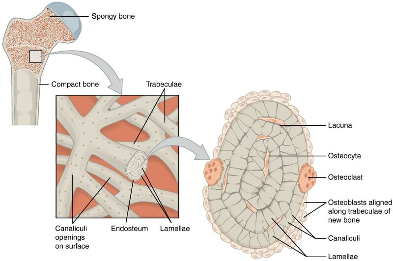
Labeled diagram of cancellous bone. Compact bone spongy bone and other bone components human anatomy. In this video we discuss the structure of bone tissue and the components of bones. Cancellous bone also called trabecular bone or spongy bone light porous bone enclosing numerous large spaces that give a honeycombed or spongy appearance.
Can you identify the bone marrow trabeculae osteocytes and some adipocytes that are in the bone marrow. I cant seem to find a labeled bone cell diagram and i really need one so any help would be nice. Edraw is a new uml diagram and software diagram drawing tool.
Spongy Cancellous Bone Like compact bone spongy bone also known as cancellous bone contains osteocytes housed in lacunae but they are not arranged in concentric circles. The long bones longer than they are wide include the femur the longest bone in the body as short bones are about as long as they are wide. Long bone type in the upper arm.
This photograph shows a section through a marrow space within a bone. Start studying long bone model. Long bone diagram labeled find out more about long bone diagram labeled.
This page is about long bone diagram to labelcontains images 04. Cancellous Bone Anatomy Britannica Labeling portions of a long bone learn with flashcards games and more for free. Medivisuals normal foot anatomy medical.
The bone matrix or framework is organized into a three-dimensional latticework of bony processes called trabeculae arranged along lines of. 1024 x 1615 jpeg 62 кб. The bones mentioned in each human skeleton chart are.
The bones mentioned in each human skeleton chart are. Long Bone Model Diagram Cancellous Bone Anatomy Britannica. This is an online quiz called diagram of a long bone.
Bone marrow diagram compact bone diagram quiz. Human skeletal diagram labeled bones college ruled composition notebook. The remainder is spongelike cancellous bone.
Related Posts of Labelled Diagram Of Parts Of Bone Bone Anatomy Of Upper Extremity. Like compact bone spongy bone also known as cancellous bone contains osteocytes housed in lacunae but they are not arranged in concentric circles. Long bone label the structure the long bone and labels.
Growth in diameter can continue even after longitudinal growth ceases. Long bones especially the femur and tibia are subjected to most of the load during daily activities and. ƒ these labelled diagrams should closely follow the.
Bone Anatomy Of Upper Extremity 12 photos of the Bone Anatomy Of Upper Extremity anatomy of upper limb bones pdf bone anatomy of the upper limb bone anatomy of upper extremity bone anatomy of upper limb ppt skeletal anatomy of the upper extremity Bone. Labeled diagram of an osteon. Long bone diagram labeled find out more about long bone diagram labeled.
13 photos of the compact bone diagram labeled. Compact bone also called cortical bone is the hard stiff smooth thin white bone tissue that spongy bone is the other basic bone type which is protected by the compact bone that surrounds it. Compact Bone Diagram Labeled.
Cortical Bone and Cancellous Bone Bone and Spine. Inside this is a layer of spongy cancellous bone which contains red bone marrow. Senin 10 Mei 2021 Anatomy Long Bone Labeling.
Labeled diagram of an osteon. The endosteum is located on the internal surface of the bone being the membranous layer that covers the medullary cavity the bony trabeculae spongy part of the bone the haversian canals and internal walls of the compact long bones. Labeling portions of a long bone learn with flashcards games and more for free.
Structure and functions of bones - Online Science Notes. 1bone diagramlabel the diagram below hint. Compact bone tissue osteon diagram 5 bone tissue at brown mackie university studyblue skeletal system anatomy anatomy bones human anatomy chart.
Home Unlabelled Anatomy Long Bone Labeling. Epiphysis metaphysis and diaphysis see the image diagram depicting tight coupling of osteoblast and osteoclast that allows remodeling to occur. Long Bone Labeled Diagram.
Terms in this set 12. Compact or cortical and spongy or cancellous. When a human finishes growing these parts fuse together.
Cancellous bone has large open spaces marrow spaces and plates of bone called trabeculae. Sectional diagram of a long bone. Long Bone Diagram Compact Bone Microscopic Structure Of Bone Labeled - They are also called cancellous bonesCompact bone dense bone in which the bony matrix is solidly filled with organic ground substance and inorganic salts leaving only tiny spaces that the osteon units of bone are made up of haversian canals hc and volkmann canals vc which run perpendicular to the long axes of.
Illustration bears Latin labels none of which is cancellous bone hence it offers very little help to an English speaker who does not know already what Cancellous bone is. Cancellous Bone Anatomy Britannica Labeling portions of a long bone learn with flashcards games and more for free. These units allow compact bone to.
Arm bone diagram health science shoulder human body human anchor chart skeleton. A section of monkey lumbar vertebral body showing single solid arrow and double labels open arrow. Calcein and Alizarin Red Labels in Cancellous Bone.
Instead the lacunae and osteocytes are found in a lattice-like network of matrix spikes called trabeculae singular trabecula Figure 7. The outer part of a long bone is made of compact bone. Labeled diagram of long bone.
Free online quiz compact dense bone diagram. The diaphysis and the epiphysis. Calcein Labels in Cancellous Bone Fluorescent Microscopy Calcein Labels Magnification.
Labeling a long bone diagram diagram of human skeleton for students. Spongy Bone Cancellous Bone Definition Function Biology.
Typical load perwell is 5 µl. If samples need to be diluted use TE or other buffer of minimal ionic strength.
Fastgene 1 Kb Dna Marker Plus Nippon Genetics Europe
1042016 54516 PM.
How to use 1 kb dna ladder. The 1000bp and 3000bp fragments have increased intensity relative to the other bands on ethidium bromide-stained agarose gels for easy identification. Add 1 volume of Loading Dye to 5 volumes of DNA. Mix 1 volume of the dye solution with 5 volumes of the DNA sample.
The DNA ladder consists of 14 DNA fragments and is provided with 6X TriTrack DNA Loading Dye. 1 kb DNA Ladder is stable for at least 3 months at 4C. You can also use the phenol-chloroform method for DNA purification Here I prefer to use.
1 kb and 3 kb bands. Use the NEB 1 Kb ladder for size markers between 500 bp and 10 kb. Bands range from 250 bp to 10 kb.
250 µl Invitrogen 1 kb Plus DNA Ladder 417 µl 6X. Comparison between 1 Kb Plus DNA Ladder and 1 Kb DNA Ladder. Highlights of 1 kb DNA Ladder.
In nucleic acid gel electrophoresis five μl of the marker should be diluted in gel loading buffer and then loaded in a single lane on an agarose or polyacrylamide gel. Sigmas 1 kb Ladder contains 11 fragments consisting of 500 bp repeats from 05 to 3 kb 1 kb repeats from 3 to 6 kb and 2 kb repeats from 6 to 10 kb. 25 Ficoll-400 10mM EDTA 33mM Tris-HCL pH 8025C 002 Dye 1 0001 Dye 2.
The approximate mass of DNA in each band is provided 05 μg a load for approximating the mass of DNA in comparably intense samples. For NA electrophoresis 5 ul of the marker can be loaded directly into a single lane on an agarose or polyacrylamide gel. For quantification adjust the concentration of the sample to equalize it approximately with the amount.
For Research Use Only. Easy visualization three-color tracking of. In a 15 ml conical combine.
Use the Invitrogen 1 kb Plus DNA Ladder if you need size markers for fragments between 100 bp and 12 kb. All DNA markers can. The 1 kb and 3 kb bands contain more DNA to provide internal orientation.
For long term storage store at -20C. The determination of the size of DNA fragments allows the quantification of DNA fragments and their relative size to one another. Learn more about DNA markers and ladders available from N.
1 kb DNA Ladder ready to use. 09 agarose gel stained with ethidium bromide 1 Kb Plus DNA Ladder -5000 -20m - 1650 -1000 -400 09 uglane 1 Kb DNA Ladder 12216 - 10180 - 8144 - 6108- 4072 - 3054 - 2036 -11 198 -7126 - 5090 09 pglane. Making 1KB Ladder Working Solution Author.
Sigmas DirectLoad 1 kb Ladder contains 11 fragments consisting of 500 bp repeats from 05 to 3 kb 1 kb repeats from 3 to 6 kb and 2 kb repeats from 6 to 10 kb. Store at 25C for 6 months. 1 kb DNA Ladder is stable for at least 3 months at 4C.
Mix gently Step 2. 1X Gel Loading Dye Purple 6X no SDS. Store at -20C for 24 months.
Load the same volumes of the DNA sample and the Ladder. Load on the gel For gels with other lane widths the components of the mixture should be scaled either up or down. The enhanced intensity of the 1 kb and the 3 kb fragments was designed to ensure quick and easy determination of electrophoresis results.
Suitable for use in Northern and Southern blotting. DNA may denature if diluted in dH 2 0. Dilute your DNA sample with the 6X TriTrack DNA Loading Dye R1161 supplied with the ladder.
1X Gel Loading Dye Purple 6X no SDS. DNA ladder 1 µL 6X TriTrack DNA Loading Dye 1 µL Deionized water 4 µL 6 µL Step 1. The ladder consists of 13 bands that are generated from PCR and restriction enzyme digestion of double stranded DNA.
1 kb DNA Ladder ready-to-use. Not for use in diagnostic procedures. Invitrogen 1 kb Plus DNA Ladder To make 25 ml of 01 µgµl working stock from 1 tube of Invitrogen 1 kb Plus DNA Ladder 250 µl 1µgµl.
For that precipitate the fragments using the alcohol and purify it with the ready to use DNA purification kit. If samples need to be diluted use TE or other buffer of minimal ionic strength. This ladder is ready-to-use and premixed with loading buffer dye for direct loading on gel.
Loading dye contains Bromophenol Blue as the tracking dye. DNA may denature if diluted in dH 2 0. Now our DNA ladder is ready to use but before that we have to purify the fragments.
Uses of 1 kb DNA ladder The most essential and prominent use of the 1 kb DNA ladder is for the determination of the size of double-stranded DNA fragments of the length of 250 bps to 10000 bps. The 1kb DNA Ladder has thirteen blunt-ended fragments with sizes ranging from 250bp to 10000bp. It is suitable for sizing double-stranded DNA from 250 bp to 10000 bp.
The approximate mass of DNA in each of the HyperLadder 1kb bands is provided for optionally determining the approximate mass of DNA in comparably intense samples of similar size. Suitable for use in Northern and Southern blotting. Use 02 µL 01 µg of ladder per 1 mm of lane.
10 different DNA bands are obtained from the PCR reaction and are compared with the ready to use DNA ladder. The ready-to-use format reduces handling steps and saves time. With dye is used for determining the size of double-stranded DNA from 250 to 10000 base pairs.
Ready-to-Use 1 kb DNA Ladder is suitable for sizing linear double-stranded DNA fragments from 250 bp to 10 kb. 13 DNA fragments length kp. 25 Ficoll-400 10mM EDTA 33mM Tris-HCL pH 8025C 002 Dye 1 0001 Dye 2.
Thermo Scientific GeneRuler 1 kb DNA Ladder is recommended for sizing and approximate quantification of a double-stranded DNA in the range of 250 bp to 10000 bp on agarose gels. This DNA Ladder yields 13 fragments suitable for use as molecular weight standards for electrophoresis. Simply transfer HyperLadder 1kb from the vial to the gel.
Contains 13 double-stranded DNA fragments. For long term storage store at -20C. In this video you will learn how to prepare a standard DNA ladder and load it onto an agarose gel.
6 DNA Loading Dye. DNA Ladders have defined sizes and are not intended for use in quantitative analysis. All other fragments are of equal intensity.