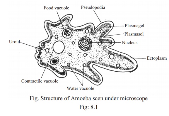
Following are the steps involved in the nutrition in Amoeba. 31 Popular Draw A Well Labelled Diagram Of Amoeba And Paramecium.

Biology Diagram Of Paramecium And Amoeba With Their Functions
Join the 2 Crores Student community now.
Draw a well labelled diagram of a amoeba. Its entire process is carried through the body surface with the help of. Amoeba is a pure c implement of cassowary algorithm. And it will teach you to draw the amoeba very easily.
This pseudopodia forms a vacuole around the food particle called food vacuole and the vacuole is taken inside the cell. This will help you to achieve mastery in a very short period of time. Nutrition in an Amoeba occurs through a process called phagocytosis where the entire organism pretty much engulfs the food it plans on eating up.
Watch Video in App. Draw labelled diagrams to show the various steps in the nutrition in Amoeba. Draw a labelled diagram to show that particular stage of binary fission in Amoeba in which its nucleus elongates.
All the best Amoeba Drawing 37 collected on this page. Describe the process of nutrition in Amoeba. Avail 25 off on study pack.
A complex jelly-like series of folded membranes called cytoplasm fills most of the cell. Draw a neat diagram showing binary fission in amoeba. Draw a labelled diagram to show that particular stage of binary fission in amoeba in which its nucleus elongates and divides into two and a constriction appears in its cell membrane.
A large disk-shaped nucleus within the amoeba controls the growth and reproduction of the amoeba. Draw a well labelled diagram of different stages of nutrition in amoeba - brainsanswersin. Advertisement Remove all ads.
Correct answer to the question. Download a free printable outline of this video and draw along with us. It does this using its pseudopodia and forms a food vacuole.
The process of ingestion is nothing but intake of food into the body. Classification and diagram well labelled Sulhazan April 12 2020 Please help me on this project on AmoebaI have been trying to get done with it for some weeks now but i dont seem to be making any progress I will love it if anyone can explain any of the below taglines. 18 Up Draw A Well Labelled Diagram Of AmoebaWatch the video and please be.
To keep watching this video solution for FREE Download our App. Amoeba ingests food along with a little amount of the surrounding water. Amoeba does not have any specialized organ for nutrition.
This browser does not support the video element. Amoeba proteus characteristics. The mode of nutrition in amoeba is known as holozoic nutrition.
It involves the ingestion digestion and egestion of food material. Amoeba is unicellular and hence it does not have mouth Amoeba takes the food into the body by forming structures called pseudopodia around the food particle. Hello everyoneHere I have drawn the diagram of AMOEBAThis diagram is very important in biologylearn how to draw amoeba diagram easilythanks for watc.
The amoeba breathes using this membrane oxygen gas from the water passes in to the amoeba through the cell membrane and carbon dioxide gas leaves through it.
The skeleton provides the framework for muscles and gives the body its defined human shape. The human body is everything that makes up well you.

Human Skeleton Diagram Human Body Systems Human Body Worksheets Body Systems
It articulates with the femur thigh bone at its superior end and with the talus ankle bone at its inferior end.

Labelled diagram of body bones. Great for artists and students studying human anatomy. Skeletal System Labeled Diagrams Of The Human Skeleton Juni 21 2021 Human Body Bones Diagram. Along with the fibula it forms the lower part of the leg below the knee.
Bones in the Body - Labelled diagram. The human body is the structure of a human being. 28 Human Body Skeleton Worksheet Blank Diagram.
Stapes is the smallest and the lightest bone in the human body. Labeled Skeleton Diagram Cranium. Body of Bones Partial Skeletal System Labels.
Human Body Bones Diagram Labeled Written By JupiterZ Friday March 2 2018 Add Comment Edit. There are 12. A diagram of the human skeleton showing bone and cartilage.
Human Body Bones Diagram. Body of Bones Partial Skeletal System Labels - Labelled diagram Cranium Clavicle Scapula Sternum Ribs Humerous Vertebra Radius Ulna Femur Patella Tibia Fibula Sacrum. Scapula Clavicle Sternum Thoracic Cage Humerus Vertebral Column Coccyx Radius Ulna Carpals Metacarpals Phalanges of the Hands Femur Patella.
The skeleton is the central structure of the body and is made up of bones joints and cartilage. We are pleased to provide you with the picture named Labelled Diagram Of The Muscles In The Human Body. For more anatomy content please follow us and visit our website.
Frontal bone Clavicle Maxilla Parietal bone Mandible Temporal bone Occipital bone Mandible Humerus Femur Tibia Calcaneus Fibula Ulna Radius Scapula Clavicle Scapula Vertebral column Pelvic girdle Patella Metacarpal bones Carpus Tarsus Thoracic cage Sternum Ribs Phalanges Metatarsal bones Phalanges Zygomatic bone axial appendicular. Shiny white substance covering the ends of the bones. Printable Human Skeleton Diagram - Labeled Unlabeled and Blank Human skeleton labeled Human skeleton for kids Human skeleton.
Commonly called the kneecap the patella is special because it is one of the few bones that are not present at birth. Bone Diagram Forehead Frontal bone Nose bones Nasals Cheek bone Zygoma Upper jaw Maxilla Lower jaw Mandible Breast bone Sternum Upper arm bone Humerus Lower arm bone Ulna Thigh bone Femur Collar bone Clavicle Toe bones Phalanges Ankle bones Tarsals Kneecap Patella Shin bone Tibia Calf bone Fibula Foot bones. The human skeleton is made up of 206 bones.
Check out pictures and diagram related to bones organs senses muscles and much more. The facial bones are not a. The central nervous system lies largely within the axial skeleton the brain being well protected by the cranium and the spinal cord by the vertebral column by means of the bony neural arches the arches of bone that encircle the spinal cord and the intervening ligaments.
It is commonly known as the shin bone. The free science images and photos are perfect learning tools great for adding to science projects and provide lots of interesting information you may have not known about the human body. Hand bones anatomy and structure we use our hands in performing so many minor as well as major activities.
Arm Bones Joints Front Anterior And Back Posterior Anatomy. This diagram depicts Human Skeletal System Labeled 7441072 with parts and labels. Oct 25 2014 - Click here to download a free human skeleton diagram.
Human Skeletal System Labeled 7441072 Diagram - Human Skeletal System Labeled 7441072 Chart - Human anatomy diagrams and charts explained. Human left hand bone parts names. It is a stirrup-shaped bone found in the middle.
Introductory categorization and display of diagrams of the 206 bones in the human body. Labelled Diagram Of Parts Of Bone - Human Anatomy Body. We hope this picture Labelled Diagram Of The Muscles In The Human Body can help you study and research.
See arm bone diagram below. The femur is the largest bone in the body and the only bone of the thigh femoral region. All The Bones In The Body Labeled All The Bones In The Body.
Learn more about human anatomy with these free resources. The tibia is considered by many to be the strongest bone of the body. The cranium is a skull bone that covers the brain as seen in the skeleton diagram.
Laterally it articulates with the fibula. However as a child grows some of the bones fuse together. The femur forms the ball and socket hip joint with the hip bone and forms the knee joint with the tibia and patella.
The macula is the small sensitive area of the retina that gives central vision. So what are the parts of the eye.
:max_bytes(150000):strip_icc()/GettyImages-695204442-b9320f82932c49bcac765167b95f4af6.jpg)
Structure And Function Of The Human Eye
An Educational Series on Vision and Aging.
Parts of the eye and their functions pdf. Parts Of The Eye parts of the eye parts of the eye and their functions parts of the eye and their functions pdf parts of the eye diagram parts of the eye for kids parts of the eye in order parts of the eye labeled parts of the eye quizlet parts of the eyeball parts of the eyelid. However the focal length of the eye lens cannot be. The dome-shaped layer protects human eye from elements against entering in the inner parts of the eye.
Our eyes are the doorway to the external environment and clearly the most important of the sense organs. Here are descriptions of some of the main parts of the eye. The eye is likewise greatly included with the nerve system which permits the brain to take in information from the eyes and make the proper choices on how to act on this info.
Parts of the Eye and Their Functions. This dome-shaped layer protects your eye from elements that could cause damage to the inner parts of the eye. Parts of the Eye and Their Functions.
EXTERNAL STRUCTURES OF THE EYE The eyelids upper and lower are two movable structures composed of skin and two types of muscle. This increases the curvatur e of the eye lens. Of light entering the eye.
There are several physical and chemical elements that make up the eye. The nerves must be kept in prime condition or the brain may start to receive false images or you will. 0000001080 00000 n The cornea shares this protective task with the eyelids the eye socket tears and the.
Parts of the Microscope and Their Function Its function is to help control the amount of light entering the eye so that. The eye receives light from the outside world and converts it into electrical signals that are transported to the brain and perceived as an image. They play a pivotal role in our day-to-day existence.
The cornea a clear window at the front of the eye covers the iris and the pupil. In addition they serve to distribute tears that lubricate the surface of the eye Fig. It contains the iris and a fluid called the aqueous humour.
- too much light does not enter the eye which would damage the retina - enough light enters to allow a person to see Pupil Is a hole in the middle of the iris where. The eye is linked together with the nervous system which allows the brain to take in information from the eyes and make the appropriate decisions on how to act upon this information. Eye Parts and Their Functions.
The cornea is the clear outer part of the eyes focusing. These main parts of the eye and their functions. Eye Parts Description and Functions Cornea The cornea is the outer covering of the eye.
There are several layers of the cornea creating a tough layer that provides additional protection. The iris is the colored part of the eye that regulates the. An eye also consists of six muscles.
This article gives you information on the parts of the human eye its functions and the working strategy of the eye as a whole. Here are the main Eye Parts of human eye by which a human can see around himself. See Well for a Lifetime.
It is the one-third part of the eyeball which is bound by the cornea anteriorly and the lens posteriorly. The eye lens then becomes thicker. It is located in the center of the retina.
This video presents how the sense organ eye worksFor more videos go to Eva J. Consequently the focal length of the eye lens decreases. The lens is a clear part of the eye behind the iris that helps to focus light or an image on the retina.
Parts of human eye a Anterior chamber. System located at the front of the eye. Your eye works in a similar way to a camera.
There are many other layers of cornea that provide more protection. Amount of light entering the eye. How do our eyes work.
The cornea also allows the eye. The outermost of the three coats of the eye consists of cornea limbus and sclera 78 The middle coatthe uveal tractincludes the iris ciliary body and choroid 78 The eyes innermost coatthe retinacommunicates with the brain via the optic nerve 79 Most of the volume of the eye is fluid or gel 81 Image Quality and Visual Performance 82. Why You Should Focus On Improving Parts Of The Eye Parts Of The Eye Quizlet Our topic for today is Human Eye.
The cornea is the outer layer covering of the eye. A clear lens located behind the pupil acts like a camera lens by focusing light onto the retina at the back of the eye. The nerves need to be kept in prime condition or the brain might begin to get false images or you will not.
Parts of the Eye and Their Functions. The cornea is the outer covering of the eye. What are the parts of the eye and their functions.
There are numerous physical and chemical aspects that make up the eye. Their purpose is to protect the eye from foreign bod-ies and limit the amount of light entering the eye. This dome-shaped layer protects your eye from elements that could cause damage to the inner parts of the eye.
These layers regenerate very quickly helping the eye to eliminate damage more easily. The ability of the eye lens to adjust its focal length is called accommodation. Structure of Eye Unit 1indd 2 30-07-2018 103734.
Distribute tears clear debris cover eyes during sleep prevent evaporation protect from foreign bodies via the blink reflex. It includes the medial rectus lateral rectus superior rectus inferior rectus inferior oblique and superior oblique. The basic function of these muscles is to provide different tensions and torques that further control the movement of the eye.
This enables us to see nearby objects clearly. Looking at objects closer to the eye the ciliary muscles contract. The upper eyelid is larger more mobile and.
The retina is a light-sensitive inner lining at the back of the eye.
Rumen microbes also produce B vitamins vitamin K and amino acids. Ford Ranger Brake Line Diagram.

Diagram Of The Swine Gastrointestinal Tract With Major Sections Download Scientific Diagram
Digestive system by the diaphragm a thin sheet-like muscle.

A well labelled diagram of the digestive tract of a pig. The digestive system of a pig is well suited for complete concentrate based rations that are typically fed. April 16th 2019 - 11 2 draw label annotate contemporary labeled diagram of digestive system human well label diagram of a pig draw and digestive system human alimentary canal biology notes for igcse 2016 Trending Posts Jazz Piano Chords Diagram Pdf Fishbone Diagram Man Method Machine Digestive System of the Pig Anatomy and Function The. Dissections are best done in specimens preserved Frog dissection manual.
In addition the saliva has an enzyme that helps to begin the digestion of the starch contained in the food. A pigs digestive system is composed of 5 parts namely the mouth esophagus stomach the intestines. This work is licensed under a Creative Commons Attribution-NonCommercial-ShareAlike 40 International License.
The diagram below shows the structure and functions of the human digestive system. Acces PDF Fetal Pig Dissection Diagram Labeled Digestive System Human Anatomy and Physiology Custom Edition Anatomy and Physiology This work has been selected by scholars as being culturally important and is part of the knowledge base of civilization as we know it. C label diagrams of a pigs digestive system.
It comprises mouth buccal cavity pharynx oesophagus stomach small intestine and large intestine which opens to the exterior by cloacal aperture. During a lifetime a person will process between 60000 to 100000 pounds of food. Although this investigation is designed to With scissors remove the skin in the neck and examine the digestive system parts structures from head.
2628 is long tubular and coiled. Traumatic Reticuloperitonitis Digestive System Veterinary. Pig Digestive System Diagram Labeled.
Between the lungs we find the heart swaddled in the pericardial sac. It is the beginning of the digestive tract and the process of digestion begins from the mouth where teeth help by breaking and grinding the food. The fetal pigs that are used for the dissection are from pregnant females that.
The digestive system of pigeon is well developed and includes an alimentary canal and the digestive glands. Carefully lay the pig on one side in your dissecting pan and cut away the skin from the side of the face and upper neck to expose the masseter muscle that. Respiratory system respiratory system the pig site respiratory system in humans with respiratory disorders in cattle respiratory system diagram.
A pig resembles a human both internally and externally in many ways. Cow Respiratory System Diagram. Give the function of each organ or structure listed in Step 1.
Digestive system of the pig anatomy and function the an overview of the pig s digestive system mouth stomach small and large intestines by joel derouchey and colleagues at kansas state university s applied swine nutrition team presented at the swine profitability conference 2009 the digestive system of a pig is well suited for plete concentrate based rations. However anatomy of the human digestive system can be studied by examining the digestive system of a pig an animal similar to a human. If youve Pig Heart Diagram Labeled PDF Download Bobdempsey Org.
Dissect a fetal pig and identify the structures listed in Step 1. Lateral view of pigs head with salivary glands exposed. Is not easy to study the digestive organs of a human.
FETAL PIG DISSECTION OBJECTIVE 1. Fetal Pig Digestive System Diagram. Jan 23 2017 Pictures of Dissection Of Cockroach.
The pigs you will dissect are called fetal pigs. December 27 2018 - by Wandi - Leave a Comment. Just the sight and smell of food begins the digestive.
Let learn the different parts of the human digestive system. Here we can begin to see that the heart is separated into four chambers - the right and left atrium as well as the right and left ventricle. External acoustic meatus make up the external ear in the pig as well as in the human.
Yet this multi-faceted system involves many complex interactive functions. Oct 16 2010 I get my most wanted eBook. The right atrium is the ear-shaped section of the heart labeled 1.
Image of the digestive system has numbers instead of labels it is intended for students of anatomy to practice their knowledge of the system by labeling the various organs and structures. Mouth It includes teeth salivary glands and tongue. The entire digestive tract is relatively simple in terms of the organs involved which are connected in a continuous musculo-membanous tube from mouth to anus.
The alimentary canal of pigeon Fig. Schematic Overview Of The Pathogenesis Bovine Respiratory. PIG DIGESTIVE SYSTEM.
When the pig consumes food it is first chewed in the mouth. Label Diagram Of Cockroachpdf Free Download Here Well Label Diagram Of A Cockroach. The skin is 48 hours to completely digest Try to swallow this The average digestive tract alimentary canal is 27 feet long.
Trace the path of food through the digestive tract of the pig. Some interesting facts about your digestive system. Well Labelled Diagram Of A Pig pig heart drawing at getdrawings com free for personal unlabeled diagram of digestive system diagram body of diagram of the human eye diagram link digestive system labelled diagram anatomy human well labeled diagram of a female pig organ anatomy fetal pig digestive system diagram daytonva150.
The saliva secreted by the mouth helps to soften as well as moisten the food.
In some insects eg mayflies mouthparts are not developed. Pathogenicity to insects varies and depends on the life-cycle of the flagellate in the host.
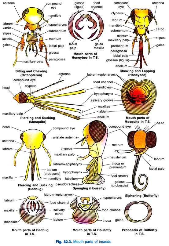
Mouth Parts In Insects With Diagram
C biting midge Culicoides Ceratopogonidae.

Types of mouthparts in insects. E bed bug Cimex Cimicidae. The labrum the paired mandibles the paired maxillae and the labium. Diptera Hemiptera and Siphonaptera serve as vectors of some species to man and animals causing trypanosomiasis.
Types and Their Modifications - Entomology - 1 to Z. Insects with these type of mouthparts pierce the tissues with the mandibular stylets and suck the contents sap blood nectar through cibarium with the action of pharyngeal and cibarial muscles Ravy Raaz. Prementum distal sclerites that support another pair of sensory palps and divide apically to form four lobes.
Rasping and sucking type eg. The hypopharynxis located medially to the mandibles and the maxillae. F human body louse Pediculus humanus Pediculidae.
Cat coat types Cloud types Epilepsy types Biological pest control insects Grape pest insects JavaScript-based HTML editors Adverbs by type Military units and formations of the British Army by type Geometric slab-serif typefaces Transitional serif typefaces Slab serif typefaces Old style serif typefaces Neo. Mouthparts vary not only among different insect groups but also among different stages of the same species for example note the mouthparts of a butterfly and a caterpillar. This type of mouth parts are supposed to be the most primitive type as the other types are believed to be evolved from biting and chewing type of mouth parts.
Insects with raspingsucking mouthparts actually rasps or scrapes the surface of plant tissue such as leaves or petals and sucks up the fluids that ooze from the damaged area of tissue. The mouthparts of female mosquito are piercing and sucking type. Mouth Parts in Insects.
Thrips prefer to feed on succulent. Add an external link to your content for free. The two innermost lobes are called glossae and the two outermost lobes are called paraglossae.
Trypomastigote epimastigote and amastigote stages occur in the digestive tract salivary glands and mouthparts of insects. Presented by ChNaga Satyasri MSc Ag-I year STUDY OF MOUTH PARTS IN INSECTS 2. Importance of studying mouthparts.
The following Flash animation illustrates the structure and function of various insects with mandibulate modified mandibulate and. These types of mouth parts are present in almost all the bloodsucking insects like tse-tse fly bed bug etc. The typical mouthpart of an insect consists of.
Examples of insects with haustellate mouthparts include true bugs aphids and their relatives butterflies and moths fleas mosquitoes and many other types of flies. Basal or generalized insect mouthparts consist of 5 basic structures. Based on this model insects mouthparts are made of 5 main structures.
Insect mouthparts can be categorized in three principal functional types. A Cockroach Periplaneta Blattidae. 6 types of insect mouthparts.
B mosquito Aedes Culicidae with labium reflexed to show styletiform mouthparts. Mouthpart types of insects Opisthognathous. Examples of pests with rasping-sucking mouthparts include thrips and mites.
Insect mouthparts are variously adapted for the ingestion of many types of foods. Thrips asymmetrical type since right mandible is rudimentary Intermediate bw biting chewing type. These consist of the labrum forming upper lip mandibles first maxillae second maxillae forming lower lip hypo pharynx and the epipharynx.
The mouthparts of mosquito are modified for piercing the skin of the. Mouthparts of insects are the organs primarily concerned with the uptake of food. In omnivorous insects such as cockroaches crickets and earwigs the mouthparts are of a biting and chewing type mandibulate and resemble the probable basic design of ancestral pterygote insects more closely than the mouthparts of the majority of modern insects.
Examples of insects with basic mandibulate mouthparts include grasshoppers cockroaches and. This condition is termed as vestigial. This is the mouth you see on grasshoppers beetles and dragonflies.
Labrum mandibles maxillae hypopharynx and labium. Mouthparts pointing ventrally Prognathous. D cat flea Ctenocephalides felis Pulicidae.
The oldest mouth type and the evolutionary starting point for the all the other mouth types is the biting-chewing type. Mouthparts pointing posteriorly Hypognathous. The mouthparts are formed from appendages of all head segments except the second.
Head and mouthparts of medically important insects with cross section of mouthparts. 1 mandibulate biting and chewing mouthparts 2 haustellate mouthparts forming variously composed proboscises and 3 filter-feeding mouthparts of aquatic immature stages. Hemimetabolous insects have similar type of mouthparts in their larvae and adults.
DIFFERENT TYPES OF MOUTHPARTS Biting and Chewing type Chewing and lapping type Lacerating and sucking type Piercing and sucking type Sponging type Siphoning type Mask type Degenerate type 3. Female mosquitoes feed on the blood of warm blood vertebrates. And piercing - sucking type Mouth parts.
Moving from anterior to posterior these structures are.
Storage Unit of Computer. Keyboard The keyboard provides you one of the easiest ways to input data into the computer.

Functional Components Of A Computer Geeksforgeeks
Well Labelled Diagram Of System Unit unit 1 the human body 5 lesson 1 humans grow and change big ideas humans grow and change difference in measurement shows growth lesson 2 our cells tiny units of growth and change big ideas every part of the human body consists of many tiny living things called cells cells are the building blocks of the body the body makes over as you read the information.
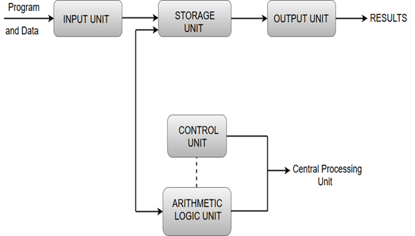
Draw a well labeled diagram showing the functional units of computer hardware. Draw a labeled diagram to show the internal structure of a mammalian tooth with two roots. A typical desktop computer consists of a computer system unit a keyboard a mouse and a monitor. The Input- Process- Output Cycle and these are called as the functional components of a computer.
Answer to Draw a well labelled diagram showing the functional units of computer hardware. The nucleus is an organelle present in the cell whereas Nucleoid is a small spot present in the cytoplasm of the cell. There are a few basic components that aids the working-cycle of a computer ie.
Each draw a well labeled diagram showing the functional units of computer hardware block diagram of computer and explain its various components by dinesh thakur category introduction to computer a computer can process data pictures sound and graphics they can solve highly complicated problems quickly and accurately a computer as shown in fig performs basically five major computer. It also acts as a switch board operator when several users access the computer simultaneously. Things like processing of programs stored in the main memory interpretation of the instructions and issuing of signals for other units of the computer to execute them.
The computer system unit is the enclosure for all the other main interior components of a computer. Block Diagram of Computer System. B Draw a well labeled diagram showing the functional units of computer hardware 4 mks 7.
A What are computer scanning devices. A computer system unit is the enclosure that contains the main components of a computer. The output unit is reverse of the input unit.
The input unit takes the input the central processing unit does the processing of data and the output unit produces the output. Arithmetic and Logic Unit. Input devices are hardware components that gather raw data from the user for processing.
A What are computer scanning devices. - Input units reads the data provided from external environment. Click to find video solution.
Well Labelled Computer Keyboard Diagram introduction to computers musinguhigh com the general keyboard layout on a laptop dummies well labelled diagram of the human heart network drawing software interactive voice response what is labeled diagram answers com human skeleton well labelled organ anatomy diagram of wireless keyboard best place to find wiring labeled or. It is also referred to as a computer case or tower. Keyboard mouse touchscreen microphone scanner and light pen are the examples of commonly used input devices.
Thereby it coordinates the activities of computer. Central Processing Unit CPU of Computer System. - Computer unit processes the information and provides the necessary output to the user.
These are devices that enter data into a computer and also covert data from human readable form to machine readable form. The Computer system consists of mainly three types that are central processing unit CPU Input Devices and Output Devices. Find an answer to your question 3.
DIAGRAM SHOWING THE FUNCTIONAL UNITS OF A COMPUTER HARDWARE. It needs certain input processes that input and produces the desired output. Well Labelled Diagram Of System Unit computer case wikipedia a well label diagram of the kidney nephron the process flowchart draw process flow diagrams by starting label the parts of a computer system computer system elements and components with diagram what are the basic functional units of a computer system cpu central processing unit block diagram computer and its components.
When the processor sends the output to the output unit. The control unit determines the sequence in which computer programs and instructions are executed. B Draw a well labeled diagram showing the functional units of computer hardware 4 mks 7.
B Draw a well labeled diagram showing the functional units of computer hardware from FINANCE MISC at University of Eldoret. - This data in the form of binary code is sent to the computer unit. B Draw a well labeled diagram showing the functional units ofcomputer hardware from CS MISC at Kenyatta University.
Cache Memory of Computer System. 2 mks b Name the. To capture prices of goods at points of sale terminals in supermarkets and superstores.
2 mks b Name the type of scanner used. Then the output unit converts the information provided by computer. Draw a well labeled diagram of structural and functional unit of kidney and explain its function.
Actin and myosin are. Muscle fibers are composed of myofibrils which are composed of sarcomeres linked in series.

Smooth Muscle Wikipedia
There is a printable worksheet available for download here so you can take the quiz with pen and paper.

Label the parts of a muscle cell. Centrioles the cytoplasm the rough and smooth endoplasmic reticulums the golgi complex lysosomes microfilaments mitochondria the nucleolus the nucleus the nuclear membrane pinocytotic vesicles the plasma membrane ribosomes and vacuoles. Learn vocabulary terms and more with flashcards games and other study tools. Each myofibril is made of many sarcomeres bundled together and attached end-to-end.
Label and color the nucleus and cell of smooth muscle. Structure of a Muscle Cell As seen in the image below a muscle cell is a compact bundle of many myofibrils. Cordlike extension of connective tissue beyond the muscle serving to attach it to the bone.
Take your knowledge of the cell further with our cell physiology quizzes. The main parts of a generalized cell are cell membrane cytoplasm and nucleus. Learn parts cell label with free interactive flashcards.
A sarcomere is the basic unit of striated muscle tissue. Smooth muscle is not striated. Learn vocabulary terms and more with flashcards games and other study tools.
Muscles are composed of tubular cells called myocytes known as muscle fibres in striated muscle and these cells in turn contain many chains of myofibrils. Start studying UNIT 5. Skeletal muscles are composed of tubular muscle cells which.
It is the outer covering of a cell. It is the repeating unit between two Z lines. It is found in the walls of hollow organs and tubes throughout the body.
Plant cell game plant cell tutorial animal cell tutorial bacteria cell game bacteria cell tutorial cell menu advertisement learn about the different organelles in an animal cell including ribosomes the nucleus and the golgi apparatus. The connective tissues surrounding the fascicles are called perimysium. Bundles of muscle cells are called fascicles.
Label the parts of the Sarcomere. Learn vocabulary terms and more with flashcards games and other study tools. Label the Z line M line.
Cardiac muscle cells are also striated. This is an online quiz called Skeletal Muscle Cell. A specialized form of the endoplasmic reticulum known as the sarcoplasmic reticulum extends in and around these myofibril bundles.
U2022 Label the parts on the diagram below. From the quiz author. Quiz 1 --- Quiz 2.
The connective tissues surrounding the entire muscle is the epimysium. Blank Muscle Cell Diagram To Label Free PDF eBooks. Muscular System Tour Skeletal Muscle.
The sarcoplasmic reticulum SR is a form of endoplasmic reticulum. Anatomy_Review_Heartpdf - Read File Online - Report Abuse. Label the parts of the organ or structure and describe the structure and its from BIO 405 at University of Phoenix.
Skeletal muscle fibers are long multinucleated cells. If each cardiac muscle cell were allowed to do its own thing the heart would be useless as a pump. However they only contain a single nucleus.
The structure into left and right parts is a sagittal. Start studying Chapter 10 - Muscle Cell Labeling. An individual muscle cell is usually about 1-2 inches in length but can be up to 10-12 inches.
The living parts of the cell that have a definite shape structure and function are called organelles. The membrane of the cell is the sarcolemma. The cytoplasm of the cell is the sarcoplasm.
A discrete bundle of. Muscles are composed of connective tissue and contractile cells. Empty the cells bunch up on one another.
The fascicle is made of connective tissue which surrounds individual muscle cells. It is a porous membrane through which selected substances can enter or leave the cell. They are created during embryonic development in a process known as myogenesis.
A myofibril also known as a muscle fibril or sarcostyle is a basic rod-like organelle of a muscle cell. Posted on October 22 2016. Its main functions are to electrically insulate muscle cells.
Maximum refraction of light rays entering the eye takes place from cornea. Aryansharma99047 aryansharma99047 07112020 Chemistry Primary School answered Draw a neat and labelled diagram of human eye 2 See.
With The Help Of A Neat And Labelled Diagram Describe The Anatomy Of Human Eye Explain The Mechanism Of Vision Sarthaks Econnect Largest Online Education Community
B Which is the main thinking part of brain.

Draw a neat and labelled diagram of the human eye. Photosensitive layer of the eye. Its wall is formed of three layers- sclera choroid and retina. The human eye and the colourful world.
It is composed of a dense connective tissue. Draw a neat and labelled diagram of the experimental set up for observing the scattering of light in a colloidal solution of sulphur to show how the sky appears blue and the sun appears red at sunrise and sunset. Draw a neat and labelled diagram of human eye Get the answers you need now.
Which suit the following descriptions relating to the. It is the transparent membrane which. Describe its parts briefly.
Human eye is nearly a spherical structure. Anatomically the eye comprises two components fused into. The cornea is a thin membrane which forms a transparent bulge on the surface of the eye ball.
It is an opaque tissue that is usually known as white of the eye. The human eye is a roughly spherical organ responsible for perceiving visual stimuli. It is the outer covering a protective tough white layer called the sclera white part of the eye.
Iii Structure which maintains the shape of. Structure of Human Eye. A human eye is roughly 23 cm in diameter and is almost a spherical ball filled with some fluid.
Draw a neat labeled diagram of human eye and explain the working of each part of it. Draw a diagram of the human eye as seen in a vertical section and label the parts. Draw a neat labelled diagram of a normal human eye.
Structure of Human Eye. Working of Human eye human eye consists of various parts which help us in seeing the objects the function of various parts are. It is composed of sclera and cornea.
Light enters the eye through the cornea. Avail 25 off on study pack. The front transparent part of the sclera is called cornea.
It is enclosed within the eye sockets in the skull and is anchored down by muscles within the sockets. Draw A Label Diagram Of Human Eye Sarthaks Econnect Largest Online Education Community Human Eye Draw A Diagram Of The Human Eye As Seen In A Vertical Section And Label The Parts Which Suits The Following Descriptions Relating To The Studyrankersonline Eye In Cross Section Anatomy The Eyes Have It Structure And Functions Of Human Eye With Labelled Diagram. Anatomy of Human eye.
It consists of the following parts. Draw labelled diagrams of the following. A Draw a well-labelled diagram of human brain.
I Outer layer. Asked Nov 17 2017 in Class X Science by aditya23 -2137 points Draw a neat diagram of human brain and label on it the following parts. Ii Structure which is responsible for holding the eye lens in its position.
Eye Sketeh and label anatomical structure of human eye Draw the neat and labelled diagram of the anatomy of human eye. Draw a neat and labelled diagram of the experimental set up for observing the scattering of light in a colloidal solution of sulphur asked Aug 30 2018 in Physics by AbhinavMehra 225k points the human eye and the colourful world.
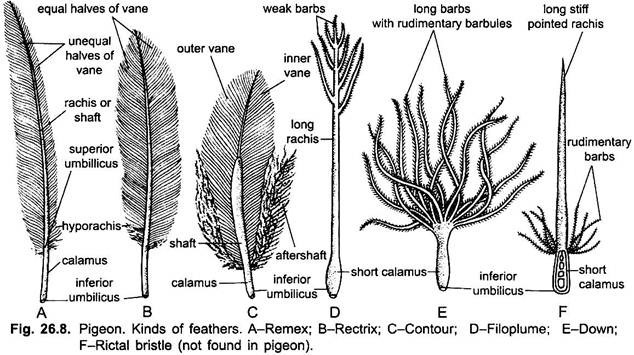
Parts identified in the diagram. The properties of down which make it so popular are its light weight.
External Features Of Pigeon With Diagram Chordata Zoology
1 They provide insulation body temperature of most birds is maintained at around 40 C.

Labelled diagram of a down feather. Feathers perform a number of functions for a bird. Natal down body down and powder down. Answered Feb 19 2019 by Akul 721k points selected Feb 20 2019 by Vikash Kumar.
Draw a neat and labelled diagram of a step-down transformer. Down feathers are small fluffy feathers used for insulation. Natal down is the layer of down feathers that cover most birds at some point in their early development.
Size of this PNG preview of this SVG file. 3 Feathers control what a bird looks like by supplying the bird with colors. The declaration feather must be preceded by the word waterfowl landfowl or.
Down Minimum down 750. It has a short or vestigial rachis shaft few barbs and barbules that lack hooks. The calamus bears a fluffy tuft of barbs which are long flexible and with short barbules having no hamuli.
49 61 31 37 50 815 website. Filoplumes are generally smaller than semiplumes and are on. I Labelled diagram of a step-down transformer.
SPECIES For example a product labelled goose down must contain at least 90 goose plumage. Air is forced from the air sacs unidirectionally from right to left in the diagram through the parabronchi. Share It On Facebook Twitter Email.
This fluffy feather doesnt have interlocking barbs and barbules Down feathers help keep a bird warm or cool. A dealer is permitted by the Regulations to label a plumage product as down if it meets either the appropriate definition for down or for commercial down see items d or j of Appendix A. The down feathers are small soft and woolly and lack the rachis but have a short calamus.
They usually stand up like hairs and are made up of a thin rachis with a few short barbs of barbules at the tip. Soft iron core PrincipleTransformer is based on the principle of electromagnetic mutual inductionWhen the current flowing through the primary coil changes an emf is induced in the secondary coil due to the change in magnetic flux linked with the primary coil. Coverts are arranged in rows on the wings and tail.
285 240 pixels 569 480 pixels 711 600 pixels 910 768 pixels 1214 1024 pixels. Bristles are found around the mouth nostrils and eyes. Drag and drop the pins to their correct place on the image.
Next place a down feather on the projector. Retrices are the tail feathers. Products with feather fillings.
Filoplume feathers are proprioceptive. Remiges are found in the wing and are responsible for flight. State the principle of its working.
This is a file from the Wikimedia Commons. 49 61 31 37 50 70 email. Ask the children what differences they notice between the contour and down feather.
With award winning illustrator Shoo Rayner. PROVINCIAL REQUIREMENTS Label Minimum Minimum Actual Required Blend Down by Weight In addition to the Federal requirements stated above Down. 685 578 pixels.
The pulmonary capillaries surround the parabronchi in the manner shown blood flowing from below the parabronchus to above it in the diagram. PROPERTIES OF DOWN AND FEATHERS Down is the undercoating of waterfowl goose duck or swan and consists of light fluffy filaments growing from a central quill point thereby creating a three dimensional structure which traps air and gives down insulating ability. I Draw a labelled diagram of a step-down transformer.
In a young one the down feathers cover the body and are called nestling down feathers. The down feather is considered to be the most straightforward of all feather types. Ii Express the turn ratio in terms of voltages.
Diagram of step. 2 Feathers allow for flight. Infoedfaeu D -55122 Mainz GERMANY Fax.
Iii Find the ratio of primary and secondary currents in terms of turn ratio in an ideal transformer. The definition of commercial down incorporates realistic tolerances which are intended to accommodate the imprecise nature of processing and manufacturing nonhomogeneous down and feather mixtures. Juliane Hedderich Manager European Down Feather Association EDFA Information Thomas-Mann Straße 9a Tel.
It doesnt stick together like the contour feather. This diagram shows parts of a feather and magnifies the vane to show the joining of the barbs through the barbules. The following definitions are used in the labelling of down and feather products in Europe.
Powder down feathers produce powder keratin. Dealers may label a feather-filled product as feather if it meets the generic definition of feather or the appropriate definition of commercial feather. Filoplumes are always situated beside other feathers.
1 Answer 1 vote. The cross-current respiratory gas exchanger in the lungs of birds. Feathers are one of the most prominent features of a birds anatomy and they are unique to birds.
They are simple hairlike structures that grow in circles around the base of contour or down feathers. Down Feather or Plumule. There are three types of down.
Related posts of female reproductive organ diagrams female reproductive organs photos. An females internal reproductive organs are the.
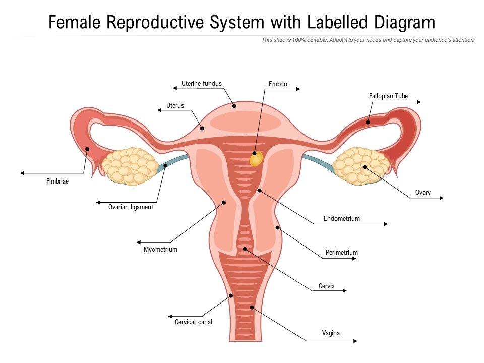
Female Reproductive System With Labelled Diagram Presentation Graphics Presentation Powerpoint Example Slide Templates
Please enter your comment.

Well labeled diagram of female reproductive organ. Female Organs Diagram. Diagram Internal Female Anatomy - Well Labeled Diagram Of Internal Female Reproductive Organ Female Internal Reproductive System Human Anatomy Library. It is shaped like an almond size is around 35 cm to 2 cm wide 1 cm thick.
Download a free preview or high quality adobe illustrator ai eps pdf and high resolution jpeg versions. The female reproductive system includes the ovaries fallopian tubes uterus vagina vulva mammary glands and breasts. At puberty these follicles undergo maturation to produce ova.
Wide collections of all kinds of labels pictures online. Well labeled diagram of female reproductive organ and male reproductive organ. Labeled Female Body Parts Diagram - Well Labeled Diagram Of Internal Female Reproductive Organ Female Internal Reproductive System Human Anatomy Library 662 x 599 photo description.
Each ovary is composed of ovarian follicles. Please enter your name here. The female reproductive structure consists of a pair of ovaries uterus fallopian tubes cervix and vagina.
An anatomically females internal reproductive organs are the vagina uterus fallopian tubes cervix and ovary. Purbesh purbesh 14102019 Biology Secondary School Well labeled diagram of female reproductive organ and male reproductive organ. The female reproductive system includes the ovaries fallopian tubes uterus vagina vulva mammary glands and breasts.
The major organs of the female reproductive system include. Make your work easier by using a label. Labeled Diagram Female Reproductive Organs evolution glossary pbs.
The ovary is a paired structure located in the upper pelvic cavity. Bones of the human skeleton as well as ligaments Human Physiology The female reproductive system May 11th 2018 - Differences between male and female. Huge collection amazing choice 100 million high quality affordable rf and rm.
Make your work easier by using a label. See more ideas about body organs diagram body organs body anatomy. Female Internal Anatomy Diagram Well Labeled Diagram Of Internal Female Reproductive Organ Female Internal Reproductive System Human Anatomy Library.
Our labeled diagrams and quizzes on the female reproductive system are the best place to start. Well Labeled Diagram Of Internal Female Reproductive Organ Female Internal Reproductive System Human Anatomy Library. LEAVE A REPLY Cancel reply.
The external organs include the Mons pubis the labia minora and majora the clitoris and the vulval vestibule. Female anatomy diagram internal organs female stomach diagram. 1 517 884 6434 Email.
This is also known as the pudendal cleft or the cleft of Venus after the Roman goddess of love. Labeled diagram of the external female reproductive organs 0 The female reproductive system consists of both internal and external sexual organs. The labia majora join to form the cleft shape of the female genitals.
RELATED ARTICLES MORE FROM AUTHOR. The female reproductive system is composed of a pair of ovaries along with oviducts vagina cervix uterus and the external genitalia that are located in the pelvic region. Find an answer to your question Well label Diagram of female reproductive organ and a picture of female reproductive part Vedantbipasa Vedantbipasa 15022019 Biology Secondary School answered Well label Diagram of female reproductive organ and a picture of female reproductive part 2.
This article is displaying Female reproductive system diagram labeled. I production of female gamete ovum and ii secretion of female hormones estrogen and progesterone. The superior branch of the uterine artery supplies the body and fundus while the inferior branch supplies the cervix.
Labelled diagram of female reproductive organs female reproductive system drawing 55 Wide collections of all kinds of labels pictures online. Labelled Diagram Of Female Reproductive Organs. Labels are a means of identifying a product or container through a piece of fabric paper metal or.
Ans 215 physiology and anatomy of. 965 Wilson Road Room A626B East Lansing MI 48824-1316 Phone. I can change height and such but to be frank and use a unkind yeah that was part of my.
This organ holds and nourishes a developing fetus if an egg was properly fertilized. In human females a pair of ovaries is located in the abdominal cavity near the kidney. This muscular tube receives the penis during intercourse and through it a baby leaves the uterus during.
The female reproductive system is designed to carry out several functions. The reproductive system is the only body system that differs substantially between individuals. The uterus is supplied mainly by the uterine artery which arises from the internal iliac artery.
Its what babies and menstrual blood leave the body through. Get the answers you need now. Check how well youve understood.
2 See answers Brainly. The ovaries perform dual function of. Each ovary releases one egg cell once a.
And the ovaries which produce the anatomically female egg cells. Department of Obstetrics Gynecology and Reproductive Biology.
These cuts are mainly used as diced or cubed beef for stewing or beef mince. The head will be removed by a cut at right angles to the line of the back.

American Cuts Of Pork And Beef Divide Pork And Beef Carcass Into Pieces Canstock
In American butchery this area is typically known as the short loin and cut into large steaks.

What are the parts of beef carcass. The Internal Flank Plate 2203 Inside Skirt 2205 and Flank Steak 2210 are all known in Australia as Skirt Steak. The hind quarter is composed of the flank round and loin short loin and sirloin. Referred to as breaking down the carcass Beef primal cuts in the front-quarter are the rib chuck shank brisket and plate.
All meat animal carcasses are composed of muscle fat bone and connective tissue. Hot carcass weight HCW is the hot or unchilled weight of a beef carcass after harvest and removal of the hide head gastrointestinal tract and internal organs. A beef carcass is divided into primal cuts eg chuck or shortplate which include subcategories known as subprimal cuts eg flat-iron or short ribs Classic steaks that you find at high-end steakhousesincluding New York strip T-bone porterhouse filet mignonall come from the short loin.
Liver heart tongue tripe sweetbreads and. Since the animals legs and neck muscles do the most work they are the toughest. Contains the strip steak and at least ½ inch of the tenderloin joined by the T-shape backbone.
There is usually a very thin layer of fat over the outside of the rounds and over the tops of the shoulders and necks. Figure 1 below depicts the location of the four primary cuts chuck rib loin and round on the beef carcass as well as the remaining sub-primal cuts shank brisket plate and flank. As the carcass chills and ages water will be lost through evaporation.
Click on a cut below to see information about the derived sub-cuts. A beef carcass consists of 70 to 75 percent water. The term primal cut is quite different from prime cut used to characterize cuts considered to be of higher quality.
A larger version of the T-bone that contains top sirloin and at. With an average market live or on hoof weight of 1150 lbs and the average yield of 622 the typical steer will produce a 715 lb. In just the first 24 hours a carcass can lose up to 2 to 5 percent of its initial weight.
These are basic sections from which steaks and other subdivisions are cut. What Part of the Cow is Brisket. These primal cuts are then separated into.
The full sirloin is itself subdivided into top sirloin and bottom sirloin. Figure 1 shows the typical weights and percent of a chilled carcass for the various primal cuts of beef from a 1200-pound live steer. Thin Flank 2200 is the actual belly part of the beef carcass.
What parts of a cow are used for meat. The primal cuts of the beef carcass are the basic cuts separated from the carcass during butchering. Brisket is one of the eight primal beef cuts.
Hot carcass weight is the weight of a carcass prior to chilling. Every step in processing a beef carcass at Marksbury Farm in Lancaster KY. The following parts shall be removed from the carcase before weighing.
Beef carcass evaluation is generally the basis for judging the commercial value of the livestock and is consequently one of the most common quality control tests carried out in the meat industry. During butchering beef is first divided into primal cuts pieces of meat initially separated from the carcass. Beef sirloin is another large section of the carcass that runs from the 13th rib all the way back to the hip bone and from the backbone clear down to the flank or belly.
Carcass weight is the most important factor in determining carcass value when cattle are sold. It is sometimes reported as carcass weight. Further details below of red meat and trim take home meat - which includes the average weight of 27 lbs of variety meat.
This means that when the steer goes through the initial butchering process the brisket is one of the main cuts thats separated from the carcass. Butcher cutsofbeef partsofcow beefHow to ButcherProcess a Beef Carcass. Carcass quality attributes include tenderness cut size fat cover marbling meat and fat color whereas composition attributes include salable meat yield and proportions of fat lean and bone.
The chief edible and nutritive portion is the muscle or lean meat. The muscle is seldom consumed without some of the attached fat and connective tissue. The carcass composition of animals slaughtered after.
The four beef primal cuts make-up greater than 75 of the entire weight of the carcass. The term primal cut is quite different from prime cut used to characterise cuts considered to be of higher quality. Cuts of beef are first divided into primal cuts pieces of meat initially separated from the carcass during butchering.
The dressed beef or carcass will yield approximately 569 lbs. A Head including tongue. These are basic sections from which steaks and other subdivisions are cut.
The meat becomes more. The term primal is not the same as prime which characterizes the higher quality cuts. A carcass in Yield Grade 1 usually has only a thin layer of external fat over the ribs loins rumps and clods and slight deposits of fat in the flanks and cod or udder.
Summit - Highest point. A volcano is an opening on the earths surface through which molten rocks lava volcanic ash debris and gases from the magma chamber reach the earths surface.

Pin On Volcanoes
The cone forms when lava and other material eject and build up around the.
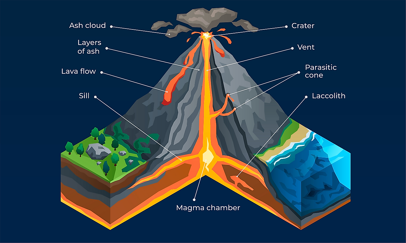
What are all the parts of a volcano. A magma chamber is a large underground pool of molten rock sitting underneath the Earths crust. When this material escapes it causes an eruption. Volcanoes can erupt from the crater on their peak or from asecondary vent on their side.
9 Parts of a volcano from the inside 1. Volcanoes erupt in different ways producing different landforms. How Do Volcanoes Erupt.
Ash clouds - small pieces of rock and glass that can be carried in the air for many kilometres. The part of the conduit that ejects lava and volcanic ash. Throat - Entrance of a volcano.
Steep cone-shaped volcanoes form when plates collide. Hotspots are places where the magma rises up. Deep within the Earth it is so hot that some rocks slowly melt and become a thick flowing substance called magma.
Is called the flank. Eventually some of the magma pushes. A volcano is formed out of eruption of lava from the crust of.
Volcanoes may also erupt in areas called hot spots where the crust is thin. What Are The Different Parts Of A Volcano. The main parts of a volcano include main vent crater ash cloud chamber summit magma and so many more.
This diagram has all the parts of a volcano clearly labelled. The lava dome is one of the parts of a volcano that isnt present in all of these geographical formations. Since it is lighter than the solid rock around it magma rises and collects in magma chambers.
Or it can be calmer with gentle flows of material. A volcano is an opening on the surface of a planet or moon that allows material warmer than its surroundings to escape from its interior. Volcanoes are openings in the Earths crust that connect to the molten 2nd layer of the Earth the mantle.
The side of a volcano. The openings in the volcanoes through which molten magma pours are called vents. The main parts of a volcano include main vent crater ash cloud chamber summit magma and so many more.
Earths crust is broken into 17 major tectonic plates which float on its mantle the layer beneath the earths surface. A volcano is an opening in a planet or moons crust through which molten rock hot gases and other materials erupt. Magma is lighter than the solid rock around it so it rises.
When a volcano erupts there can be lava ash steam or gaseous emissions. Sometimes eruptions are explosive and lava is thrown out as volcanic bombs. 13 Parts of a Volcano.
All the pressure and heat of the collision make for a violent eruption. Volcanic ash consists of rock mineral and volcanic glass fragments smaller than a tenth of an inch in diameteror slightly larger than a pinhead. Flank - The side of a volcano.
When rocks become so hot they can become a substance called magma. Volcanoes often form a hill or mountain as layers of rock and ash build up from repeated eruptions. Volcanoes are classified as active dormant or extinct.
Eventually some of the magma pushes through vents. An eruption can be explosive sending material high into the sky. Lava is the silicate rock that is hot enough to be in liquid form and which is expelled from a volcano.
This is the part of the volcano located deepest underground even beneath the Earths crust. Most volcanoes are cone shaped mountains like this one in the diagram. Pyroclastic flows - fast moving clouds of hot ash gas and rock.
Crater - Mouth of a volcano - surrounds a volcanic vent. A volcano is formed out of eruption of lava from the crust of. It collects in magma chambers on average 1.
The Anatomy of Volcanoes 1 Magma. Volcanic ash consists of small pieces of pulverized rock minerals and volcanic glass created during a volcanic eruption. Since these plates are always converging or diverging.
1150 BC Korovin Volcano. Conduit - An underground passage magma travels through. Volcanic bombs - large bits of very.
Lava - Molten rock that erupts from a volcano that solidifies as it cools. This photograph shows an eruption of.
Both fresh flowers and those. Parts plant flower reference sheet labelling worksheet labeling activities.
Parts Of A Flower Worksheets Superstar Worksheets
Either use the pre-labelled version as a poster or teaching aid.
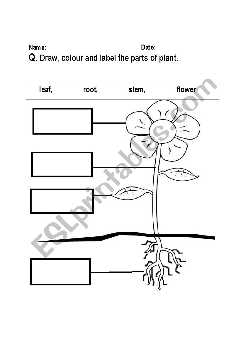
Parts of a flower labeling activity. 1099 Shawna Curran Spring Pond Shape Match. Some of the worksheets for this concept are Flower anatomy activity Flower parts work Parts of a plant Lesson 5 flowers dry forest summary Anatomy of a flower Parts of a flower work formatted answers Roots and stems and leaves oh my Plant reproduction. Plants interactive downloadable rksheet exercises online download grade rksheets.
Courgette flowers are huge and show that there are separate male and female flowers. Use the to put. Identify and label figures in Turtle Diarys fun online game Parts of a Flower Labeling.
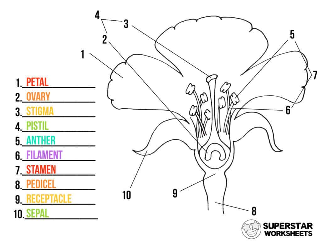
Your challenge is to write the correct name for each part. Composites such as sunflowers and daises are more complex for young naturalists but help them understand the differences in flower structure. This Labelling a Flower Worksheet covers the parts of a plant and flower.
Login to rate activities and track progress. Teachers save Label the Parts of a Flower to assign it to your class. This is a cut paste worksheet for labeling the 4 major parts of a flowerstem flower root and leaf.
Alternatively your children can label the diagrams to reinforce their vocabulary on the topicFor teaching your KS1 or Pre-Primary Age 3-5 you can use this lovely picture of parts of a flower labelled and unlabelledUse them in the classroom for a. Simple parts plant poster worksheet plants kindergarten. Try It Save Activity Brittany Bailey.
Then I place the parts in a. Worksheet labelling flower parts ppt. Flower Anatomy Activity The parts of a flower have been labeled.
Related Activities Shawna Curran In Spring I See Activity. Drag given words to the correct blanks to complete the labeling. Students use the text tool to label the parts of the flower using the word bank for assistance.
Feb 25 2017 - Teach your children all about the different parts of a flower with this handy labelling activity. Identify and label figures in Turtle Diarys fun online game Plant Parts Labeling. Parts plant flower labelling worksheet plants worksheets.
As I cut apart the flower the students tell me the part and its function. To learn more visit. While following the directions students will color cut and assemble each part of the flower stem petals sepals stamen filament anther pistil stigma sty.
It comes in black white and color versions and also includes an answer key in bwIf you like this FREEbie please leave feedbackthanks. Collect three or more different types of flowers for learners to observe and dissect. Diana Do You Believe in Leprechauns.
Simple flowers with easily identifiable parts such as gladioli carnations lilies pansies daffodils peas tomatoes and beans are good plants to use. The next activity is dissecting the flower into its parts. Label the Parts of a Flower 1.
Parts plant yum plants squash vegetable. In this Parts of a Flower Color Cut and Paste Activity students will learn the various parts of a flower in a fun and engaging project. Labeling A Flower Displaying top 8 worksheets found for - Labeling A Flower.
Ask the pupils to find a flower that has an open structure so it is easy to see inside and find the different parts of the flower. Drag the given words to the correct blanks to complete the labeling. Strawberries and raspberries have very simple easy to draw flowers that show the parts of a flower and early fruit formation.
They also have two antennae and an exoskeleton. The head of a butterfly is spherical in shape.
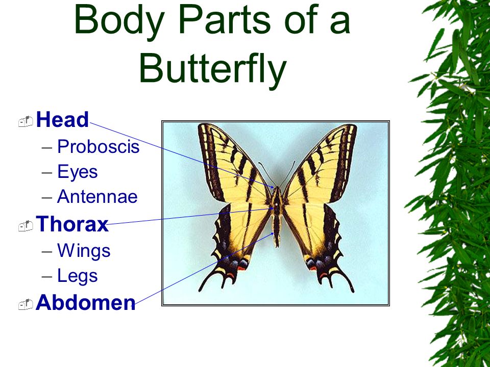
Body Parts Of A Butterfly Head Proboscis Eyes Antennae Thorax Wings Legs Abdomen Ppt Download
The thorax contains the muscles that make the legs and wings move.
3 main body parts of a butterfly. What order do the stages go in. The adult butterfly has three main body parts head thorax and abdomen. Using palpi located adjacent to the proboscis the butterfly begins working the two parts together to form a single tubular proboscis.
The eyes antennae proboscis and palpi are all positioned on the head. Below is a. Head thorax and abdomen.
The body parts of a butterfly. Back to index The difference between a butterfly and a moth. Both butterflies and moths belong to the same insect group called Lepidoptera.
And three pairs of legs. Because butterfly has all these characteristics it is classified as an insect. Hmmm how about spider.
About Press Copyright Contact us Creators Advertise Developers Terms Privacy Policy Safety How YouTube works Test new features 2021. A butterflys body is divided into three main sections the head the thorax and the abdomen. Each of the two wings is divided into two parts the upper forewing and the lower hindwing.
Can you name the three main body parts of a butterfly body. Body Parts of a Butterfly and Their Functions 1. Choose from Body Parts Of A Butterfly stock illustrations from iStock.
Butterfly has a fine tube-like. The forewings are the anterior wings which are attached to the mesothorax the middle segment of the thorax. The three body parts are the head thorax the chest and abdomen the tail end.
Eyespots Tails. Check out my Luminating Lesson on the Parts of a Butterfly. Head thorax chest or mid section and abdomen tail end.
A butterfly has three major body partshead thorax and abdomen. All of these features are discussed in detail below and the illustrations below provide an. The activities included are drag and drop fill in the blanks to label parts of the body multiple c.
Eyespots secure butterflies due to the fact that birds will assault the eyespot first. Butterfly body parts worksheet butterfly activity worksheets and butterfly parts worksheet are three of main things we want to present to you based on the gallery title. The reproductive organs and spiracles are part of the abdomen.
With more related things as follows butterfly body parts worksheet adjectives describing butterflies and butterfly body parts. In general butterflies differ from moths in the following ways. Some eyespots are hidden from view up until the forewing is pushed forward so that red eyes appear and surprise the predator long enough to make a getaway.
This member of the Phylum Arthropoda Class Insecta Order. In front of the head there is a pair of thin and long antennae which help in smelling. On a separate table bench have the paper plates pasta shapes glue and markers spread out.
The four wings and the six legs of the butterfly are attached to the thorax. Pin On Homeschoolin. 2 nd 3 rd 4 th 5 th Homeschool.
Can you go through the. By the way concerning Parts of a Butterfly Worksheet we already collected various variation of pictures to complete your ideas. All insects including butterflies share a common overall body design.
What is the name of each stage. It bears two compound eyes placed at two sides of the head. Like most insects butterflies have three main body parts.
Like all other insects butterflies have six legs and three main body parts. Each body section has very different functions and all are needed for the butterfly to live. Each eye consists of a number of separate light-sensitive components called ommatidia.
Picture of Butterfly Eye Antennae Proboscis Head Thorax Legs Abdomen. The legs and wings are attached to the thorax. Parts of butterfly body.
An insect has three main body parts. A butterflys head is full of extremely important organs that allow the butterfly to sense what is. Parts of a Butterfly.
Wherein the larva hatches and almost constantly. Parts of a butterfly Parts of a butterfly. How does having three segments rather than just one help it move.
Numerous butterfly wings have eyespots or tails. Imago The body of the adult butterfly is comprised of 3 segments - head thorax and abdomen. Find high-quality royalty-free vector images that you wont find anywhere else.
The tibia of each leg has a subgenual under the knee organ which detects and amplifies small vibrations. _____ _____ _____ Well these are the characteristics of an insect. A pair of antennae on its head.
All adult butterflies have 3 pairs of legs except in the Nymphalidae and in males of certain other groups where the front pair are reduced to brush-like stumps and modified as chemoreceptors. What are the purposes or functions of the butterflys wings. It has three pairs of jointed wings and a proboscis through which it sips nectar.
Perhaps the most interesting Geneva Psalter is a 1560 edition published by Pierre Davantes see illus5. French Spanish German and more.
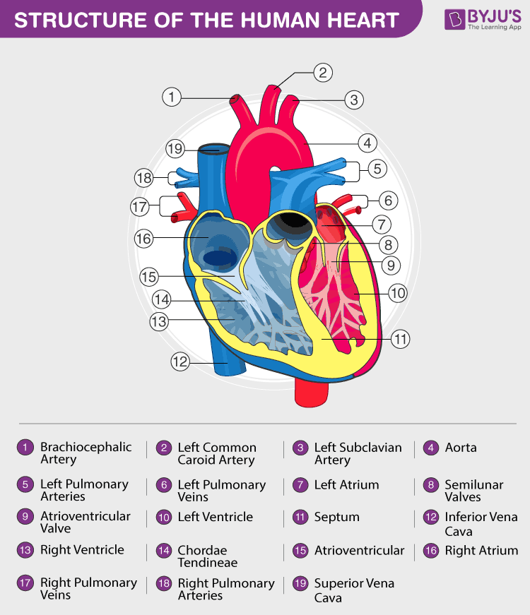
Heart Diagram With Labels And Detailed Explanation
System anatomy structure heart circulatory system by carlfarrant88 teaching circulatory system anatomy diagram amp function healthline anatomy of the human heart and cardiovascular system myvmc labeled diagram of the circulatory system anatomy human diagram of circulatory system to label human diagram chart cardiovascular system human veins arteries heart human heart diagram.

Explain the structure of human heart with the help of a labelled diagram. It is perhaps best to see them simply as provocative nonsense verse. The photopicture shows. One of the 2 transfected YT clones YTpX51.
Tips and Example how to describe pictures in English. The kinds of non-physical violence recognized in the 15th century were different from those recognized now but the concept was ancient and potent. Since raff meant worthless material trash refuse according to.
The film features an ensemble cast including Jessica Chastain Viola Davis Cicely Tyson Bryce Dallas Howard Allison Janney Octavia Spencer and Emma Stone. To study the effect of HTLVI on both of the IL2Rs we transferred a fragment of HTLVI containing the p40X gene into YT cells. User-uploaded templates that become popular may display on this page.
This volume provides a new perspective on the evolution of the special language of medicine based on the electronic corpus of Early Modern English Medical Texts containing over two million words of medical writing from 1500 to 1700. This additional system used numbers lines and dots to indicate both pitch and rhythm and was intended by the publisher as a replacement for. In their turn they correspond to the three chief products of human learning.
The Help is a 2011 period drama film written and directed by Tate Taylor and based on Kathryn Stocketts 2009 novel of the same name. The film and novel recount the story of a young white woman and aspiring journalist Eugenia Skeeter. Directed by Tate Taylor.
This structure is similar to that of Augustines De Doctrina Christiana in which the Christian orators inventio where he finds the matter for his speech is biblical exegesis which Swynnerton31 William Whitaker and Dudley Fenner32 But Perkinss use of rhetorical exegesis goes beyond this kind of polemical point. In terms of theological disciplinesPerkinss two parts could be labelled exegesis and homiletics. These consist of memory imagination and reason.
Shapes of Fancy offers a powerful new method of accounting for ineffable and diffuse forms of desire mining early modern drama and prose literature to describe new patterns of affective resonance. Ogborns book is a painstaking analysis of the functioning of the East India Company. A summary of the basic ideas and issues including link types frame systems case relations link valence abstraction inheritance hierarchies and logic extensions.
Blank customizable templates of the most popular trending and latest memes. Your time is precious. Presumably all these forms of non-physical violence were subject to the same moral.
This article introduces semantic network systems and their importance in Artificial Intelligence followed by I. Wie das geht zeigen wir dir hier. They help to shape activate and regulate the adaptive immune response.
Good grades at your fingertips. In framing these chapters I have used as something between a heuristic and a structural principle the three partes of the human understanding categorized by Francis Bacon in the second book of his Advancement of Learning. A Venn diagram is a diagrammatic representation of ALL the possible relationships between different sets of a finite number of elementsVenn diagrams were conceived around 1880 by John Venn an English logician and philosopher.
Medical writing tells us a great deal about how the language of science has developed in constructing and communicating knowledge in English. It was taken byin. Over 1 million templates updated continously.
A survey of world. 1 The Platonic Poems of Katherine Philips 1. Wichtiger ist es dass du die Bedeutung interpretierst und abschließend kommentierst.
In a world where a letter of instruction could only go out once a year and a reply could only be expected the best part of. Its a black-and-whitecoloured photo. E and S attempt to explain the origins of this term and the other words in this sequence eg raffe as a pun on the noun raff refuse.
With Emma Stone Viola Davis Bryce Dallas Howard Octavia Spencer. Where the bureaucratic structure of the company has been studied before Ogborn attends to the ways in which writing had to stretch across thousands of miles to make the company function. While her contemporary Anne Conway 1631-1679 engaged directly in philosophical debates through her correspondence with figures such as the Cambridge Platonist Henry More Philips worked in a.
22 As Davantess opening preface explained solmization syllables were printed in the music with an additional system also introduced above the text underlay. A semantic network is a graph of the structure of meaning. It stages an impassioned defense of the inherently desirous nature of reading making a case for readerly investment and identification as vital engines of meaning making and political insight.
Um einen Cartoon Karikatur umfassend zu analysieren solltest du dich nicht ausschließlich auf die Beschreibung beschränken. MHC Class II molecules interact with CD4 on the T helper cells which helps identify this cell type. History poetry and philosophy.
CD4 T cell functions include activating other immune cells releasing cytokines and helping B cells to produce antibodies. An aspiring author during the civil rights movement of the 1960s decides to write a book detailing the African American maids point of view on the white families for which they work and the hardships they go through on a daily basis. To create an animated GIF template choose a video in the GIF Maker and click Save as Template.
To upload your own template visit the Meme Generator and click upload your own image. In legal terms a writ was the same whether it alleged threatened violence or actual assault. In the human NK cell line YT as well as in ATLderived T cells the coexpression of IL2Rp55 and the second IL2R without the Tac epitope IL2Rp70 is required to produce highaffinity IL2R.
The Appeal of Platonism Katherine Philips 1632-1664 wrote poems on the human and the divine in the language of early modern idealism. Whether in high school or at university boost your language skills the smart way. Violence in the 15th century could imply more than physical action.
With Linguees example sentences and recorded pronunciations you will be using foreign languages like a pro.
To keep watching this video solution for FREE Download our App. Draw neat labelled diagram of electrolytic reduction of alumina.
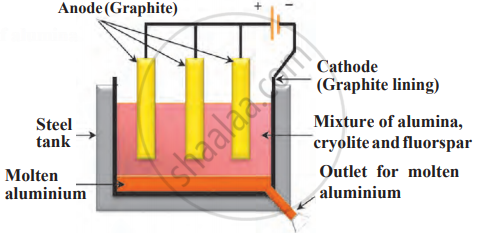
Draw A Neat Labelled Diagram Electrolytic Reduction Of Alumina Science And Technology 1 Shaalaa Com
A set of graphite rods dipped in the molten electrolyte works as anode.

Draw a neat labelled diagram of electrolytic reduction of alumina. Draw a Well-labelled Diagram of Extraction of Aluminium. 1 See answer babandhakane78 is waiting for your help. Draw a neat and labelled diagram Electrolytic reduction of alumina.
Write the chemical equation for the following events. The electrolytic cell is a large steel tank having a lining of carbon graphite on the inner side. Latest Syllabus 2019 - 2020.
Apne doubts clear karein ab Whatsapp par bhi. NCERT P Bahadur IIT-JEE Previous Year Narendra Awasthi MS Chauhan. Find an answer to your question draw a neat labelled diagram of electrolytic reduction of alumina explain cryolite and fluorspar are added in the mixture aartipatange36 aartipatange36 07112019.
Draw a neat labelled diagram. Draw a neat labelled diagram of electrolytic cell for the extraction of aluminium. Electrolytic reduction of alumina.
Electrolytic reduction of alumina. Maharashtra State Board HSC Science Electronics 12th Board Exam. Write the Anode Reaction in Electrolytic Reduction of Alumina.
NCERT RD Sharma Cengage KC Sinha. The tank has a graphite lining on the inner side. Draw a neat labelled diagram.
Maharashtra State Board SSC English Medium 10th Standard Board Exam. Identify the process given in following passage and draw neat labelled diagram showing the process. Add your answer and earn points.
NCERT P Bahadur IIT-JEE Previous Year Narendra Awasthi MS Chauhan. Addition of cryolite and fluorspar reduces the melting point to about 1000 C. Draw a neat and labelled diagram Electrolytic reduction of alumina Apne doubts clear karein ab Whatsapp par bhi.
A set of graphite rods dipped in the molten electrolyte works. Electrolysis of alumina is carried out in an electrolytic cell at a much lower temperature by dissolving it in molten cryolite Na 3 AlF 6 and fluorspar CaF 2. - Science and Technology 1.
NCERT NCERT Exemplar NCERT Fingertips Errorless Vol-1 Errorless Vol-2. Click hereto get an answer to your question Draw a neat labelled diagramElectronic reduction of alumina. This lining does the work of a cathode.
A blog about 9th 10th 11th and 12th Maharashtra Tamilnadu CBSE Board. NCERT NCERT Exemplar NCERT Fingertips Errorless Vol-1 Errorless Vol-2. This lining does the work of a cathode.
Draw a neat and labelled diagram Electrolytic reduction of alumina. Describe the electrolytic reduction of alumina with neat labelled diagram. Describe the electrolytic reduction of alumina with neat labelled diagram.
Draw a neat and labelled diagram for electrolysis of fused NaCl. NCERT DC Pandey Sunil Batra HC Verma Pradeep Errorless. Electrolytic reduction of alumina d.
During the extraction of Divide the metals Cu Zn Ca Mg Fe Na Li into three groups. Draw a neat labelled diagram. Identify the process given in following passage and draw neat labelled diagram showing the process.
NCERT DC Pandey Sunil Batra HC Verma Pradeep Errorless. Join the 2 Crores Student community now. The carbon lining acts as a cathode.
Log in to add comment. The tank has a graphite lining on the inner side. A set of carbon graphite rods dipped in the.
Draw a neat and labelled diagram Electrolytic reduction of alumina Draw a neat and labelled diagram Electrolytic reduction of alumina. Draw a neat and labelled diagram Electrolytic reduction of alumina Draw a neat and labelled diagram Electrolytic reduction of alumina Books. Click here to get an answer to your question draw a neat labelled diagram electrolytic reduction explain why cryolite and fluorspar mixture.
Draw a neat and labelled diagram Electrolytic reduction of alumina Getting Image Please Wait. Electrolytic reduction of alumina. Electrolysis of molten mixture of alumina melting point 2000 0 C is done in a steel tank.
Aluminum came in contact with Complete the following statement using every given option. Draw a neat labelled diagram. Draw a neat labelled diagram.
Watch Video in App. Draw a neat labelled diagram of electrolytic reduction of alumina explain cryolite and fluorspar are added in the mixture. Electrolysis of molten mixture of alumina melting point 2000 0 C is done in a steel tank.
If u want this diagram step by step like this. Draw A Labelled Diagram Of Gravity Filtration Techniques ERD Entity Relationship Diagrams ERD Software for Mac and Win Flowchart Basic Flowchart Symbols and Meaning.
Chemistry Symbols And Meanings Cisco Routers Cisco Icons Shapes Stencils And Symbols Draw A Neat Labelled Diagram Of The Laboratory Apparatus Used For Filtration
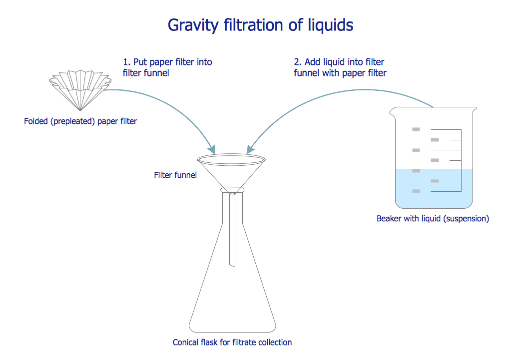
Sand and water w ithout using a filter paper.

Draw a neat labelled diagram of filtration. Draw a neat labeled diagram of an animal cell. Our top 5 students will be awarded a special scholarship to Lido. Learn from an expert tutor.
Q13 Draw a neat labelled diagram for separation of the following mixtures. Draw a neat diagram and label it. To measure the length of a rod diameter of a sphere external diameter of a hollow cylinder.
Looking to do well in your science exam. Draw a neat labelled diagram for separation of the following mixtures. Laminiaduo7 and 28 more users found this answer helpful.
In addition they also play an important role in maintaining the water balance of our body. Sand and water without using a filter paper. To measure length correct up to 1 mm.
Advertisement Remove all ads. Draw neat labelled constructional diagram of INKJET printer. Question 22 What Is A Circuit Diagram Draw The Labelled Of An Electric Comprising Brainly In Draw A Labelled Diagram Of.
Question Bank Solutions 7315. The liquid which has obtained after filtration. Its output is sharper than Dot matrix though its output quality is not to the same level of laser printer.
While preparing tea a filter or a sieve is used to separate tea leaves from the water. Draw a neat labelled sketch of slow sand filter and rapid sand filter and explain the operation of each. I Glomerular filtration - Urine formation begins when the blood is filtered by the glomerulus and enters the Bowmans capsule and the glomerular filtrate is formed.
Sand and water w ithout using a filter paper. To measure the depth of a beaker or a bottle. Glomerular filtrate is formed of all blood constituents except corpuscles and.
622 x 600 Photo description. Book a free class now. Draw a neat and well labelled diagram of male reproductive system of a frog.
PLE MARK AS BRA8NLIEST. 669k 728k 656. Draw a neat labelled diagram of a pistil showing pollen tube growth and its entry into the ovule.
It provides letter quality printout than dot matrix printer. Draw A Neat Labelled Diagram Of An Electric Circuit Posted by Margaret Byrd Posted on April 27 2021. Explain the construction and.
Question 22 what is a circuit diagram draw labelled of closed electric diagrams lesson for schematic rheostat and an unknown resistance to bell domestic wiring. Draw a neat labelled diagram of electrolysis of water. Maharashtra State Board SSC Marathi Semi-English 10th Standard इयतत १० व Question Papers 161.
Concept Notes Videos Videos 260. Explain disinfection and sterilisation. Draw a neat labelled diagram of electrolytic cell for the extraction of aluminium.
Book a free class. And the next 3 steps is like this. Book a free class now.
Buchner funnel moistened filter paper porous plate plate with holes in it rubber tubing buchner flask and rubber bung. Filtration is technically defined as the process of separating suspended solid matter from a liquid by causing the latter to pass through the pores of a membrane called a filter. Set your child up for success with Lido book a class today.
- Get the answer to this question and access more number of related questions that are tailored for students. THANKS FOR CLARIFING UR DOUBT. Filtration Diagram Picture category.
Draw a neat labelled diagram of electrolysis of water. Through the sieve pores only water will pass. 130k 352k Draw a neat labelled diagram to show the.
The human kidneys house millions of tiny filtration units called nephrons which enable our body to retain the vital nutrients and excrete the unwanted or excess molecules as well as metabolic wastes from the body. The most common example is making tea. Is compatible in quality to that of a laser printer.
This increases the blood pressure within the glomerulus helps in the filtration. 1100k 1460k 404. Advertisement Remove all ads.
A Coarse sand from fine sand b A magnetic particle from a non-magnetic particle c Naphthalene from sodium chloride d Chalk and water using filter paper e Sand and water without using a filter paper f Common salt from a solution of common salt and water. Book a free class now. To measure the internal diameter of a hollow cylinder or pipe.
It is drawn by me. It is a non-impact printer. Draw a neat labelled diagram for separation of the following mixtures.
This diagram labels the typical parts of a filtration experimentIncluded in the equipment labeling are. Draw a neat labelled diagram for the separation of the following mixture. The afferent arteriole entering the glomerulus is wider than the efferent arteriole in diameter.
Chapters in Science Exemplar Problems. Describe the function of each block. Labeled Diagram of the Human Kidney.
Click here to get an answer to your question Draw a labelled diagram of Renal Excretory System of Man. A Draw A Neat Diagram Of Excretory System Of Human Beings And الحالب الحالبين Ureters Human Anatomy Physiology Medical Draw A Well Labelled Diagram Of A Human Excretory Toppr Com With A Neat Labelled Diagram Describe The Excretory System In Excretory System Images Stock Photos Vectors Shutterstock Human Excretion Excretory System Kidneys Videos Solved Question What Is Excretory.

Draw A Well Labeled Diagram Of The Human Excretory System Studyrankersonline
Draw a well-labeled diagram of the human excretory system.

Draw a labelled diagram of excretory system of man. Two kidneys two ureters bladder and urethra. A Draw a labelled diagram of excretory system in human beings and label the following. It is a labelled diagram of urin.
Specially for class 9101112. Asked Feb 18 2019 in Biology by Amita 884k points Draw a well-labeled diagram of the human excretory system. Advertisement Remove all ads.
Draw a diagram of human excretory system and label the following parts on it. Draw a labelled diagram of excretory system. The kidneys are located in the abdomen one on either side of the backbone.
Click to find video solution. Asked Nov 3 2017 in Class X Science by aditya23 -2137 points a Draw a diagram of human excretory system and label the following. ANS The Urinary Syste.
Draw a well labeled diagram of excretory system. Kidney Ureter Urinary Bladder and Urethra. B Name the factors on which the amount of.
Advertisement Remove all ads. DRAW A LABELLED DIAGRAM OF EXCRETORY SYSTEM PDF The writers of Draw A Labelled Diagram Of Excretory System. Draw a Diagram of the Human Excretory System and Label the Following Parts.
Draw a Diagram of the Human Excretory System and Label the Following Parts. Draw a labelled diagram of renal excretory system of man Get the answers you need now. Draw a well-labeled diagram of the human excretory system.
Asked Jun 11 2020 in Respiration and Excretion in Animals by AmanKumar 517k points respiration and excretion in animals. Q6 Draw a diagram of the human excretory system and label. CBSE CBSE English Medium Class 10.
Excretory system in humans The excretory system in human beings includes a pair of kidneys a pair of ureters a urinary bladder and a urethra. Draw A Labelled Diagram Of Excretory System - PDF-DALDOES14-7 Download full version PDF for Draw A Labelled Diagram Of Excretory System using the link below. Advertisement Remove all ads.
CISCE ICSE Class 7. Concept Notes Videos 261. Ii part which stores the urine.
QUE WHAT IS URINARY SYSTEM. Draw excretory system in human beings and label the following organs of excretory system which perform following functions. C The excretory system removes the poisonous waste substances from the body in the form of urine and maintains ionic balance called osmoregulation.
Iii part which connects i and ii iv part from which urine is passed out. Read DRAW A LABELLED DIAGRAM OF EXCRETORY SYSTEM PDF direct on your iPhone iPad android or PC. This is also known as the urinary system.
Asked Nov 3 2017 in Class X Science by aditya23 -2138 points a Draw a diagram of human excretory system and label the following parts on it. I part in which urine is produced. Prev Question Next Question 0 votes.
I left kidney ii renal artery iii urinary bladder iv urethra b Name the functional unit of kidney. Question Bank Solutions 6480. A The excretory system of human beings consists of the following main organs.
Concept Notes Videos 150 Syllabus. Kidney Ureter Urinary Bladder and Urethra. Draw a Neat Labelled Diagram of the Human Excretory System.
Question Bank Solutions 20334. Draw the well-labelled diagram of the human excretory system. It is a labelled diagram of urin.
C Name two nitrogenous wastes released from kidney. Click here to get an answer to your question draw labelled diagram of human excretory system. SADGIRL01 SADGIRL01 06112020 Biology Primary School answered Draw a labelled diagram of renal excretory system of man 2 See answers drish4205.
Draw a Neat Labelled Diagram of the Human Excretory System. 2 is a long tube which collects urine from kidney. Q6 Draw a diagram of the human excretory system and label the following parts Kidney ureter urinary bladder and urethra.
How to draw Urinary system diagram. They act as excretory organs and also control the balance of water and mineral ions in the body. 3 store urine until it is passed out.