Parts correlated with the intestines are found below. Weve arranged the synonyms in length order so that they are easier to find.
Toe Wikipedia
Some languages have different names for hand and foot digits English.

Parts of the foot digits. Regions of the Foot. More recent studies conducted by Ozer et al. Arterial arch supplies the digits of the foot via the plantar metatarsal arteries including all the muscles and bones however these studies noted few morphological variations.
Synonyms crossword answers and other related words for DIGITS OF THE FOOT toes We hope that the following list of synonyms for the word toes will help you to finish your crossword today. Crossword Clue The crossword clue Number of foot digits with 3 letters was last seen on the October 22 2018We think the likely answer to this clue is TENBelow are all possible answers to this clue ordered by its rank. Middle digit of the foot.
Walk so that the toes assume an indicated position or direction. The toe refers to part of the human foot with five toes present on each human foot. Parts of your foot correlated with the stomach are found above the waistline.
One of the digits on the foot Please find below the One of the digits on the foot answer and solution which is part of Daily Themed Crossword May 19 2019 Answers. Three types of foot posture exist in mammals. Toes are the digits of the foot.
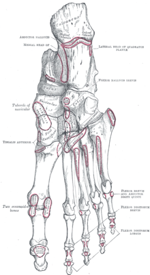
Bones of the foot dorsal view. Flexor digiti minimi brevis opponens digiti minimi These two muscles are found on the lateral side of the foot inferior to the fifth metatarsal. You can easily improve your search by.
The word that solves this crossword puzzle is 4 letters long and begins with T. And Gabrielli et al have illustrated the contributing arteries and the positioning of the plantar arch within the foot. Arabic Russian Polish Spanish Portuguese Italian Czech Tagalog Turkish Bulgarian and Persian there are no specific one-word names for fingers and toes.
They are often considered parts of the same muscle the flexor digiti minimi brevis. Five-toed organ at the end of a human leg that supports the body and is used for walking. The bottom of the foot is called the sole.
Most land vertebrates have feet and. It may surprise you to know that the foot is one of the most complicated structures of the body. Such complexity is necessary because the foot is required to do many different activities such as walking running and climbing.
The part of the foot which joins it to the leg is called the heel. The Hindfoot begins at the ankle joint and stops at the transverse tarsal joint a combination of the talonavicular and. Forepart of a hoof.
Each toe consists of three phalanx bones the proximal middle and distal with the exception of the big toe Latin. Armored from head to foot dactyl digit - a finger or toe in human beings or corresponding body part in other vertebrates. The foot is traditionally divided into three regions.
Many other animals with backbones also have five digits. Digit of the foot between the third toe and the little toe. It contains a lot of moving parts - 26 bones 33 joints and over 100 ligaments.
Quick and free help to solve your crossword clue. The bottom of your foot is connected with your pelvic area. Digit of the foot between the big toe and the third toe.
Toe - one of the digits of the foot foot human foot pes - the part of the leg of a human being below the ankle joint. Respectively finger and toe German. The hindfoot the midfoot and the forefoot Figure 2Additionally the lower leg often refers to the area between the knee and the ankle and this area is critical to the functioning of the foot.
Doigt and orteilIn other languages eg. Finger and Zeh French. Best Answer for Digits On The Foot Crossword Clue.
They both have five digits the fingers and toes. The largest of the digits of the foot situated on its inner edge. Even the basics can lead you to a very simple self-treatment.
Toe a nail golf the part of a clubhead farthest from the shaft. His bare feet projected from his trousers. 1 plantigrade in which the surface of the whole foot touches the ground during locomotion eg human baboon and bear 2 digitigrade in which only the phalanges toes and fingers touch the ground while the ankle and wrist are elevated eg dog and cat and 3 unguligrade in which only a hoof the tip of one or two digits touches the grounda.
The hallux only contains two phalanx bones the proximal and distal. A complete list of crossword puzzle answers for the clue Foot digits. She toes inwards one of the digits of the foot.
The part of footwear that provides a covering for the toes. These are called digit of the hand or. Many other players have had difficulties with One of the digits on the foot that is why we have decided to share not only this crossword clue but all the Daily Themed Crossword Answers every single day.
The thinnest part of your foot usually found towards its center is known as the waistline. Peoples hands and feet have the same shape.
Find an answer to your question e Draw a labelled diagram of Bacteriophase. The phage particle undergoes a chance collision at a chemically complementary site on the bacterial surface then adheres to that site by means of its tail fibers.

Diagram Of Bacteriophage How To Draw Bacteriophage Diagram Class 11 Biology Youtube
1 See answer sagarbhavna59 is waiting for your help.

Labelled diagram of a bacteriophage. The bacteriophage consists of a polyhedral head a short collar and a helical tail. This is a well. This is the well labelled diagram of bacteriophage.
Bacteriophage can be either infectious or non-infectious to the host cell. A diagrammatic sectional view of T-4 phage is shown in Fig. Http Www Mun Ca Biology Scarr 2014 Hershey Chase 1952 Edited Pdf.
Thousands of varieties of phages exist. This is the diagram of Bacteriophages. The head is hexagonal which consists of about 2000 identical protein subunits.
In an experiment a culture of Euglena is placed in a petri dish and a piece of cardboard with a circle cut out of its center is placed over the petri dish allowing light to enter only the center of the petri dish. The bacteriophage particle resembles a tiny sperm. Bacteriophage any of a group of viruses that infect bacteria.
The first two scientists to observe bacteriophages were Frederic Twort of England and Felix dHerelle of France. Its genome size is about 49 kb. The term bacteriophages eaters of bacteria was coined by d Herelle.
Head- The head consists of 2000 capsomeres with double-stranded DNA enclosed within. Section 19 2 Pearson School. How To Draw Bacteriophage Easy Steps.
Draw A Neat Labelled Diagram Of Bacteriophage Brainly In. Euglena gracilisis a protist that has animal and plant-like characteristics. How to draw an bacteriophages in exam is the topic.
Because of its eyespot Euglena exhibits phototaxis. Draw A Labelled Diagram Of A Bacteriophage Biology. Bacteriophages or bacterial viruses are viruses that parasitize bacteria.
They have tadpole-like structure ie with head and tail. Twort in Great Britain 1915 and Felix dHerelle in France 1917. It has a head and a tail.
Tail- The tail consists of an inner hollow tube which is surrounded by a contractile sheath with 24 annular rings. Certain types serve key roles in laboratory research. Given is a diagram of a bacteriophage.
A bacteriophage is smaller than the bacteria. The virus particle of T-even phage is about 200 to 280 m µ in length. The long helical tail consists of an inner tubular core which is connected to the head by a collar.
Draw A Neat Labelled Diagram Of Bacteriophage Brainly In. Sagarbhavna59 sagarbhavna59 08012021 Science Primary School answered e Draw a labelled diagram of Bacteriophase. At the end of tail end plate is present to which tail fibres are attached.
Tail is having hollow core end is surrounded by tail sheath. The given diagram is of bacteriophage viruses that infect the bacteria. Bacteriophages were discovered independently by Frederick W.
Bacteriophages fall into four major groups based on the genetic material they possess. Bacteriophage Transcytosis Provides A Mechanism To Cross. IMAGES Bacteriophage Diagram Images Pencil Diagram Of Bacteriophages Images Simplest Way Of Drawing Bacteriophage Diagram.
In addition there is some sort of attachment region adapted to stick to the surface of the host cell. This is the well labelled diagram. In which one of the options all the four parts a b c and d are correct.
The T4 phage is tadpole shaped and consists of head collar tail base plate and fibres figure. The genetic material of bacteriophage can be either DNA or RNA and linear or circular. The bacteriophage size ranges from 25-200 nm in length.
Nucleic acid generally DNA is present inside the head. Targeting Human Embryonic Stem.
Draw a neat and well labelled diagram of male reproductive system of a frog. ALL IMAGES VIDEOS NEWS MAPS SHOPPING.

Draw A Labelled Diagram Of A Human Male Reproductive System Cbse Class 10 Science Learn Cbse Forum
Not everyone was completely clueless and those who struggled got creative.

Well labelled diagram of male reproductive system. Well Labelled Diagram Of Male And Female Reproductive System - PDF-WLDOMAFRS20-0 Download full version PDF for Well Labelled Diagram Of Male And Female Reproductive System using the link below. Or that enormously awkward class when you learned the male and female reproductive systems. Buzzfeed recently asked a group of adults to label diagrams of the male and female reproductive systems and the results were uh nuts.
Internal organs include the vas deferens prostate and urethra. NCERT NCERT Exemplar NCERT Fingertips Errorless Vol-1 Errorless Vol-2. Male testis is known as primary sex organ and reproductive duct like epididymis vas deference and ejaculatory duct reproductive gland like seminal vesicle prostate glandcowpers gland and external genitalia penis all are known as secondary sex organ of male reproductive system.
Draw And Label Diagrams Of Male And Female Reproductive System. Read WELL LABELLED DIAGRAM OF MALE AND FEMALE REPRODUCTIVE SYSTEM PDF direct on your iPhone iPad android or PC. Labeled diagram of the male reproductive system.
Reproductive System Male Anatomy Diagram Side View First read the definitions below. Draw a neat diagram of human male reproductive system and label the parts performing the following functions. Add your answer and earn points.
Draw well labelled diagram of Male reproductive system 1 See answer priyankabharti2246 is waiting for your help. Draw a neat and well labelled diagram of male reproductive system of a frog. Labels are a means of identifying a product or container through a piece of fabric paper metal or plastic film onto which information about them is printed.
Make your work easier by using a label. Scrotum seminal vesicles vas deferens testicles testes and prostate constitute all the remaining. Each sac contains one testis.
The male reproductive organs include the testes seminal vesicles penis and some associated glands such as the prostate gland. The shaft and the glans. For some that memory seems to fade rather quickly.
Brainly User Brainly User Answer. There are two oval testes each contained in a protective bag called scrotum or scrotal sac lying outside the abdominal cavity. This leaderboard has been disabled by the resource owner.
All these organs work together to produce sperms male gamete and other components of semen. This leaderboard is currently private. Testes are the primary sex organs of the man.
- The outlet of the rectum lying in the fold between the buttocks. D Common passage for sperm and urine. Penis and Urethra are a part of reproductive and urinary systems.
In males fertility depends directly on their reproductive system both at an endocrine level so that the hormones produced trigger the production of sperms and at the anatomical level as each part is. Maths Physics Chemistry Biology. Well Labelled Diagram Of Male Reproductive System Selina Solutions Concise Biology Class 10 Chapter 13 The - 8 Given below is the outline of the male reproductive system Name the parts labelled 1 to 8 and state their functions Also name the corresponding structure of part 4 in the female reproductive system Solution.
These organs work together to produce sperm the male gamete and the other components of semen. Make a well labeled diagram of male reproductive system - 12915529. The male reproductive system includes testes scrotum spermatic ducts sex glands and penis.
Then label the male anatomy diagram by placing the correct number next to the appropriate definitions. Female male model human. NCERT P Bahadur IIT-JEE Previous Year Narendra Awasthi MS Chauhan.
In the diagram of male reproductive system above can be seen that the reproductive system of a male human includes the scrotum testes spermatic ducts sex glands and penis. Get started using our labeled and unlabeled diagrams and quizzes. How do organisms reproduce.
Show more Show less. Each testicular lobules consists of 2-3 seminiferous tubules blood vessels nerves and connective tissue. These external organs include the penis scrotum and testicles.
A Production of sperms b Gland which provide fluid c Provides low temperature for the formation of sperms. 2015 Northwestern University. NCERT RD Sharma Cengage KC Sinha.
Read WELL LABELLED DIAGRAM OF THE MALE REPRODUCTIVE SYSTEM PDF direct on your iPhone iPad android or PC. NCERT DC Pandey Sunil Batra HC Verma Pradeep Errorless. The male reproductive system is mostly located outside of the body.
Maths Physics Chemistry Biology. This leaderboard is disabled as your options are different to the resource. The penis is actually made up of two parts.
The male urethra is a common pathway for both semen and urine. The male reproductive system is responsible for sexual function as well as urination. The human male reproductive system consists of a scrotum testes testicular lobules seminiferous tubules urethra and penis.
Well Labelled Diagram Of The Male Reproductive System - PDF-WLDOTMRS18-5 Download full version PDF for Well Labelled Diagram Of The Male Reproductive System using the link below. Wide collections of all kinds of labels pictures online. The scrotum is a pouch of deeply pigmented skin divided into two separate sacs.
The most important male reproductive organ is the testis which produces sperms. Male reproductive system early Share Share by Jenniferross. Click Share to make it public.
Male reproductive system organs is formed of two type of organs primary sex organ and secondary sex organ. The penis is the male. Looking to learn the anatomy of the male and female reproductive systems.
MARK AS BRAINLIEST.
Labelled Spider Specimen Sale. We will ship all orders the same day if ordered by the USPS and fedex cut off time of 200 pm.
Label And Color The Parts Of The Spider
Spider labeled labeled spider body parts 8460.

Labelled spider parts. Use the to label the parts of the spider. Click to turn in your work. 5 creatures grow body parts explore activities fun facts kids.
We use USPS Priority for small orders as they normally deliver in the USA IN 2-3 days. Parts Spider Labeled Diagram. Students color cut paste and label the various parts of Charlotte the spider.
Labels are a means of identifying a product or container through a piece of fabric paper metal or plastic film onto which information about them is printed. Parts spider book card set learning. Look through for Labelled Spider Specimen recently.
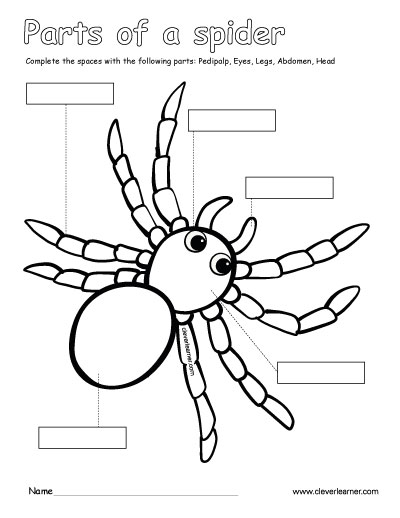
Uncover Labelled Spider Specimen and different product on sale at. 3 Tap the mic and tell facts about spider bodies and other information about spiders. The first anterior part of the body is called the cephalothorax.
1st Grade Kindergarten 2nd Grade Science. Children must then label the key parts of the insect. One is an answer sheet showing all parts of the insect with labels filled-in such as spinnerets a palp and abdomen.
Buy Labelled Spider Specimen at cost effective prices. Cephalothorax - the fused head and thorax also called the prosoma. Spiders treasure trove scientific.
A Well Labelled Diagram Of A Spider. The body of insects is divided in three major parts. Spiders home natural history museum.
Label the Parts of A Spider Science 1. Parts Of A Spider And Its Labeled Diagram There is a thin flexible pedicel that separates the abdomen from the cephalothorax allowing for the movement of its abdomen during mating and silk spinning. This page is a collection of pictures related to the topic of Spider Body Parts Label which contains Pin on Speechie Stuff Butterflies SpidersIdentifying Spider FamiliesLabel the External Spider Anatomy Diagram PrintoutLife science worksheets pdf and games for kids.
Share Labelled Spider Specimen with family and friends. Also known as prosoma the cephalothorax is a fused body part containing the head and thorax which bear the legs eyes and mouthparts. Spiders are different from insects.
691 teachers like this. Spiders habitat diet behavior facts. It contains the guts heart reproductive organs and silk glands.
Make your work easier by using a label. Label Gallery Get some ideas to make labels for bottles jars packages products boxes or classroom activities for free. This can assist visual learners and improve childrens memory for the other sheetThe latter presents all parts of the spider on the left-hand side.
Abdomen - the belly also called the opisthosoma. You should make a label that represents your brand and creativity at the same time you. Ogre faced spiders listen closely snatch bugs air science news students.
A large hand-drawn illustration of a spider is in the centre. Unlike an insect the spiders body is in two sections. There are four parts to label on this worksheet head abdomen legs and s.
Steam Enclosures Camping Shower Outdoor Solar Steam Tubs Door Wheels Shower Riser. What are the parts 1. This is a SIMPLIFIED version of our cut and paste labeling the parts of the spider worksheet to go along with EB.
The head and thorax bearing the eyes mouthparts and legs are fused together to form the cephalothorax. Spider Point - Startseite - spider-partsde. As such they can demonstrate what they have learnt what parts.
Head thorax and abdomen. The information can be in the form of hand-written or printed text or symbols. Fiat 124 Spider parts catalog Fiat X19 parts catalog.
Spider Body Parts Label. A well labelled diagram of a spider spiderpartsphoto. Label Gallery Get some ideas to make labels for bottles jars packages products boxes or classroom activities for free.
An easy and convenient way to make label is to generate some ideas first. Fiat Spider parts of Texas. You should make a label that represents your brand and creativity at the same time you.
An easy and convenient way to make label is to generate some ideas first. Wide collections of all kinds of labels pictures online. The body of spiders is divided in two major parts.
Read the definitions then label the external spider anatomy diagram below. This is joined by a slim waist pedicel to the second body section the abdomen on which are found the silk spinning organs spinnerets the reproductive openings.
The kidneys are paired organs and are located one on. Excretory System Summary Excretion is one of the most important physiological processes accomplished by the excretory system.
Draw A Diagram Of The Human Excretory System And Label The Various Parts Science Shaalaa Com
Name the parts of the human excretory system2.
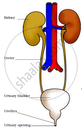
Name the parts of human excretory system. AnswerThe main excretory organs include kidney ureter urinary bladder and urethra. Only the organs specifically used for the excretion are considered a part of the excretory system. A The excretory system of human beings consists of the following main organs.
C The excretory system removes the poisonous waste substances from the body in the form of urine and maintains ionic balance called osmoregulation. The excretory activity of the kidneys is modulated by specialized hormones that. The waste substances dissolved in the blood are carried to the kidneys for filtration.
Parts of excretory system - Human Body Though there are many essential organs or parts of excretory system the most basic ones are just three namely lungs kidneys and skin. These capillaries are called glomeruli and they manage to penetrate into microscopic structures called nephrons. Name true basic step in the process of dna barcoding manishachaudhari1802 in 3 hours 0 javaab.
A pair of kidneys A pair of ureters A urinary bladder A urethra. List the parts of human excretory system and their functions. The process of excretion is done by both kidneys equally.
A dedicated system of organs that removes waste products from the human body is called the human. The human excretory system comprises of the following structures. Click here to get an answer to your question Name the parts associated with human excretory system and explain their role.
However as excretion involves several functions that are only superficially related it is not usually used in more formal classifications of. This system helps in expelling the toxic nitrogenous wastes from the body and osmoregulation. Urine passes to the urinary bladder via ureter and is expelled out of.
Human Excretory System Parts and Function Human Excretory System. Why the right kidney is positioned slightly lowerthan the. Urinary bladder stores the urine for some period of time and the urethra releases urine to the outside.
Share 0 KSUDHEER Good attempts. The excretory system in humans being gathers and drains out the wastes from the body. The human excretory system includes the kidneys and their functional unit the nephron.
Name the parts of the human excretory system which produces urine and releases the urine to the outside. Click here to get an answer to your question 1. Kidneys are the main organ of the human excretory system.
- 956491 jayak6hwa9ttiny jayak6hwa9ttiny 14122016. The human excretory system consists of a pair of kidneys a pair of ureters a urinary bladder and a urethra. The human excretory system organs include.
List the parts of human excretory system and their functions. In the narrow sense the term refers to the urinary system. Share with your friends.
Name the parts of the human excretory system which produces urine and releases the urine to the outside. Two kidneys two ureters bladder and urethra. Some other accessory parts and component organs include gall bladder liver eccrine glands urinary bladder large intestine urethra and.
Two bean-shaped kidneys two ureters one urinary bladder and one urethra. Human excrete urea which is formed in the liver. View Full Answer.
These organs work together to remove nitrogenous waste- urea from our bodies. Human Excretory System. The renal artery that carries the blood to the kidney branches into smaller and smaller vessels.
How to Identify a Honey Bee The easiest way to tell a bee from another flying insect is their general body shape hair antennae eye shape mouthparts and hind legs. To identify a honey bee look for an insect with a barrel-shaped body and short fuzzy hair.
Queen Bee Wikipedia
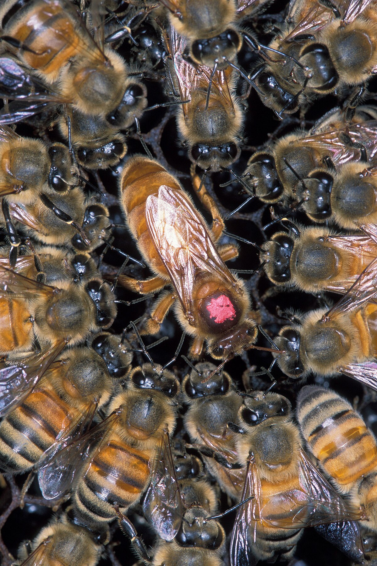
Identify the Queen Honey Bee.

How to identify a queen honey bee. So when the queen is absent eggs will be the first thing to go missing. Tips for finding the Queen Honey Bee Queen. If you suspect a bee as queen then you can gently lift the bee by its thorax the middle of its body and then hold it under a magnifying glass and inspect the stinger.
Gently lift the first of these out and scan for the queen. Her wings are short in comparison with her body and do not reach the end of her abdomen. You will recognize this area because instead of just honey you will see some cells filled with brood or cells that recently contained brood.
In many bees the sexes look totally different and as you know that is true of honey bees. You can monitor the movement of. The main reason that many of beekeepers did not register 2 queens at the same time is that when you find the queen you do not look for the second one.
If playback doesnt begin shortly try restarting your device. The queen bee is the only bee in the hive which can lay fertilized worker bee eggs. Before you even start looking for the queen prepare your brush and paint.
You can also identify a queen bee by monitoring its behavior. The size of a honey bee queen is around 2 centimeters which is about twice the length of a worker. Its really her shape that distinguishes her.
Many times after declaring a specimen to be Apis mellifera male the person asking for help becomes unhinged. Identifying the Queen Bee The queen is larger than both the drone and worker bee. How Honey Bees Recognize Their Queen.
For this reason beekeepers should always check for eggs during inspections to confirm the presence of a queen. Besides its physical appearance you can identify a honey bee. You may also notice cells filled with pollen.
Identifying Queen Bee By Behavior. Unlike wasps and other insects bees wont have a thin middle section between their thorax and abdomen. She constantly emits pheromones.
Her abdomen is long and so are her legs. A second rule for identifying bees is to determine sex first. First bees generally have very little or no obvious thinning of the body between the abdomen and thorax.
Each queen honey bee has a unique smell. Theyll usually nest either in trees or other hollow spaces. Videos you watch may be added to the TVs watch history and.
I have also included photographs of some of these queens. Her thorax is slightly larger and her stinger has a much smoother and curved look. Despite the fact that queen bee is the largest it is still difficult to spot one among thousands of worker bees.
Similarly to the drone bee the queen does not have pollen baskets or wax glands. Queen Bees Smell Funny. These are like our hormones but on the outside These pheromones are passed throughout the colony and tell the colony that all is well.
How to Find a Virgin Honey Bee Queen in Your Hive. Under her you will find the female workers who keep the hive in tip-top shape and the droneswho dont do much more than mate with the queen before they die. It is not a single doubt because queen bee carries ovaries in her long abdomen to produce a lifetimes quantity of eggs.
Next time you look for the queen and find one try to spot a second one who knows maybe you find her. If playback doesnt begin shortly try restarting your device. Slightly larger thorax mid section long tapered abdomen.
So slide the frames one-by-one into the empty space until you get to the edge of the brood nest. Gentleness Spring Buildup Pollen Hoarding Honey Production Temperament of the queen on the frame Brood pattern Colony size out of spring Resistance to Varroa and tracheal mites Resistance to disease. Despite these differences queen breeders generally breed for some of these characteristics below.
A couple of special characteristics help distinguish the queen bee. Okay let me phrase it another way. Queen can vary in size but she is usually only slightly larger than a worker bee.
This is the easiest way to tell a bee from a wasp. The gist of it thoughwhich is the standard and based on the structure of honeybee coloniesis that the queen is meant to be the mother of all bees within the colony.
Draw A Diagram Showing The External Features Of A Toppr Com. This interactive poster explores the external anatomy of a bony fish.

New Page 1
We are going to t ake a look at the external outside anatomy of bony fishes.
Draw and label the external features of a bony fish. Water passes through the mouth and over the gills of the fish. Dissection labeled images the biology corner external body parts of a bony fish lutjanidae well labeled diagram of frog and toad booklection com diagram of frog anatomy huge color image lab 23 dissection frog logos science lab kits how to draw a toad free download here pdfsdocuments2 com lancelet wikipedia show a well labelled diagram. Bony fish represent the largest and most diverse class of fishes with well over 20000 species.
The bony operculum often has another bony flap called the preoperculum overlaying it Fig. Structure And Function Fish Manoa Hawaii Edu. Click hereto get an answer to your question Draw a diagram showing the external features of a fish and label any two parts.
Note the swim bladder and lateral line two distinct qualities of an osteichtyan. Students will label the external features of a bony fish. The Operculum is the bony plate that covers fishes gills.
THE ANATOMY OF A BONY FISH Ninety percent of all fish are bony fish which are fish that have a skeleton made of bone. _____ hope this may help u. Bony fish include swordfish tuna flounder and salmon.
They are either single fins along the centerline of the fish such as the dorsal back fins caudal ta il fin. Parts Of A Cow Useful Anatomy With Pictures 7 E S L. Experiment To Observe Adaptive Features In Animals With Pictures.
Label Fish Diagram Example Wiring Diagram. Diagram Of The Perch Wiring Diagram T5. Fins Each fin on a fish is designed to perform a specific function.
There are many kinds of fish. Fish Drawing Labeled Images E993 Com. Fish Anatomy Wikipedia.
Bony-Fish Pictures and Diagrams. Tilapia Fish Drawing At Getdrawings Free Download. In chimeras and bony fishes the operculum covers the posterior end of the head which protects the gill openings.
Sea horses are significant among both osteichthyes and the overall animal kingdom. Features of some selected animals study of external body parts a cow both external and internal parts of a cow useful anatomy with. Students will describe adaptations of bony fish for life in the aquatic environment.
Draw And Label The External Features Of A Cow. Outcomes Students will identify the characteristics of bony fish. Animal Kingdom Fishes Bony Fish Anatomy Of A Perch Image.
Fins are appendages attachments used by the fish to maintain its position move steer and stop. Anatomy Of Beef Cattle Beef2live Eat Live Better. Fishes are a large and varied group of aquatic animal superbly designed for underwater life.
External body parts of a bony fish Lutjanidae April 19th 2019 - External body parts of a bony fish Lutjanidae Ichthyology 2 Morphological Characters INTRODUCTION This lab is concerned with the external structures and organization of cartilaginous and bony fishes As one might expect when examining a group with. Shark Anatomy Facts Key Functions Diagram Pictures. Here is the l abeled diagram of bony fish.
Com external body parts of a bony fish lutjanidae external features and body wall of toad with diagram echinoderm form and function of external features frog wikipedia form and structure lichen website botanical web portal well labeled diagram of frog and toad booklection com well labelled diagram of a toad pdf download wfs340 frogtoad id university of tennessee frog diagram it would. Draw A Labelled Diagram Of Bony Fish And Write The Function Of The Rainbow Fish Diagram Available Free To Download At My Blog Flickr Diagram Tilapia Fish Label Diagram Full Version Hd Quality Label Label A Fish Diagram Parts Of A Fish Labeling Goldfish This Diagram Is Of A Bony Fish What Parts Of The Fish Are Labeled Vertebrates Quiz 1 Fish Diagram Quizlet Fish Eggs Diagram. Most bony-fish reproduce by externally by egg-laying.
Parts Of A Cow Useful Anatomy With Pictures 7 E. External body parts of a bony fish lutjanidae labeled diagram non fiction text structure literacy well labelled diagram of a toad pdf download 4 identifying cane toads cvcia landcare diagram of frog anatomy huge color image animal label me printouts enchantedlearning com channa punctatus lata taki fish external features 1 7. Draw A Labelled Diagram Of Bony Fish And Write The Function Of The.
Alternatively if students have not completed the Lesson. As the water passes over the gills. Some fishes also have a strong spine or spines that project back from the preoperculum or operculum.
Bony Fish Diagram Wiring. The significant external structures in the head region of almost all fishes consist of a pair of eyes a pair of nares nostrils singular naris a mouth cephalic head lateral line canals and some sort of gill opening. This Diagram Is Of A Bony Fish What Parts Of The Fish Are Labeled.
Some have bones but others like sharks and rays have no bones only cartilage. The gill cover called an operculum protects the gills. April 3 2019 - by Wandi - Leave a Comment.
Duration 30-45 minutes Preparation This lesson uses the fish print created in the Lesson. Dissection labeled images the biology corner structure of fungi offwell external anatomy of a frog diagram of a frog cane toad essay 1243 words studymode com form and structure lichen website botanical web portal lab 23 dissection frog logos science lab kits diagram of frog anatomy huge color image frog diagram it would be nice to have a version of the external features and body wall. These spines are usually.
Bony Fish Diagram Wiring. Gyotaku or have used their. Structure Of Gills In Fishes With Diagram.
The anatomy of a trout a typical example of an aquatic osteichthyan of the ray-finned variety.
Label the diagram below with the following choices. Each nucleotide is itself make of three subunits.
/what-are-the-parts-of-nucleotide-606385-FINAL-5b76fa94c9e77c0025543061.png)
3 Parts Of A Nucleotide And How They Are Connected
Draw And Label The Three Parts Of A Nucleotide Best Label Ideas 2019 Nucleotide Wikipedia Nitrogenous Base An Overview Sciencedirect Topics Dna Function Structure With Diagram Article Khan Academy.
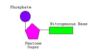
Label the parts of the nucleotide below. Both deoxyribonucleic acid DNA and ribonucleic acid RNA are made up of nucleotides which consist of three parts. Cytosine thymine and uracil are pyrimidines. A group a five carbon anda base.
For the dna diagram below label the three parts of a. List the three parts of a DNA nucleotide. Adenine and guanine are purines.
A nucleotide is an organic molecule that is the building block of DNA and RNA. Course Title BIOL MISC. он One strand half of a double-stranded DNA molecule is drawn below.
School Austin Community College. Nucleotide phosphate sugar nitrogen base hydrogen bond covalent bond purine pyrimidine hydroxyl group the 5 end the 3. They also have functions related to cell signaling metabolism and enzyme reactions.
Which of the following answers correctly labels each part of the DNA nucleotide shown below. Labeled Parts Of A Nucleotide - Fun for my own blog on this occasion I will explain to you in connection with Labeled Parts Of A NucleotideSo if you want to get great shots related to Labeled Parts Of A Nucleotide just click on the save icon to save the photo to your computerThey are ready to download if you like and want to have them click save logo in the post and it will download. Label the parts of the drawing below.
Purines and pyrimidines are the two categories of nitrogenous bases. The above structure is a nucleotide. Each nucleotide contains a.
Label the following parts of a DNA polymer Nitrogenous base adenine Nitrogenous base quanine Nitrogenous base cylosine Sugar phosphate backbone Nitrogenous base thymine Nucleotide Hydrogen bond Reset Zoom. Label the parts of the drawing below. Label the parts of the nucleotide below.
What amino acid is coded for with GUC. For the DNA diagram below label the three parts of a nucleotide Circle the part. What is the start codon.
Include all of these terms. Identify the sugar shown. Pages 8 This preview shows page 3 - 6 out of 8 pages.
A nucleotide is made of three parts. What are the three stop codons. Station 1 Draw The Nucleotide Below And Then Label The Three Parts.
Include all of these terms. Draw And Label The Three Parts Of A Nucleotide Pensandpieces. Label the parts of the drawing below.
It consists of a. Number the five carbons of the sugar subunit. Label the individual parts subunits of the nucleotide.
A phosphate group a 5-carbon sugar and a nitrogenous base. Is made up of individual DNA units called nucleotides. A nucleotide is drawn below.
Use the below to answer the following. A five carbon sugar called deoxyribose Labeled S A phosphate group a phosphorous atom surrounded by four oxygen atoms Labeled P. Draw in the other half.
Include all of these terms. The four nitrogenous bases in DNA are adenine cytosine guanine and thymine. Which of the following answers correctly labels each part of the DNA nucleotide shown below.
Nucleotide Deoxyribose Phosphate group Base pair Hydrogen bond Nitrogenous base 7. Biology questions and answers. Solved A Nucleotide Is Drawn Below Label The Individual What Are The Three Parts Of A Nucleotide Albert Io Dna Structure Howstuffworks 13 06 01 Imagine The Unimaginable Harnessing The Power Of Dna.
That make up the. A nucleotide is made up of three parts. What amino acid is coded for with AGU.
The boxed area at the lower left encloses one nucleotide. Nucleotide phosphate sugar nitrogen base hydrogen bond covalent bond purine - 10277760. Find an answer to your question Use the drop-down menus to label the parts of a nucleotide.
The frontal lobe is important for cognitive functions and control of voluntary movement or activity. Each side of your brain contains four lobes.

Lobes Of The Brain Brain Facts Occipital Lobe Problem Solving
The frontal lobe is involved in reasoning motor control emotion and language.

What are the four lobes of the brain and what are their functions. The four lobes of the brain are the frontal parietal temporal and occipital lobes Figure 2. Frontal lobe parietal lobe temporal lobe and occipital lobe. The four lobes of the brain are the frontal parietal temporal and occipital lobes Figure 2.
The frontal lobe is involved in reasoning motor control emotion and language. The frontal lobe is located in the forward part of the brain extending back to a fissure known as the central sulcus. The frontal lobe is involved in reasoning motor control emotion and language.
The occipital lobe is primarily responsible for visual processing. It is near the neck and has no concrete functionThe occipital lobe is a bit like a transit road on which the majority of our mental processes circulate organize and connect. Wikimedia Although we now know that most brain functions rely on many different regions across the entire brain working in conjunction it is still true that each lobe carries out the bulk of certain functions.
Terms in this set 4 frontal lobes. Learn how the brain lobes function to support our thoughts and reactions. What are the 4 lobes of the brain and their function.
Traditionally each of the hemispheres has been divided into four lobes. Traditionally each of the hemispheres has been divided into four lobes. Each of the hemispheres is divided into four parts.
The parietal lobe processes information about temperature taste touch and movement while the occipital lobe is primarily responsible for vision. The parietal lobe processes information about temperature taste touch and movement while the occipital lobe is primarily responsible for vision. One of their main functions is to allow us to make sense of things we touch for example whether an object is smooth or sharp firm or soft.
Cerebral Cortex Lobes Function. This is why its important to understand that there are no internal barriers between them. The cerebral cortex is located in the division of the brain known as the forebrain.
Modulation of emotions modulation of visceral and autonomic functions learning memory. The Parietal Lobes are located behind the frontal lobes and above the temporal lobes. Of the four brain lobes the occipital lobe is the smallest.
The human brain contains the frontal occipital temporal and parietal lobes. Frontal parietal temporal and occipital. It is your brains thinking crown your bodys ultimate control and information-processing center.
Subcallosal cingulate parahippocampal gyri Function. The frontal lobe is located in the forward part of the brain extending back to a fissure known as the central sulcus. Each side of your brain contains four lobes.
Subsequently one may also ask what are the 4 lobes of the brain and their function. The four lobes of the brain are the frontal parietal temporal and occipital lobes Figure 2. The frontal lobe is located in the forward part of the brain extending back to a fissure known as the central sulcus.
The frontal lobe is located in the forward part of the brain extending back to a fissure known as the central sulcus. It contains the primary and. The parietal lobes also tell us.
Each side of your brain contains fourlobes. Lobes of the Brain. The frontal lobe is involved in reasoning motor control emotion and language.
The four lobes of the brain are the frontal parietal temporal and occipital lobes Figure 2. Frontal parietal temporal and occipitaloccipitalThe occipital lobe is the smallest of the four lobes of the cerebral hemisphere. It is present posterior to the parietal and temporal lobes.
The frontal lobe is important for cognitive functions and control of voluntary movement or activity. Most of the actual information processing in the brain takes place in the cerebral cortex. Thus it forms the caudal part of the brain.
He participates in the processes of perception and visual recognition. The frontal lobe parietal lobe occipital lobe and temporal lobe have been associated with different functions ranging from reasoning to auditory perception. At the medial surface of each hemisphere and around the corpus calosum Gyri.
But it is not less interesting. At the back of the frontal lobe near the central sulcus lies the motor cortex. The four large areas that make up the brain always work in harmony and share information constantly.
On the other hand although each. The cerebral cortex is divided into four sections or lobes. They include the frontal lobe parietal lobe occipital lobe and temporal lobe.
It is divided into four lobes that each have a specific function. Although most of our brain functions rely on multiple regions communicating in conjunction with one another each lobe of the brain is thought to carry out the bulk of a certain set of functions. Processing and integration of taste sensation visceral and pain sensation and vestibular functions.
Cerebral lobes and their functions. Positions and Functions of the Four Brain Lobes. The parietal lobe processes information about temperature taste touch and movement while the occipital lobe is primarily responsible for vision.
For example there are specific areas involved in movement and sensory processes vision hearing somatosensory. When we think of cerebral lobes we tend to make the mistake of imagining separate structures that are independent of one another. The frontal lobe is important for cognitive functions and control of voluntary movement or activity.
What are the 4 lobes of the brain and their function quizlet. The human brain is the most complex organ in the body. This lobe is located at the front of the brain and is associated with reasoning motor skills higher level cognition and expressive language.
On ventral side are called Sternum. There are 10 segments.

Cockroach Illustration Labeled Educational Body Structure Scheme Royalty Free Cliparts Vectors And Stock Illustration Image 138666431
The peripheral cells have the ability to move ions and water into the glands.

Labelled diagram of cockroach. 1 Answer 1 vote. Draw labelled diagram of heart in cockroach and explain its physiology. The body of the cockroach is elongated and segmented.
Share It On Facebook Twitter Email. The body cells in cockroach discharge their nitrogenous waste in the haemolymph mainly in the form of. Answered Jun 12 2020 by RajeshKumar 507k points selected Jun 12 2020 by.
Hold the specimen Fig. Draw a labelled diagram of alimentary canal of a cockroach. It is the lobe which overhangs the mouth and is used as a wedge to force open cracks in the soil.
Hello friendsIn this video we will Draw the labelled diagram of cockroach and classification of cockroach Cockroach classification in phylum ArthropodKingdo. Morphology of Cockroach with well labeled diagram September 30 2020 by Akshay Shape size and colour. With the help of neat labelled diagram explain circulatory system in cockroach.
Please scroll down to see the correct answer and solution guide. Cockroach body is narrow elongated and bilaterally symmetrical. Out of them 3 are found in the thorax 10 are situated in the abdomenThe heart is.
The diagram would also include the legs and antenna of the cockroach. Announcing Numerades 26M Series A led by IDG Capital. Diagram of a Cockroach grasshopper lizard - well labelled is required 1 answer below a very well labelled diagram of a cockroach lateral and dorsal view grass hopper lizard and grasses Jan 28 2021 0426 AM.
The heart has 13 chambers. April 13th 2019 - A diagram of a cockroach would include labeledsegments of theinsect including the head and thorax The diagram wouldalsoinclude the legs and antenna of the cockroach A well labelled diagram Diagram of a Cockroach grasshopper lizard well. On dorsal side or notum are called Tergum.
Draw a labelled diagram of alimentary canal of a cockroach. It is the first body segment or buccal segement. Cockroach Salivary Glands.
61 with your left hand and clip the wings. Female reproductive system consists of primary and secondary reproductive organs. Cockroaches have salivary glands as well as an esophagus for easier digestion.
Cut the lateral membrane pleura between the terga and sterna of the thorax and abdomen with a pair of fine scissors. Draw a neat diagram of alimentary canal of cockroach. Draw labelled diagram of heart in cockroach and explain its physiology.
A diagram of a cockroach would include labeled segments of the insect including the head and thorax. Draw a labelled diagram of the alimentary canal of a co. Draw a labelled diagram of the alimentary canal of a cockroach.
Label any six parts. With help of neat labelled diagram describe female reproductive system of cockroach. Primary reproductive organs are ovaries.
The exoskeleton is thick and hard made up of calcareous plates called sclerites. A Nauphoeta Cinerea has salivary glands with 4 types of cells that include the central cells peripheral cells non-secretory duct cells and secretory duct cells. It is dark brown or reddish brown in colour.
It has a tubular pulsatile heart which is found mid- dorsally in the pericardial sinus. Fix the specimen in a dorsal position on a dissecting tray with the help of pins passing through abdominal sterna and coxa of legs. There is a pair of large ovaries lying laterally in the 2nd to 6th abdominal segments.
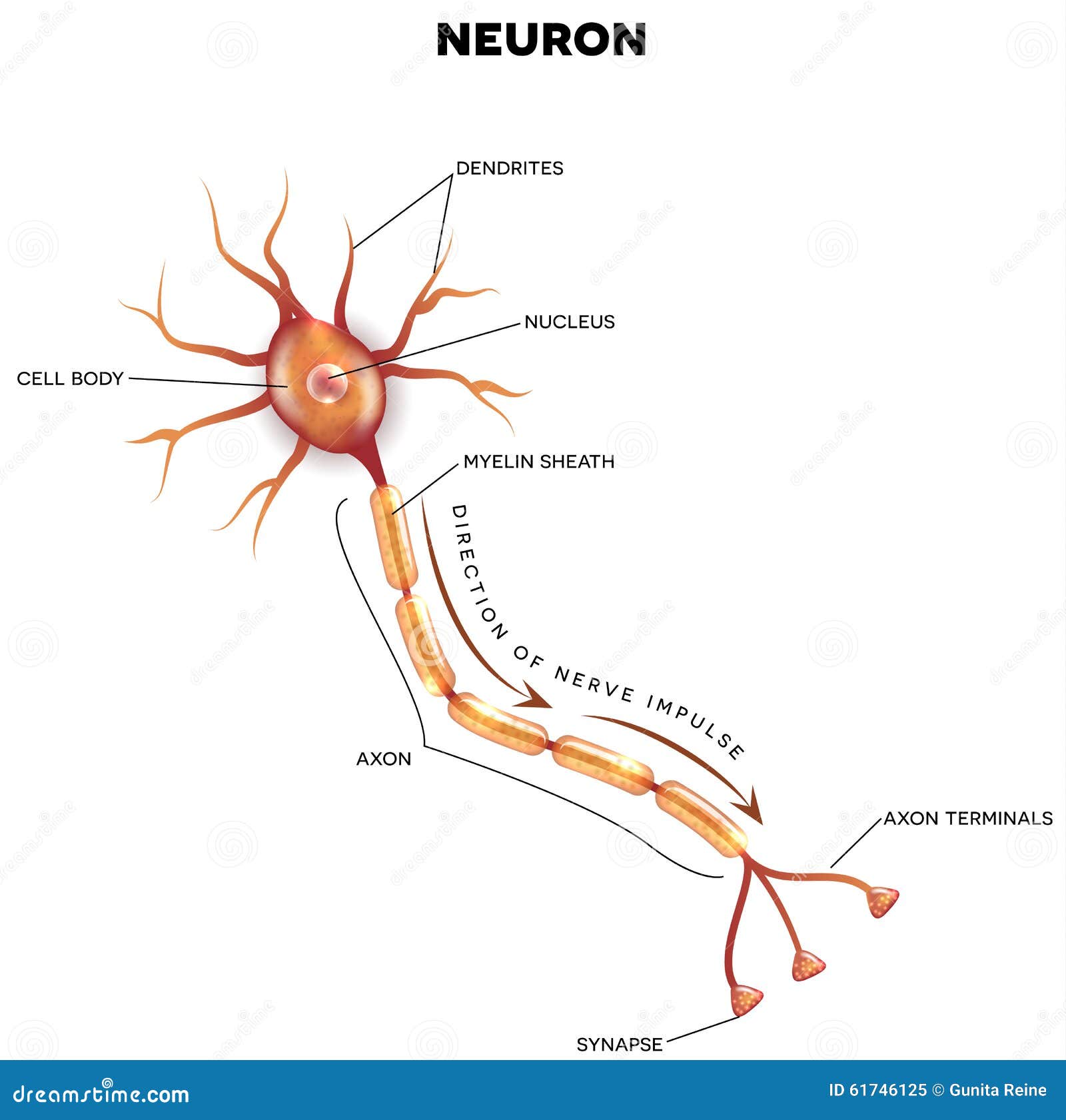
A group of neurons forms a nerve. Human neural system has about 100 billion neurons.

Labeled Neuron Diagram Science Trends
The nervous system and sense organs.
Labelled diagram of a neuron. It is in the CNS that all of the analysis of information takes place. The neuron is a specialized and individual cell which is also known as the nerve cell. Parts of a Neuron Diagram.
Asked Jan 9 2019 in Class X Science by navnit40 -4938 points Draw well labeled diagram of a Neuron and name the following parts. Hope it is useful for you. Draw a labelled diagram of a neuron.
They are found in the brain spinal cord and the peripheral nerves. I Draw a well labelled diagram of a Neuron showing the following parts. M agnesium is a chemical element with symbol Mg and atomic number 12.
Dendrites receives messages from the surrounding and sends it to the cell body. A group of neurons forms a nerve. A neuron is a structural and functional unit of the neural tissue and hence the neural system.
All neurons have three main parts. Draw a labelled diagram of a neuron. Complete neuron cell diagram.
Perikaryon Dendrites Axon Node of Ranvier and Myelin sheath. Draw a labelled diagram of a neuron. Neurons Nerve Cells.
Complete neuron cell diagram ensvg. Axon transmits the message away from the cell body and pass it to the the next receiving neuron. Draw well labeled diagram of a Neuron and name the following parts.
Dendrites A branch-like structure that functions by receiving messages from other neurons and allow the transmission of messages to. Well Labelled Diagram Of A Neuron - Electrical Work Wiring Diagram - labeled diagram of the neuron nerve cell that is the main part rh rf com well labelled diagram of a typical neuron draw a well labelled diagram of theIB Biology Notes - Nerves hormones and homeostasisIB Biology Notes - Nerves hormones and homeostasis. Draw a labelled diagram of a neuron.
In vertebrate animals neurons are the core components of the brain spinal cord and peripheral nerves. Question 3 Adapted from March 2012 NSC P2 Question 22. Thus neurons with longer processes projections are the longest cells in the body.
A neuron is also known as the nerve cell. While they have the common features of a typical cell they are structurally and functionally unique from other cells in many ways. Draw A Labelled Diagram Of A Neuron Class 12 Solved Question paper 2020 Class 10 Solved Question paper 2020.
Question 2 List the functions of the spinal cord. 1 Node of Ranvier 2 Nissil granules 3. Draw a labeled diagram of a myelinated neuron.
Structure of a neuron. A neuron is a specialized cell primarily involved in transmitting information through electrical and chemical signals. Click to rate this post.
Neurons are the fundamental unit of the nervous systemThe neuron is a specialized and individual cell which is also known as the nerve cell. The peripheral nervous system PNS which consists of the neurons and parts of neurons found outside of the CNS includes sensory neurons and motor neurons. Neurons also known as neurones and nerve cells are electrically excitable cells in the nervous system that process and transmit information.
It is a shiny gray solid which bears a close physical resemblance to the other five elements in the second co. B Draw labelled diagrams to illustrate the general structure of a neuron c Illustrate the differences between neurons based on their structure. Certain neurons may almost equal the length of body itself.
Neurons are the fundamental unit of the nervous system. The central nervous system CNS consists of the brain and the spinal cord. Although they have a characteristic elongated shape they vary widely in size and properties based on their location and type of functions they perform.
Cell body consists of nucleus mitochondria and other organelles. Neuron comprises of dendrite axon and cell body. D Distinguish between the different neurons based on their functions.
Digestive System Chapter Notes Class 11 Biology Class 11. I Absorption of digested food.
Draw A Well Labelled Diagram Of Human Alimenntary Canal And La
Ii Absorption of water.

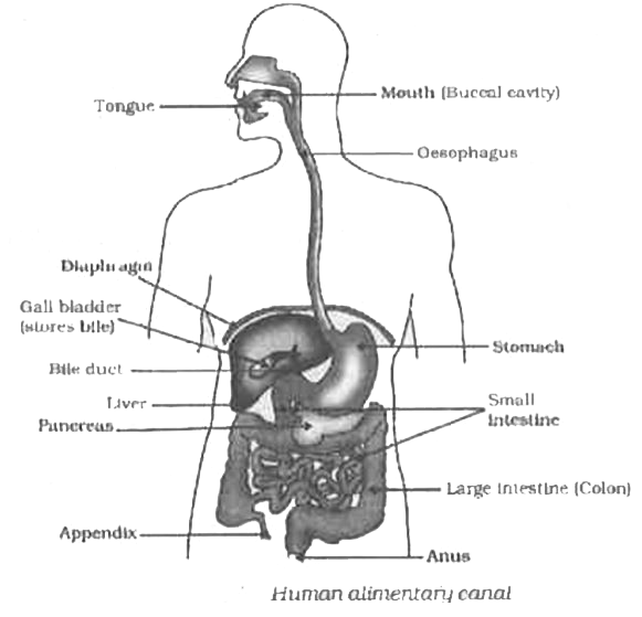
Draw a well labelled diagram depicting human alimentary canal. Draw a labelled diagram of alimentary canal of a cockroach. I Liver ii Pancreas iii Small intestine iv Large intestine. 300 700 227.
Dorjeeminduk dorjeeminduk 02102020 English Secondary School answered 13. The alimentary canal in human beings measures about 8 to 10 meters in length. Click hereto get an answer to your question a Draw a labelled diagram of human alimentary canalb Explain the digestion of fat in our body.
I Absorption of digested food ii Absorption of water. Diagram Of Digestive System. If you do not find the exact resolution you are looking for go for Original or higher resolution.
Download this image for free in High Definition resolution using a download button option below. Draw a neat diagram depicting human. You can also save this page easily so you can view it.
Draw a diagram depicting human alimentary canal de laalimentary canal and lable the componengall bladder liver and pancreas in it state the role of liver and pancrease1 name the organs perform the following functiuons in humansa absorption of digested food b absorption of digested food. Asked Oct 12 2019 in Biology by Suchita 664k points life processes. Draw The Diagram Of Alimentary Canal Of Man And Label The.
Describe the alimentary canal of man. Iv Part where digested food is absorbed. I Liver asked Jul 18 2018 in Chemistry by Anukriti bharti 381k points life processes.
Alimentary Canal And Action Of Enzymes. Draw A Labelled Diagram Of Alimentary Canal Of A Cockroach. Draw a well labelled diagram of human alimentary canal and label the following parts.
1 answer a Draw a diagram depicting Human Alimentary Canal and label on it. Let learn the different parts of the human digestive system. A Draw a well labelled diagram of human alimentary canal and label the following parts.
Asked Jul 18 2018 in Chemistry by Anukriti bharti 381k points life processes. Draw Labelled Diagram Of Alimentary Canal is important information accompanied by photos and HD images sourced from all websites in the world. Draw a diagram depicting Human Alimentary Canal and label on it.
Draw a well labeled diagram of human alimentary canal 2 See answers. Human Digestive System With Diagram. A Draw a well labelled diagram of human alimentary canal and label the following parts.
What Is The Length Of The Alimentary Canal In Human. Digestive System Of Frog With Diagram Vertebrates. C Name the organ which performs the following functions in humans.
Draw a labelled diagram of Alimentary canal system of cockroach. Asked Oct 12 2019 in Biology by Suchita 664k points life processes. It is one among the few important topics which are repetitively asked in the board examinations.
Alimentary Canal Pmg Biology. B What are villi. Where does this process take placec What is the role of saliva in the digestion of food.
Click here to get an answer to your question 13. Draw a well labelled diagram of human alimentary canal and label the following parts. Gall bladder Liver and Pancreas.
The diagram of the human digestive system is useful for both Class 10 and 12. 10 2 The Digestive System Lessons Tes Teach. B State the roles of Liver and Pancreas.
Draw A Labelled Diagram Of Alimentary Canal Of A Cockroach. Draw a well labeled diagram of human alimentary canal Get the answers you need now. Draw a well labelled diagram of human alimentary canal and label the following parts.
Alimentary Canal Structure And Functions Of Alimentary Canal. Where does this process take placec What is the role of saliva in the digestion of food. Iii Part where HCl is produced.
Gall bladder Liver and Pancreas. Iii Small intestine iv Large intestine. A Draw a diagram depicting Human Alimentary Canal and label on it.
Draw a well labeled diagram of human alimentary canal. Click hereto get an answer to your question a Draw a labelled diagram of human alimentary canalb Explain the digestion of fat in our body. Life Processes Class 10 Important Questions Long Answer Type.
B State the roles of liver and pancreas. Draw A Well Labelled Diagram Of Human Alimentary Canal And. With The Help Of Well Labelled Diagram Explain Alimentary.
Draw a labelled diagram of a portion of human alimentary system showing the location of liver pancreas and gall bladder and their associated ducts. A Draw a diagram depicting human alimentary canal and label on it gall bladder liver and pancreas. Gastrointestinal Tract Wikipedia.
The diagram below shows the structure and functions of the human digestive system. Openstax Anatomy And Physiology Ch23 The Digestive. Figure 23 1 Alimentary Canal And Related Accessory Digestive.
Gall bladder Liver and Pancreas. Mouth It.
Certain neurons may almost equal the length of body itself. Draw a well-labelled diagram of a neuron.
Draw The Structure Of Neuron And Label Cell Body And Axon Studyrankersonline
I Neuron ii 1 Seat of memory.

Draw a neat labelled diagram of nerve cell neuron. Here Ill draw Daigram. 1 Seat of memory 2 Coordinates muscular activity iiiMention any three major activities of WHO. Effect of Nervous system List-2 Effect of Nervous system.
Of the cells genetic material Axon - A long fibre of a nerve cell a neuron that acts somewhat like a fiber-optic cable carrying outgoing efferent messages. Ovary as shown in labelled diagram of neuron draw a neatly labelled diagram of a neuron it contains the nucleus also called the soma discribe the structure of neuron with neat labelled diagramexplain how the nerve impulse is transmitted across a synapses describe the structure of a neuron with help of a neat labelled. They are found in the brain spinal cord and the peripheral nerves.
Easy method to draw the diagram of Neuron Nerve Cell Watch later. To Identify Striped Muscle Fibers And Nerve Cells In Animals Lab. 1 Node of Ranvier 2 Nissls granules 3 Cyton ii Name the part of the human brain which is associated with the following.
The structure of neuron. An axon can be over 20 cm a foot in length which for. Nerve cell is functional unit of nervous systemIt has mainly two parts the Cyton and AxonCyton have several dendrites emerging from surface.
Labelled Diagram of a Nerve Cell. A neuron is a structural and functional unit of the neural tissue and hence the neural system. Neurons are the fundamental unit of the nervous system.
If playback doesnt begin shortly try restarting your device. Each nerve cell has one axon. Cerebrum 2 Coordinates muscular.
Asked Nov 17 2017 in Class X Science by aditya23 -2137 points a Draw the structure of neuron and label cell body and axon. Nerve Cell aka Neuron. The neuron is a specialized and individual cell which is also known as the nerve cell.
Add your answer and earn points. The cell body contains cytoplasm and certain granular bodies called Nissles granules which contain a group of ribosomes. Draw two triangles.
The neuron sends electrical impulses from its cell body through the axon to target cells. Axon of nerve cells sometimes extend to 1 mt length in nerves of leg. I where the information is required.
Draw A Well Labeled Diagram Of A Typical Action Potential Of A Nerve. A neuron is a specialized cell primarily involved in transmitting information through electrical and chemical signals. Function of Nerve Cell.
Also write where in the body does the myelinated and non-myelinated nerve fibres are commonly found. The nervous system - Long answer type Page 136. The Structure And Function Of Sensory Relay And Motor Neurons.
A typical neuron has one large fiber an. Cyton gives rise to Axon from a triangular structure called axon hillock. Cyton something looks like a star so it can be drawn from a basic star shape.
Neurons are the structural and functional units of the nervous system. Find an answer to your question Draw a neat labelled diagram of neuron Lovelysowmya Lovelysowmya 30092020 Biology Secondary School answered Draw a neat labelled diagram of neuron 1 See answer Lovelysowmya is waiting for your help. It is irregular in shape or polyhedral.
Neurons Nerve Cells. Click to rate this post. They are odd-looking cells with many finely branched fibers extending from the main cell body.
A group of neurons forms a nerve. Labeled Diagram Of The Neuron Nerve Cell That Is The Main Part. B Name the part of neuron.
Ii through which information travels as an electrical impulse. Thus neurons with longer processes projections are the longest cells in the body. Draw a labelled diagram of a neuron.
Well labelled diagram of neuronStructureofneuronbiologyofdiagrams nervecelldiagramHi there Welcome to my channel Biology Daigrams. These are connected to each other by thin wirelike threads that carry electrical signals and some extend for more than 3ft 1m. A neuron is also known as the nerve cell.
Heartbeat to normalcy 5. Foreman Describe the basic characteristics of an action potential including what causes it to fire in the first place and how helps it to travel even faster and farther. Draw Structure Neuron Diagram Labelled Cell Nerve Posted in Worksheet February 2 2021 by Kimberly R.
Neurons form the bodys living wiring system. Explain how neurotransmitters help Label the does the letter a represent preview this quiz on. Nerve cells or neurons are the structural and functional units of the nervous system.
Powered by Create your own unique website with customizable templates. The nervous system is made up of billions of cells called neurons nerve cells. Human neural system has about 100 billion neurons.
It consists of three major parts namely Cell body dendrites Axon. Neuron Or Nerve Cell and Its Types - Structure of the Neuron Report Error Is there an error in this question or solution. Chapter 8 The Nervous System and Sense Organs Sketch and Label the Diagram Q 3.
Draw a neat diagram of a neuron and label the following parts. A group of neurons forms a nerve. The structure of a neuron varies with their shape and size and it mainly depends upon their functions and their location.
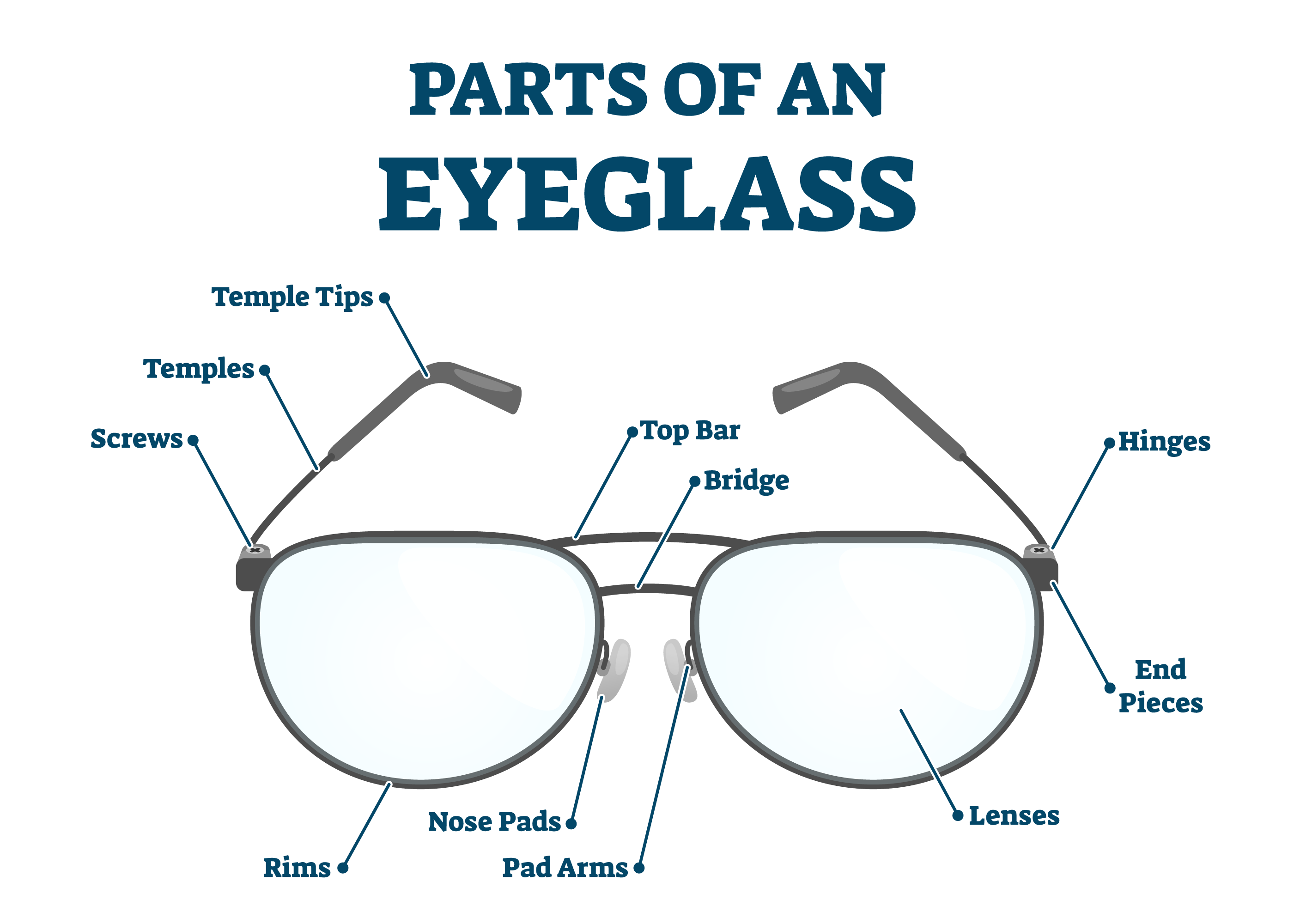
There are many different issues that can arise with your glasses breaking having missing pieces or needing small parts replaced. Plastic or silicone coatings called earpieces cover the ends of the temples to ensure comfort and relieve the pressure of temples on the top of your ears.
Parts Of Eyeglasses Anatomy Of Glasses Smartbuyglasses
The eyepieces hold the lenses and connect to the temples by hinges and the bridge is the part.

Eyeglasses parts diagram. Often called the arm this is the piece of the frame that extends over the ear to help hold the sunglasses in place. Attaching these more complex components is usually a job for an expert with specialized tools but the screws and other small jobs can easily be completed by an individual with no formal eyeglass repair training. Parts of glasses for Eyeglass Repair nose pads glasses screws rimless screws temple tips and lens interliner.
85 343Count Get it as soon as Wed Jul 7. If you are lucky this sunglasses repair kit with a massive amount of replacement parts. Eyeglasses and Sunglasses are completely different types of products although the names of all the parts are exactly the same.
Save 5 with coupon. Q uite often t hese things affect the shape color and strength of your glasses. Eyeglasses parts diagram Part identification.
Connects the end piece to the temple and allows the temple to fold inward. Assemble together different parts of the frame using flexible or fixed components. Eyeglasses Parts Diagram Diagram.
We offer the most common replacement parts for glasses including nose pad replacement parts and temple tips. The frame front is composed of two eyepieces connected by the bridge. Virtually any part of your eyeglasses can be replaced.
Sustain the frame on the ears. Just make sure you know. Jan 30 2019 - Learn the glasses frame parts and terminology of your reading glasses and eyeglasses.
LMP Optical Supply 2 Pairs of Replacement 12 mm x 6mm Square Slip in Silicone Nose Pads for Sunglasses and Eyeglasses. To buy correct replacement parts for your Ray-Ban shades please follow our instructions carefully. Its shape and thickness will make the look of the frame.
For most repair works you will need universal sunglasses spare parts kit aff. Diagram of Glasses Parts and Descriptions. Sunglasses parts diagram Part identification.
88 607100 g 5 coupon applied at checkout. Eye WiresRimsFrame Front. Usually at our store customers have come with these problems.
They also share how all these parts. Any parts you need for eyeglass manufacturingrepairing or designing even DIY you could find them here. They also share how all these parts work togeth.
Thank you for visiting our website. Front part of the glasses frame that accomidates the lenses bridge and. 2 Count Pack of 1 45 out of 5 stars.
You can find the different parts of your model by searching for them through the search engine filtering by model or in our best-sellers. 45 out of 5 stars. As you know eyeglasses can experience hits folds and they are subjected to various humidity levels and temperatures.
Eyeglass frames have two basic parts. Maintain the frame in a right position on your nose. Eyeglasses frames have several components and the names of the different frame parts are useful to learn.
Check the video below for how to repair your eyeglasses the fast easy way or the professional way. Some hinges called spring hinges allow for hyperextension of the temple for a better fit. ZOYE supplies all kinds of eyeglass partssunglasses parts such as eyeglass hinges eyeglass templesbridges pad arms nose pads spring inserts etc.
In this video All American Eyeglass repair explains the different parts of a pair of Silhouette eyeglasses. Eyeglass Parts Eyeglass Hinges ZOYE Eyeglass Parts Coltd. Browse our variety of sizes.
Upgrade Version Eyeglass Repair Kit1500 Pcs More Complete Glasses Screws Kit and Nose Pads with 6 Pcs Screwdrivers and 3 Pcs Tools for Glasses Eyeglasses and Sunglasses Repair. Also called the arms of a pair of eyeglasses temples are the stems that extend from the sides of the frame to the ears theyre named for the part of your face they rest closest to. More importantly the earpieces help to hold the glasses.
The frame front that holds the lenses and the temples that hold the frame from falling off your face. Today our team are delighted to declare our company have actually uncovered an very fascinating niche market to be examined that is actually eyeglasses parts diagramA lot of people looking for details regarding eyeglasses parts diagram and also certainly one of all of them is you is actually not it. In this section we will explain what each part does so you have a better understanding of how glasses come together.
The kit includes a precision screwdriver and wide assortment of replacement parts nose pads screws etc for sunglasseseyeglasses that will fit most Ray-Bans. The first step in finding a perfect pair of readers is knowing how they work. 03 Select it Once you found it it is very important that you select the correct colour or size.
It cost under 12.
Mapped into the vertical z left-hand diagram the net mass balance profile bz is negative at. Knowing that these ice figures have so much more than meets the eye.

Glacier Diagrams
These geography worksheets look at the features and vocabulary used when studying glaciers and go on to label a diagram of a glacier.
Labelled glacier diagram. A diagram of what a glacier is and how it works. Here we look at the shrinking world of glaciers. Geo 101 Final Comprehensive Exam Geology 101 With Ander.
Glaciers cut distinctive U-shaped valleys with a flat floor and steep sides. Water Cycle Diagram To Label - Atkinsjewelry They will glue in the information as i write and the other students copy. Parts Of A Volcano Worksheet Printable Pdf Learning How.
Is the ablation zone near the upper or lower region of a glacier. Introduction To Glaciers Suncups Piedmont Glaciers. A glacier with a much larger average accumulation area is growing while one with a larger ablation area is a glacier thats shrinking and could eventually disappear.
As more and more snow falls it is compacted so the bottom layers become ice. Glacier diagram labeled glacier diagram moraine glacier diagram worksheet glacier formation diagram retreating glacier diagram. SVGErstellung Der Quelltext dieser SVG-Datei ist Diese Vektorgrafik wurde mit Inkscape erstellt.
Glacial Landforms And Coastal. You should make a label that represents your brand and creativity at the same time you shouldnâ t forget the main purpose of the label. Explore more than 947 Glacier Labelled resources for teachers parents and pupils.
Diagram of an alpine glacier. Hello Guys In this Video Im going to teach you guys How to Draw Structure of a cell Labeled DiagramSubscribe to the Channel and also share this video. Dies ist eine exzellente Datei bei Wikimedia Commons Exzellente Bilder und wird als eine der hervorragendsten Bild-Dateien.
Recent trends suggest that many of the worlds glaciers are shrinking at alarming rates source. The measurements show that the glacier has been retreating since 1780 and that in the last 40 years the rate that the glacier. Glacier diagram labeled accumulationzone Label Gallery Get some ideas to make labels for bottles jars packages products boxes or classroom activities for free.
Lastly the fourth glacier worksheet is an activity which combines learning about glaciers with geographical. When the two areas are roughly equal its considered a stable glacier. Top 9 US Senate Races.
Friday February 19 2021 Edit. Volcano diagram parts worksheet worksheets ks2 science printable planbee label volcanoes children different inside pdf labels projects geography ks1 labelled. No comments posted yet.
Type Of Glaciers Vector Illustration Labeled Alpine Or. An easy and convenient way to make label is to generate some ideas first. The steepening of glacier mass balance gradients with northern hemisphere warming.
Most of the times. Glacier Diagram Written By admin. - PREV NEXT PellucidWombat.
This page is a collection of pictures related to the topic of Correctly Labeled Glacier which contains Glaciers. Figure 82 Schematic diagrams of a glacier white in mountainous topography gray showing accumulation and ablation areas on either side of the equilibrium line. Dont have an account.
Draw a diagram of shearing at the side of a glacier. Election Gazette Election 2020 News Links and More. 1 Votes Log in to vote.
Climate change can affect glacier stability over a long term. Fileimage Piedmont Glaciersvg Wikimedia Commons. The solid-liquid curve labeled BD shows the temperatures and pressures at which ice and liquid water are in equilibrium representing the meltingfreezing points for water.
This is normally at the start of a glacier in a highland area. This one shows the Gangotri glacier which feeds into the mighty Ganges river. Glacier Waters - Knitted sweater with saddle shoulders in.
Ice moves downhill due to the force of gravity. The Gangotri glacier is one of the largest glaciers in the Himalayas and it has had accurate measurements taken of it since 1780. Snow K-3 Theme Page at.
You should make a label that represents your brand and creativity at the same time you shouldnt forget the main purpose of the label. On Feb 23 2011 759 pm. Label Gallery Get some ideas to make labels for bottles jars packages products boxes or classroom activities for free.
Examinations of glacier-mice interiors have found springtails tardigrades and nematode worms. Base your answers to questions 86 and 87 on the block diagram below and on your. There are many examples of diagrams like this one which record the retreating glaciers.
We see how glaciers are formed and then identify the landforms created by glaciers. Alpine Ice Extent At The Last Glacial Maximum Lgm With. GEOG 203 Study Guide 2014-15.
You can click on the image to enter full-screen mode.
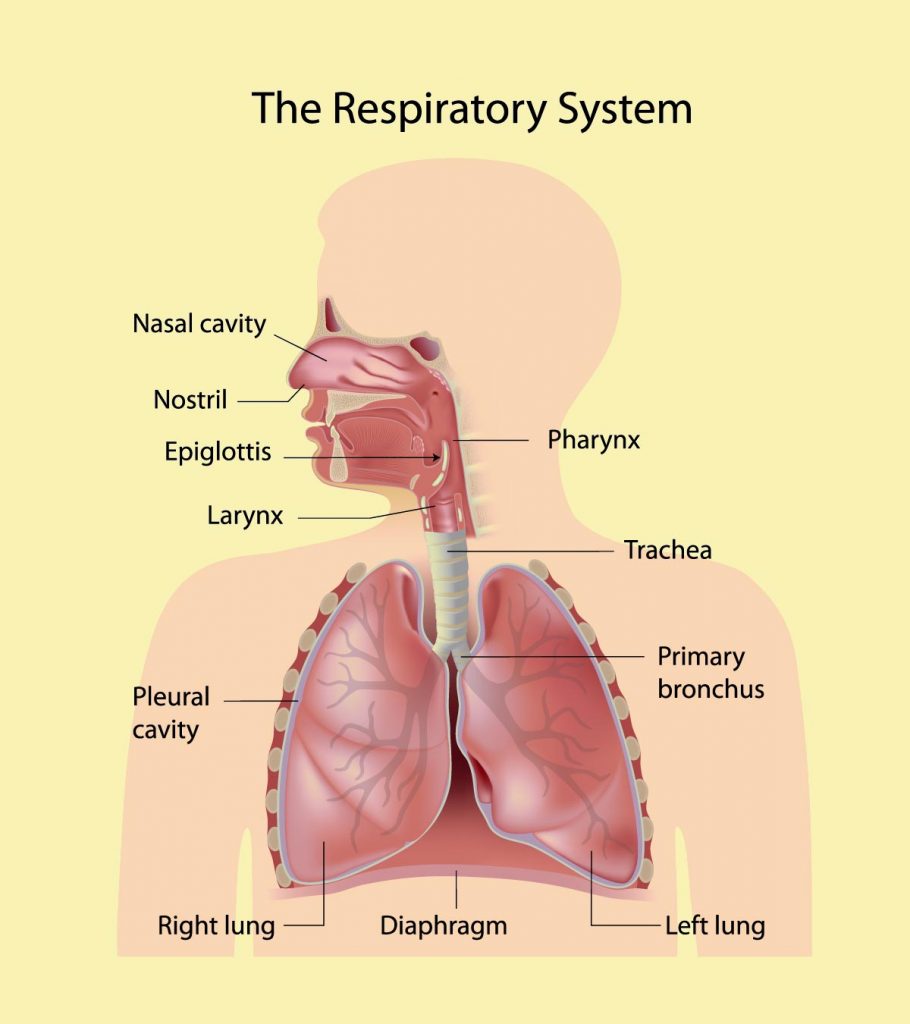
There are three essential parts to your respiratory system. Write a situation where you are applying a conceptual skill a human and a technical ski.
30 Facts And Information About Respiratory System For Kids
The larynx trachea bronchi bronchioles and alveoli make up the lower respiratory tract.
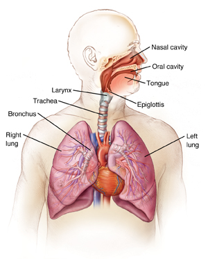
Name the parts of the breathing system. Parts of the Respiratory System. Lets learn in more detail about the lungs. Human respiratory system the system in humans that takes up oxygen and expels carbon dioxide.
All these parts together form the respiratory tract that starts from the external nostrils and nasal chamber and goes up to the lungs. Tuo in the space providedSituationAnswerar dided to preserve 1. The nose nasal cavity and pharynx make up the upper respiratory tract.
The muscles that power your lungs are also part of the respiratory system. The lungs are responsible for passing oxygen into your body while also removing carbon dioxide. What are the parts of breathing system - 1490277 1.
The respiratory system is the network of organs and tissues that help you breathe. A breathing system is an arrangement of tubes and other components that transports gases between the anaesthetic machine and the patient. Free Identity what processed product being cured and submerged inthe given altuation below.
Write antem lonmanis toolne selted apteBantanited Nah. Other facets of the respiratory system are the larynx epiglottis alveoli and the blood vessels that carry oxygen away from the lungs. Learn about the anatomy and function of the respiratory system in this article.
These parts work together to move oxygen throughout. Your respiratory system includes your. Neurons in the medulla send signals to the diaphragm and the intercostal muscles to regulate the contractions which initiate the breathing process.
The entire passageway to the lungs is also known as the air tub e. The one you probably think of most is the lungs. The autonomic system of the PNS controls involuntary processes such as breathing.
Throat pharynx Voice box larynx Windpipe trachea Diaphragm. Here we explain the anatomy of. A very common breathing system used in anaesthesia is the circle breathing system and I will introduce its functioning to you.
It includes your airways lungs and blood vessels. Parts of the human respiratory system. One end of the body is connected by the shortest possible means to the patient.
This network consisting of bronchi bronchioles and alveoli forms the lung. It also receives waste Carbon Dioxide from the blood and exhales it. Breathing is a part of the respiratory process.
Nose and nasal cavity. Main parts of the human respiratory system include the mouth nasal cavity lungs bronchial tubes diaphragm and trachea according to National Heart Lung and Blood Institute. Meanwhile the airways like the mouth nose larynx and pharynx help you bring air and oxygen into the lungs through breathing.
The volume of this limb makes up apparatus dead space The other end is connected to a length of tubing that acts as a reservoir. The nasal cavity is the uppermost part of the respiratory system divided into two by the nasal septum. Give one example for each skill.
The other parts of the respiratory system include the nose larynx pharynx trachea or the windpipe bronchi lungs blood vessels the airways for the passage of air and the muscles that support the breathing. The major organs of the respiratory system include the nose pharynx larynx trachea bronchi lungs and diaphragm. The function of the human respiratory system is to transport air into the lungs and to facilitate the diffusion of oxygen into the bloodstream.
The main body of the T-piece is within the breathing system and must therefore be of adequate diameter. Kimmyboytiguelp9wg9u kimmyboytiguelp9wg9u 07062018 Science Junior High School. It is the best entrance for outside air as hairs and mucus line the inside wall and operate.
The medulla oblongata of the brain regulates breathing.
Which modern techniques are available to overcome this problem. Describe the structure of a human kidney with the help of a labelled diagram.

Describe The Structure Of A Leaf With The Help Of A Labelled Diagram Science Nutrition In Plants 10219117 Meritnation Com
It is the stalk that connects a leaf to the stem of the plant it is made of complex conducting tissues called vascular tissues.

Describe the structure of leaf with the help of labelled diagram. 1 Petiole- the stalk that supports a leaf in a plant and attaches the leaf blade to the stem. Dorsiventral leaves are present in dicot plant and have three main parts. With the help of labelled diagrams describe the structure of human sperm and unfertilized ovum.
Midrib divides the surface of the lamina into two. Describe the structure of a leaf with the help of a labelled diagram class seven long answer question. I Nucleus ii Centrosome.
Describe the structure of the following with the help of labelled diagrams. I Nucleus ii Centrosome. Describe the internal structure of a dorsiventral leaf with the help of labelled diagrams.
I Nucleus ii Centrosome. Asked Feb 16 2018 in Class XI Biology by vijay Premium 539 points excretory products and their elimination. Parts of a Leaf Diagram.
A leaf diagram representing the parts of a leaf. Answered Jan 14 by MoniKumari 520k points selected Jan 14 by Sakil01. Describe the internal structure of a dorsiventral leaf with the help of labelled diagrams.
The unit of life. 1 petiole 2 leaf base and 3 leaf blade or lamina each performing specific functions. Asked Jan 14 2020 in Biology by Sakil01 509k points Describe the internal structure of a dorsiventral leaf with the help of labeled diagrams.
A leaf has two main parts. NCERT Class 11 - Biology. The vein that runs along the middle of the lamina is called midrib.
These veins and veinlets give rigidity to the leaf blade and help in the transportation of water and other substances. Describe the structure of the following with the help of labelled diagrams. Anatomy of Flowering Plants.
How does the internal anat 0932. Epidermis -The outermost covering of the leaf on the upper side called adaxial epidermis and on the lower side it is known as the abaxial epidermis. 2 Lamina- the green flat part of a leaf that is specialized for photosynthesis.
The unit of life. Describe the internal structure of a dorsiventral leaf with the help of labelled diagrams. I Nucleus ii Centrosome.
A waxy thick layer cuticle is present and distinct. Asked Jan 14 2020 in Biology by Sakil01 509k points cell. Describe the structure of nucleus and centrosome with the help of labelled diagram.
Anatomy of flowering plants. 1 Answer 1 vote. Describe the structure of a leaf with the help of labelled diagram - eanswersin.
Solution For Describe the internal structure of a dorsi-ventral leaf with the help of labelled diagram. Describe the internal structure of a dorsiventral leaf with the help of labeled diagrams. Anatomy of Flowering Plants Ask Eguru Up Teachers Skip to content.
Describe the internal structure of a dorsiventral leaf with the help of labelled diagrams. Describe the structure of the following with the help of labelled diagrams. Asked Apr 23 2020 in Biology by Devanshi 672k points cell.
Share It On Facebook Twitter Email. Correct answer to the question. A typical leaf shows three main parts.
With the help of a labelled diagram describe the internal structure of human heart. Share It On Facebook Twitter Email. Describe the structure of the following with the help of labelled diagrams.
Describe The Internal Structure Of A Dorsiventral Leaf With Help Labelled Diagrams Sarthaks Econnect Largest Online Education Community Exploring Photosynthesis In A Leaf Chloroplasts Grana Stroma Thylakoids And Other Parts Of Structure Of Leaves The Epidermis Palisade And Spongy Layers Lesson Transcript Study Com Draw The Labelled Diagram Of Parts A Simple Leaf. 1 Answer 1 vote. A couple is unable to conceive.
Describe the structures. You must be signed in to discuss.
Required tractor power HP 35 HP Above. Frames Width mm 1850.
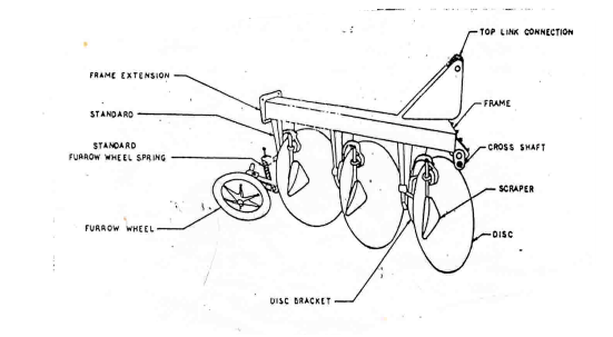
Types Of Disc Ploughs
By continuing to browse the ConceptDraw site you are agreeing to our Use of Site Cookies.

Well labelled diagram of a disc ridger. Solution for Draw a well labelled diagram showing parts of a precise tilting level. Disc Ridger View Specifications Details Of Disc Ridger. Diagram Of A Well Labelled Headpan.
Diagram Of A Well Labelled C P U. It is composed of those parts necessary for the rigid structure required to cut lift turn and invert the soil. Economic History Of Tractors In The United States.
Parts of the mouldboard plough bottom are. Jaw crusher schematic diagram labeled well labelled diagram of a jaw crusher well labelled diagram of a jaw crusher a well labelled diagram of a ball mill is widely used in stone production we can produce various types of crushersjaw read more labeled diagrams of universal milling machines beltconveyers a typical diagram of a well label. Click hereto get an answer to your question Draw a well labelled diagram of structure of choroplast.
It consists of many carbon steel discs and sometimes longer-lasting boron steel discs which have many varying. Description Of Disc Plough Tilt Angle And Disc Angle. Draw A Well Labelled Diagram Of A Tractor December 08 2018 Harrow Tool Wikipedia.
One Row 2 Disc Two Rows 4 Disc No. Share It On Facebook Twitter Email. - These platters are spun at speeds in excess of 4000 RPM nowadays 7200 RPM based hard disks are common.
Labelled Diagram Of A Disc Plough Qqhm can i get a labeled diagram of a wooden plough labelled diagram of a disc plough qqhm labelled diagram of a disc plough pdfsdocuments diagram. Answer Nucleus Nucleoid 1. A wide variety of diagram of disc plough options are available to you There are 129 suppliers who sells diagram of disc plough on Alibaba mainly located in Asia The top countries of suppliers are India China from which the percentage of diagram of disc plough supply is 3 96 respectively.
Draw a well labelled diagram of structure of choroplast. ConceptDraw DIAGRAM is the ideal choice for network engineers and. Found in prokaryotic cell.
Labelled Diagram Cordless. A disc harrow is a harrow whose cutting edges are a row of concave metal discs which may be scalloped set at an oblique angle. It is mainly used for ridging in the field of potatobeansvegetables after tillageit has the advantagess of convenient adjustment in ridging spacingridging heightridging rows and anglewide range of matched tractorsgood ability of.
Weight Kg 200 Kg. Products Solutions Samples Buy This site uses cookies. Chloroplasts have double membrane envelop.
Please insert diablo 2 cd labelled expansion disc labelled white blood. Draw And Label A C P U. Labled diagram of nventional grinding machine.
Draw a well labelled diagram of an eukaryotic nucleus. Explain briefly the structure of a hard drive. 1 Answer 1 vote.
Label Diagram Of A Jaw Crusher And Principlesin. How is it different from nucleoid. Found in eukaryotic cell.
Prev Question Next Question 0 votes. Draw And Lable A Cpu. Download ConceptDraw PRO Free 21 Trial for Mac and PC.
APlough bottom The part of the plough which actually cuts lifts pulverizes and through the soil out of the furrow. Cloud Computing Architecture Diagrams Martin ERD Diagram. Labelled Diagram Of Cnc Grinding Machine JUMBO.
Working Depth mm 350. All local area network devices can use the shared printers and disk storage. 60 HP Above.
Sexual reproduction in flowering plants. Well Labelled Diagram Of A Laboratory Layout Plan. Disc Ridger with Roller.
- In the inside of a hard disk there is the spindle which is used to hold circular magnetic disks also known as platters. It is an agricultural implement that is used to till the soil where crops are to be planted. They are 10 micro meter in length and 2 to 4 micro meter in diameter.
Maximum width between ridges mm 1010 mm. They are usually made up of a non magnetic material aluminum alloy. It is also used to chop up unwanted weeds or crop residue.
Asked Oct 10 2018 in Biology by Supria 639k points Draw a well labelled diagram of a typical anatropous ovule. Genetic material in the form of single chromosome. Chloroplasts in higher plants are oval or disc shaped.
Labelled world map continents oceans labelled compound light microscope well labelled diagram reproductive. Draw a well labelled diagram of a typical anatropous ovule. 660 x 6 Born steel.
Learn How To Draw A Tractor Agricultural Step By Step. ConceptDraw DIAGRAM is a perfect network diagramming software with examples of LAN Diagrams templates and predesigned vector objects. X thickness mm 660 x 6 Born steel.
This site uses cookies. By continuing to browse the ConceptDraw site you are agreeing to our Use of Site Cookies. Agriculture Free Full Text Safety Critical Manuals For.
Having heavy-duty body frame and Boron steel discs these Disc Ridgers have adjustable fittings which make the process of ridging more well-organized and sweat-free in every type of soil condition. Genetic material enclosed in nuclear envelope Genetic material not enclosed in nuclear envelope 3 Nucleolus and one or more chromosomes present.
Zea mays 31 results Scanning electron microscope image of corn leaf surface. Maize m eɪ z MAYZ.
Maize Zea Mays Biological Drawing Resources For Biology Teaching By D G Mackean
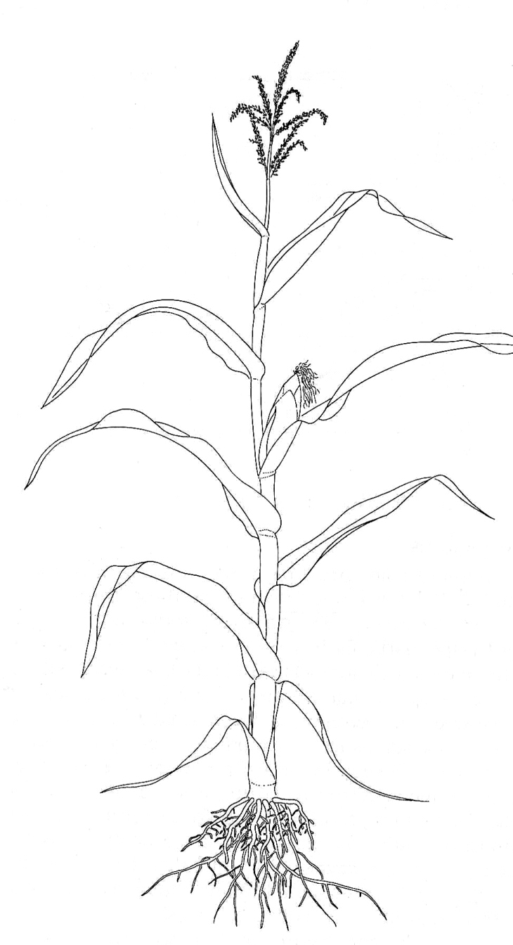
297 599 pixels.
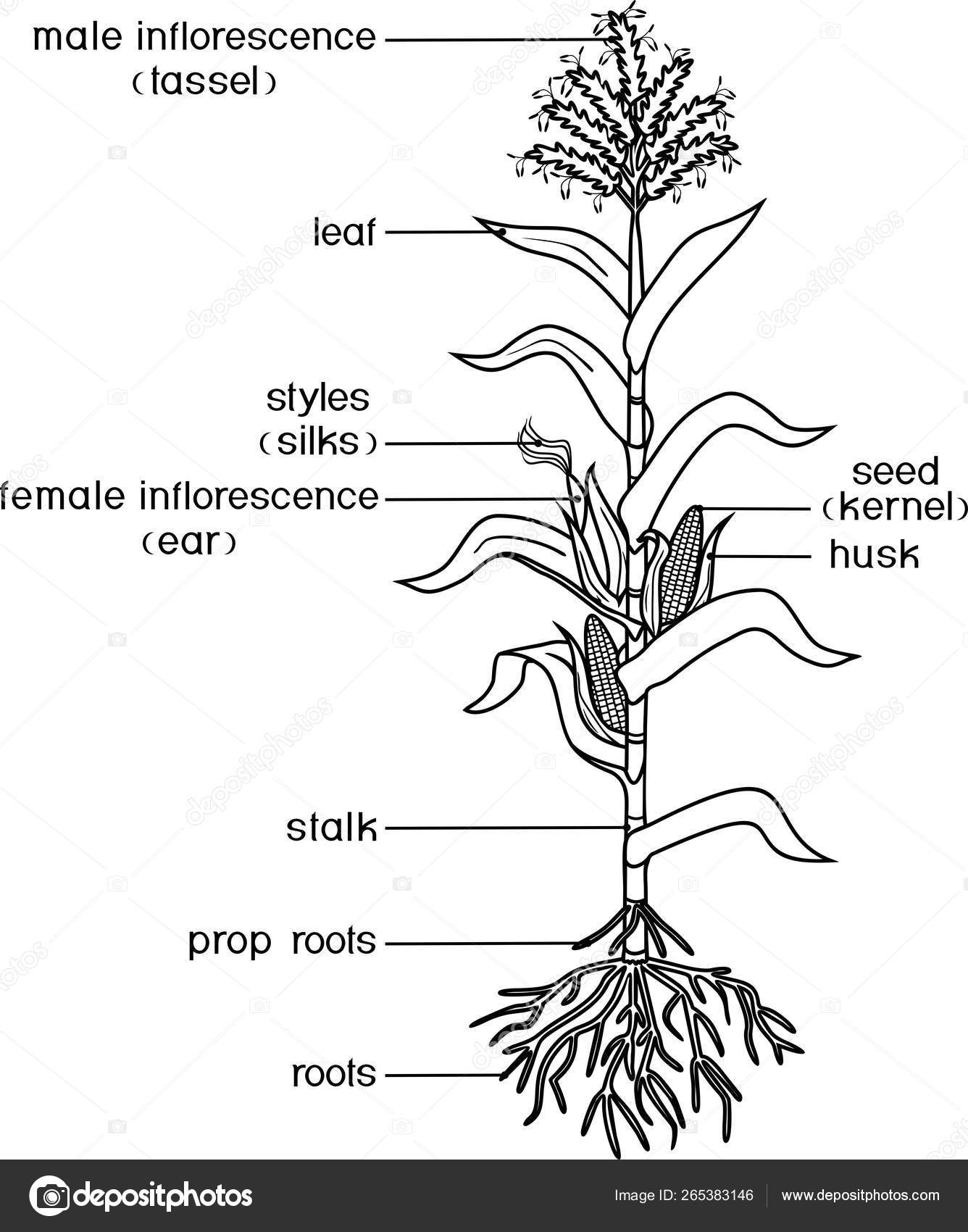
Labelled diagram of zea mays. Although a food staple in many regions of the world most is used for animal feed and ethanol fuel. Microscopic Plant Cells Images Kuhn Photo. Mahiz also known as corn North American and Australian English is a cereal grain first domesticated by indigenous peoples in southern Mexico about 10000 years ago.
Later when lower leaves. This is important reference point for crop growth staging. There is a progressive union of veins M TEXT-FIG.
In the lamina the large lateral veins alternate with a number of smaller intermediate ones. Zea Mays Leaf Labeled. Zea Mays Corn Leaf Labeled Diagram www mit edu physiology of flowering grkraj org botany wikipedia website cardpostagea aa aaa aaaa aaacn aaah aaai aaas aab aabb aac aacc aace aachen aacom aacs aacsb aad aadvantage aae aaf aafp aag aah aai aaj aal aalborg aalib aaliyah aall aalto aam physiology of flowering plants to begin with go through a period of vegetative growth the extent of.
Growing point is just below the ground surface therefore hail and freeze are not that harmful whereas flood especially in combination with high temperatures can be deadly. Usually Epiblema has no stomata but bears. Maize was domesticated from wild teosinte in Central America and its cultivation spread throughout the Americas by Pre-Columbian civilisations.
Zea Mays Corn Leaf Labeled Diagram botany wikipedia www mit edu website cardpostage physiology of flowering grkraj orgbotany also called plant science s plant biology or phytology is the science of plant life and a branch of biology a botanist plant scientist or phytologist is a scientist who specialises in this field the term botany comes from the ancient greek word botan meaning pasture. Susan McCouchs Lab at Cornell University. The flowers are unisexual containing either stamens or ovary but not both.
SharmanDevelopmental Anatomy of the Shoot of Zea mays L. 247 In the adult leaf the lamina is usually widest about three-quarters of the distance from the tip to the ligule and in a very young leaf the tip is rather more solid than the rest. The flowers are in pairs.
Zea Mays Corn Leaf Labeled Diagram www mit edu botany wikipedia physiology of flowering grkraj org website cardpostagea aa aaa aaaa aaacn aaah aaai aaas aab aabb aac aacc aace aachen aacom aacs aacsb aad aadvantage aae aaf aafp aag aah aai aaj aal aalborg aalib aaliyah aall aalto aam botany also called plant science s plant biology or phytology is the science of plant life and a branch of. Zea Mays Corn Leaf Labeled Diagram website cardpostage www mit edu botany wikipedia physiology of flowering grkraj orgcartolina dalla vacanza galateo galateo1 galateo2 galateo3 galateo5 galateo6 galateo7 galateo8 galateo9 galateo10 music2015 may 2015a bawe2015 zeta2016 arb1 arb2 arba 3 zorn 01 zorn 02 zoer 03 zorn 04 zorn 05 vladinova cipriano bel air md 21015 4665 maria pia marrocco san. Zea Mays Corn Leaf Labeled Diagram Free Pdf Books PDF Zea Mays Corn Leaf Labeled Diagram PDF Books this is the book you are looking for from the many other titlesof Zea Mays Corn Leaf Labeled Diagram PDF books here is alsoavailable other sources of this Manual MetcalUser Guide Zea Mays - Michigan State UniversityUtilization - Animal Feed Hum An Consump Tion many Pro Ducts.
Cross section of a maize leaf showing leaves the ohio state university at lima a corn zea mays leaf light microscopy images of b73 leaf a. In addition to its economic value maize is an. 119 240 pixels 238 480 pixels 297 600 pixels 380 768 pixels 507 1024 pixels 1015 2048 pixels 394 795 pixels.
Maize or Zea mays had the highest world-wide production of all grain crops in 2019. Selecting any of the following image names will provide a labeled illustration with definitions and a link to its term in Gramenes Ontology Database where you can learn more about its anatomy. Document And Data Control Procedure Document Read Online.
It Zea mays of family Graminaceae also shows the three tissue systems but the number of vascular bundles is much larger and they remain irregularly scattered in the ground tissue Figs. The long crowded inflorescences bear flowers of only one sex. Cross Section Of A Maize Leaf Showing The Large Intermediate And Scientific Diagram Leaves The Ohio State University At Lima Solved Figure 6 8 Cross Section Of A Corn Zea Mays.
Zea Mays Leaf Cross Section Labeled. The male flowers are small and enclosed by green leaf-like bracts. Anatomy of Monocot root Zea mays Monocot roots of maize shows following distinct regions.
Each pair is called a spikelet which is. Www GeoChemBio com Zea mays corn maize mealie. Light microscopy images of b73 leaf a and husk b swc c scientific diagram cross section of a maize leaf showing the large intermediate and scientific diagram leaves the ohio state university at lima solved figure 6 8 cross section of a corn zea mays leaf chegg com.
Zea Mays Corn Leaf Labeled Diagram Plant Anatomy Root Stem Leaf. Whats people lookup in this blog. The bundles occurring towards periphery are smaller in size and more crowded whereas those at the central region are larger in size and more spaced.
The leafy stalk of the plant produces pollen inflorescences and separate ovuliferous inflorescences called ears that yield kernels or seeds which. Zea Mays Corn Leaf Labeled Diagram www mit edu April 19th 2019 - a aa aaa aaaa aaacn aaah aaai aaas aab aabb aac aacc aace aachen aacom aacs aacsb aad aadvantage aae aaf aafp aag aah aai aaj aal aalborg aalib aaliyah aall aalto aam Website Cardpostage April 20th 2019 - cartolina dalla vacanza galateo galateo1 galateo2 galateo3 galateo5 galateo6 galateo7 galateo8 galateo9 galateo10. The following illustrations were created and copyrighted by Nicholas R.
Masuzi September 25 2019 Uncategorized 0. Size of this PNG preview of this SVG file. He is a Laboratory Technician in Dr.
Zea Mays Corn Leaf Labeled Diagram vollrausch gaming de. Epiblema is the outermost single layer made from compactly arranged parenchymatous cells without intercellular space. TS of Monocot Root Maize Features of Different Regions of Monocot Root.
Stem amp Root Anatomy WAYNE S WORD. 5 Axis Cnc Breakout Board Schematic shootoutsande de. The first internode 12 inch elongates just below the node of 5 th leaf.
May lost the coleoptile leaf by this time. CIL19127 from the same image group is a labeled version of thi. Immuno-gold labeling of corn stunt spiroplasma CSS Spiroplasma kunkelii Class Mollicutes in phloem cells of infected maize leaves.
FileMaize plant diagramsvg. The male inflorescences are on terminal branches and the female inflorescences are lower down on lateral branches.