In phylum Cnidaria the polyp present in either colony or individual. Earthquake crosssection labelling activity earthquake cross section save resource.
Obelia Life Cycle With Labelled Diagram

In this heart diagram labelling quiz youll review the different chambers valves and passageways that make up the heart and improve your understanding of how they are associated with the lungs and other parts of the body.
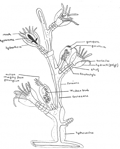
Diagram of obelia with labelling. Like most hydrozoans it has both polyp and medusa stages. This site uses cookies. Habitat Structure and Diagram.
Click and drag to make a text box and in the area you want the first label and the size you want it to be. Both are of same thickness. This shallow water is up to 80 meters in depth.
It consists of branched filaments approximately the thickness of fine sewing cotton. See more ideas about anatomy anatomy and physiology human anatomy and physiology. Obelia occurs as a delicate whitish or light brown almost fur-like growth on seaweeds or timber.
It is the slide of obelia which is a marine colonial and arborescent animal. Mar 21 2019 - Explore Summer Ekelunds board Anatomy Labeling Diagrams followed by 235 people on Pinterest. To save a resource you must first join or sign in.
Mitochondria Pictures With Labels Mitochondria Labelled Diagram. All the individuals in a colony are attached through branches to a horizontal hydrorhiza. It is found in different colors like brown black blue green etc.
Labeled Diagram Of A Bulldozer. Need module 3 earthquakes tsunamis activity 1. Obelia is sedentary marine colonial form found attached on the surface of sea weeds molluscan shells rocks and wooden piles in shallow water up to 80 metres in depth.
Draw A Neat Labelled Diagram Of Power House Of A Cell What Is The. Diagram of an earthquake with labelling. Obelia is a typical example of a marine colonial hydrozoan.
You can use this diagram of a volcano labelling quiz as a helpful guide for studying the anatomy of a volcano the different structures found above and beneath the surface and their role in Earths evolution as a planet. FileDiagram of human eye without labelssvg. It is a branched fixed colony Fig 2012.
The human eye diagram displayed at right shows all of the major features of human ocular anatomy. The growth of the colony is sympodial. It consists of vertical branching stems are called as hydrocauli and the root like branches are called hydrorhiza.
A lot of people have been using net for finding information guidelines reports or any other reference for their purposes. Place a commercially prepared slide of Obelia medusae on the stage of the compound microscope and focus at 40X. A common example of it is Obelia geniculata.
Ö Select Insert Menuà Text Box. Learn vocabulary terms and more with flashcards games and other study tools. 8 2 Applications And Skills 8 2 2 Perspectives On Mitochondria.
Some of the horizontal branches anchoring the colony on some support are called Hydrorhiza while other branches are vertical and known as Hydrocaulus. Diagram of an earthquake with labelling. Labeling Diagrams with Text Boxes LABELED DIAGRAM Ö Place and resize the diagram or picture in your word document.
The upper body of polyp is surrounded by the tentacles and from the lower body it is attached to the solid surface. These are animals which belong to the class of Hydrozoa and Phylum Cnidaria and consist of various species. The branches are called hydrocauli.
Habit and Habitat of Obelia. How To Draw Mitochondria Easy Step By Step Drawing Biological. The earth is made up of blocks referred to as plates which are in constant motion.
Simple Structure Of Mitochondria. The hydroid colony of Obelia is delicate semitransparent and whitish to light brown in color. In this article we will discuss about the life cycle of obelia explained with the help of suitable diagrams.
By continuing to browse the ConceptDraw site you are agreeing to our Use of Site Cookies. Make sure that you do not make it too big so that you have room for the diagram labels. A Well Labelled Diagram Of A Bulldoer.
In Obelia there are two types of a polyp Dimorphic - Gastrozooids and another one is gonozooids. It is a type of body form in phylum Cnidaria. Obelia refers to the sedentary marine colonial form which has an attachment on the surface of seaweeds rocks wooden piles and molluscan shells in shallow water.
The medusae of prepared slides are stained a variety of meaningless colors but in life they are transparent and colorless. Aerospace and Transport Draw And Label A Bulldozer. Sketch Of A Bulldozer With Labelling.
Its origin can vary according to type and size of quake. It is found attached to submerged objects like rocks and weeds etc.
B Describe the mechanism of hearing in mammals. Structure of the earth diagram activity chapter 1 plate tectonics what is the lithosphere siyavula draw the diagram of sectional view human heart and on it name draw the neat diagram showing layers of earth.

3 Draw A Diagram Of The Earth To Show Internal Structure Of Scholr
Earths magnetic field comes from the outer cores ocean of iron which is an electrically conducting fluid in constant motion.

Draw a labelled diagram to show the internal structure of the earth as we know it today. Surrounding the iron ball is an ocean of liquid iron known as the outer core This inner and outer core duo is referred to as Earths geodynamo. Label the parts of the Earth using Textables and arrows. Blood Circulatory System in Human.
Draw A Well Labelled Diagram To Show The Internal Structure Of Earth. The structure of an atom explained with the rock cycle lithosphere siyavula seimic waves and earth s interior draw a well labelled diagram structure the rock cycle lithosphere siyavula. Search for earth or globe and pull down the Earth item.
Draw a well labeled diagram of the schematic diagram showing the structure cartoon 1024 800 transp png schematic diagram showing the structure neat diagram showing layers of earth Draw The Neat Diagram Showing Layers Of Earth Brainly InDraw A Diagram To Depict The Structure Of Earth From Geography Interior Cl 11 CbseThe Structure Of Earth. Draw a neat and labelled diagram. Show Answer Show Explanation.
Interestingly there are two cores noted the inner and the outer. Name the three layers of earth. Describe the different sections of the Earth.
Draw a Labelled Diagram to Show the Internal Structure of a Mammalian Tooth with Two Roots. Hello EveryoneDiagram Of Structure Of Earth Labelled Diagram Of Earth Layers OF Earth Diagram Of Structure Of Earth Labelled Diagram Of Earth Layers. Near the surface the Earth is dominated by silicon and oxygen.
A i Draw a well labelled diagram to show the internal structure of the earth ii Describe the distinguishing characteristics of any one of the layers shown in your diagram b Outline the importance of either the hydrosphere or the biosphere to man Show Answer Show Explanation. The Earth has an outer solid layer called the crust a highly viscous layer called the mantle a liquid layer that is the outer part of the core called the. These layers are both physically and chemically different.
Draw A Labelled Diagram Of The Structure Earth. The crust is the outermost layer of the earth and the core is the innermost layer of the earth located at a depth of 2900 Km. Internal structure of the heart.
Maharashtra State Board SSC English Medium 8th Standard. Draw a labelled diagram of earths interior to show the different layers and discontinuities - 19405972 Rushque4301 Rushque4301 11072020 Geography Secondary School answered Draw a labelled diagram of earths interior to show the different layers and discontinuities 2 See. This diagram shows us how thin the crust is in relation to the rest of the Earth along with relative sizes of the mantle and core.
A Draw a well-labelled diagram to show the intreral structure of the earth b Describe the main features of any two of the parts shown in a above. Notice also how the chemical composition of the Earth varies with depth. Chapter 1 plate tectonics seismic evidence for internal earth draw a well labelled diagram structure the structure of earth marcellus Internal Structure Of The EarthThe Structure Of Earth Earthquakes Discovering GeologyDraw Neat Diagram Label Them And Explain The Interior OfWhat Are The Earth S LayersDraw The Neat Diagram Showing Layers Of Earth Brainly InEarth S Read More.
Advertisement Remove all ads. This article briefly throws light on these 3 different interior layers of the earth. Structure Of The Earth Diagram Activity.
Seismic evidence for internal earth seismic evidence for internal earth the structure of earth draw and label the layers of earth interior of the earth crust mantle The Structure Of Earth Earthquakes Discovering GeologyInternal Structure Of The Earth10 H Structure Of The EarthDraw The Neat Diagram Showing Layers Of Earth Brainly InEarth S Internal Structure. The structure of the Earth is divided into layers. Click on Image Options to change to the diagram.
Internal structure of the heart. Draw a labelled diagram to show the structure of earth. Identify and describe the parts of the Earth in a storyboard.
The interior of the earth can be divided into 3 different layers crust mantle and core. A i Draw a well labelled diagram to show the internal structure of the earth ii Describe the distinguishing characteristics of any one of the layers shown in your diagram b Outline the importance of either the hydrosphere or the biosphere to man. C State two ways of caring for the ear.
Draw a neat and labelled diagram. By Hilman Rojak August 17 2020. Internal structure of the heart.
Surrounding the core is the mantle and Earths crust forms the top layer. A Make a labelled diagram to show the internal structure of the mammalian ear.
Halogen atoms F Cl Br I Mercury Hg Selenium Se Ferrocence Fe Analogs and Derivatives of. A nucleotide is the basic structural unit and building block for DNA.

Nucleotides Bioninja
A nucleobase a five-carbon sugar ribose or deoxyribose and a phosphate group consisting of one to three phosphates.
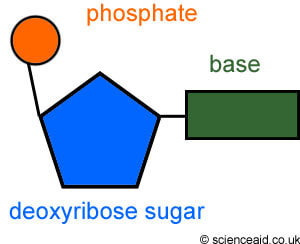
Labeled parts of the nucleotide. Nucleotide Structure Courtesy of the National Human Genome Research Institute. One nucleotide is composed of a base a deoxyribose sugar and a phosphate. Free amino group -NH 2 Redox Dyes.
Common labels used to generate nucleic acid probes include radioactive phosphates biotin fluorophores and enzymes. In addition the bioconjugation methods used for nucleic acid probe generation may be adapted for attaching nucleic acids to other molecules or surfaces to facilitate targeted delivery or immobilization respectively. Nucleotides are composed of three subunit molecules.
A nucleotide is composed of 3 parts. A nucleotide has a sugar or deoxyribose group a phosphate that is attached on the 5 carbon and an organic nitrogenous base which is attached to the 3 carbon. Draw or label a diagram of a dna molecule including the four nucleotides based on the size of the nucleotide and the number of bonds between nucleotides the.
Imaging and Ener Ics Of Single Ssb Ssdna Molecules Reveal Cell Free Protein Array. It consists of a. Vazquezk1234 3 weeks ago Biology High School 5 pts.
Draw a nucleotide and label its three basic parts. A nucleotide is made up of three parts. Five-sided sugar phosphate group nitrogenous base nitrogen containing.
Both deoxyribonucleic acid DNA and ribonucleic acid RNA are made up of nucleotides which consist of three parts. List the four nitrogen bases. Adenine and guanine are purines.
Molecules Review Worksheet Chapter 2 23 25. Name the 4 nitrogen bases on dna. Labeled Parts Of A Nucleotide - Fun for my own blog on this occasion I will explain to you in connection with Labeled Parts Of A NucleotideSo if you want to get great shots related to Labeled Parts Of A Nucleotide just click on the save icon to save the photo to your computerThey are ready to download if you like and want to have them click save logo in the post and it will download.
DNACellCycleReviewpdf - Read File Online - Report Abuse. Putting it all together. Answered Label three parts of a nucleotide.
Draw And Label The Three Parts Of A Nucleotide Best Label Ideas 2019 Nucleotide Wikipedia Nitrogenous Base An Overview Sciencedirect Topics Dna Function Structure With Diagram Article Khan Academy 3 Parts Of A Nucleotide And How They Are Connected Dna He 5 B 1 He 5 B 2 He 5 B 3. Which parts make up the sides of the ladders. The four nitrogenous bases in DNA are adenine cytosine guanine and thymine.
Correctly Label The Parts Of The Two-nucleotide Nucleic Acid Depicted Drag The Appropriate Labels To Their Respective Targets Reset Help 5 Position H2C OH In RNA Nitrogen Base Attached To 1 Position 3 Position Phosphodiester Bond Deoxyribose 2 Phosphate Base. Nucleotides are made of what three parts. Label the following parts of a DNA polymer Nitrogenous base adenine Nitrogenous base quanine Nitrogenous base cylosine Sugar phosphate backbone Nitrogenous base thymine Nucleotide Hydrogen bond Reset Zoom.
Draw And Label The Three Parts Of A Nucleotide Pensandpieces Nucleotide Drawing 3 3 Dna Structure Bioninja Ppt Dna Rna And Protein Synthesis Review Powerpoint N The Diagram To The Right Identify The Three Components Of A Nucleic Acid Dna Function Structure With Diagram Article Khan Academy What Are The Three Parts Of A Nucleotide Albert Io. Label the deoxyribose molecule. RNA contains uracil instead of thymine.
In RNA uracil is used in place of thymine. A phosphate group a 5-carbon sugar and a nitrogenous base. In RNA the.
Labeled Parts Of A Nucleotide Supramolecular Self assembly Of Nucleotidemetal Coordination Molecules Free Full Text 52 Best Dna 1 software Of Life Images On Pinterest Triglycerides are An Example Of the Function Of Lipids. Nitrogenous Base Purines and pyrimidines are the two categories of nitrogenous bases. Label three parts of a nucleotide - 16807912 1.
Phosphate molecules consist of a phosphate atom surrounded by four oxygen atoms. The double helix is held together by these weak bonds. Sketch and label a dna nucleotide.
The four nucleobases in DNA are guanine adenine cytosine and thymine. Label the nucleotide to the right. These building blocks are hooked together to form a chain of DNA.
The above structure is a nucleotide. Pentose Sugar In DNA the sugar is 2-deoxyribose.
Sterilization and aseptic processing are essential practices for healthcare product manufacture and many healthcare services. Dry heat sterilization is used on equipment that cannot be wet and on material that will not melt catch fire or change form when exposed to high temperatures.

Hot Air Oven Working Principle Uses Labelled Diagram Pharmawiki In Ppt Download
School University of Notre Dame.
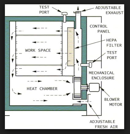
Labelled diagram hot air oven diagram. The diagram with fully label of ovenhot-air-oven WIRING DIAGRAM FOR MAYTAG GEMINI RANGE MODEL MER6770AAW Where is the model number is located on the unit of a gemini oven. Hot Air Oven Labelled Diagram Uses of HOT AIR OVEN dry heat sterilization A dry heat cabinet is easy to install and has relatively low operating costs. Diagram of hot air oven deekey de read and download neat labelled diagram of hot air oven free ebooks in pdf format holt mcdougla modern world history teacher edition diagram what is the principle behind the working of microwave oven draw neat labeled diagrams wherever necessary.
Hot air oven with labelled diagram image pdf free download here mark scheme unit g635 working waves specimen june ocr what property of oven gloves allow a baker to pick up hot bread gently heat the flask of air with the help of a labelled do not touch the bulbs or block air use a labelled ray diagram to show neff fan oven wiring diagram i need. A hot air oven is a laboratory instrument that uses dry heat to sterilize laboratory equipment and other materials. 1080x821 draw a neat labelled diagram of human ear with its each part - Eardrum Drawing.
It penetrates materials It is nontoxic and does not harm the environment. Aneorobic culture methods 8. Draw a neat labeled diagram of trophozoite of Giardia lamblia.
Hot air oven Definition. Vincent motorcycle misc information. The circulating fans and fan motor that are equipped with the.
Lab diagnosis of syphilis 7. A hot air oven is used to sterilize equipment and materials used in the medical field. Hot Air Oven Working Principle Sterilization Labelled Diagram Temperature PDF PPT is the main theme of this article.
With stainless steel interiors and fibreglass insulation between the walls to maximize thermal efficiency hot air oven effectively sterilizes glassware instruments and samples. A hot air oven is a type of dry heat sterilization. Discuss the morphology of bacterial cell with a diagram and state briefly add a note on cell wall of bacteria.
The execution of these processes in an appropriate manner is essential for patient safety. Hot air oven labelled diagram. The Hot Air Oven works on the basis of Hot Air inside the chamber which is created due to the forced air circulation.
366x254 hot air oven labelled diagram - Drawing A Diagram. These hot air oven diagram are sturdy and perform optimally saving energy bills. Neat Labelled Diagram Of Hot Air Oven port manteaux word maker onelook dictionary search.
Diagram Labelled Hot Air Oven With Labelled Diagram Image Diagram Of A Well Labelled Bean Cowpea Well Labeled Diagram Of Hammer Mill vspnijmegen nl April 15th 2019 - well labeled diagram of hammer mill youtube oct 12 2013 as a professional crushing and grinding equipments manufacturer 1 34 a well labelled grinding. Define sterilization classify various methods of sterilization and describe in detail about dry heat sterilization in dental practice. Name two specific diagnostic tests of syphilis 17.
It is a universal fact that in chambers the hot air first rises up and once it reaches the top of the instrument it came back to the bottom of the chamber. Hot air oven 5. And it is noncorrosive for metal and.
Glass observation window eliminates the need of opening the door. Philippine electrical wiring building our philippine. 306x229 draw a labelled diagram of an electric motor principle and worki - Electric Motor Drawing.
Forced air convection system maintains temperature uniformity. That equipment cannot be wet or material that will not melt catch fire or change form when exposed to high temperatures are sterilized by using the dry heat sterilization method. Thinking outside the box a misguided idea psychology today.
Discuss the different structures of bacterial cell with a neat-labelled diagram. Applying systems thinking to computing. Course Title NURSING 1234.
Full text of new internet archive digital library of. Pathogenesis and lab diagnosis of mycobacterium tuberculi. Draw a neat labelled diagram of Immunoglobulin-A 16.
Tes Global Ltd is registered in England Company No 02017289 with its registered office. Plant and Animal Cell GCSE Recap Labelled diagram.
Animal Plant Cell Teaching Resources
They modify the original diagram to create a series of new cells.

Animal cell labelled diagram ks3. Labelling Plant Animal Cells Teaching Resources Cell wall a jelly that fills the cell chemical reactions happen here. Ribosomes are only visible with an electron. A generalised animal cell and its components.
It is easier to describe these parts by using diagrams. KS4 KS5 Science Biology. S1 animal and plant cell structures Group sort.
Explain that some of the life processes take place within individual cells respiration and photosynthesis. Mitochondrion are visible with a light microscope but cant be seen in detail. Cell wall cell membrane chloroplasts cytoplasm mitochondria nucleus vacuole.
Which of these 3 parts of a cell are only found in plant cells. State the differences and similarities between plant and animal cells by completing the table below. Plant Cell Diagram Ks 4.
Animal Cell Diagram Labeled Ks 3Kevin Brace The Plant Cell Task. Ensure that your students understand the core components of a basic animal cell with this Animal Cell Labelling activity sheetThis resource features a large-scale illustrationdiagram of an animal cell with four arrows pointing to the cell nucleus cytoplasm cell membrane and mitochondria. Plant and Animal Cell GCSE Recap Labelled diagram.
Present in plant cell yes or no. Animal Cell Labeling Labelled diagram. Part of cell Present in animal cell yes or no.
Plant and Animal Cell GCSE Recap Labelled diagram. Cytoplasm controls what the cell does. 10000 results for ks3 animal and plant cell.
This website and its content is subject to our Terms and Conditions. B2 plant or animal cell Flip tiles. Cell wall nucleus cytoplasm.
Unlabelled diagram of plant cells the parts of a plant cell and. Cells are made up of different parts. What is the name given to the part labelled S.
Please give constructive feedback d. Chloroplasts cell wall vacuole. Plant and animal cell structures.
Plant and Animal Cells KS4 Match up. Key Stage 3 activity on labelling animal plant and specialised cellsStudents are given a worksheet showing basic animal cells. Comparing Plant Cells and Animal Cells Task.
Cytoplasm controls what the cell does. Animal Cell Labeling Labelled diagram. As observed in the labeled animal cell diagram the cell membrane forms the confining factor of the cell that is it envelopes the cell constituents together and gives the cell its shape form and existence.
Label the parts of cells which appear in both types of cells on the two diagrams below. Unlabelled plant cell diagram ks3. Plant Cell Diagram Ks 4 Labeled.
We all remember that the human body is quite elaborate and one way I. A labeled diagram of the plant cell and functions of its organelles we are aware that all life stems from a single cell and that the cell is the most basic unit of all living organisms. 7 rows Animal cells usually have an irregular shape and plant cells usually have a regular shape.
Cell part Function job Nucleus covers the membrane and gives strength to a plant cell. 10000 results for label animal cell Plant and Animal Cell GCSE Recap Labelled diagram. Bacterial cells have a more simple structure compared to animal plant and fungal cells and are usually much smaller.
This is a great and easy way to assess understanding of cell structure and help students see the features that are shared between different cells. Patrick Baker Animal cells usually have an irregular shape and plant cells usually have a regular shape. KS4 KS5 Science Biology.
Learn about plant reproduction and the process of pollination with bbc bitesize ks3 science. Plant And Animal Cell Diagram Ks3 DIAGRAM Lottie Santiago An answer sheet is also included. 10000 results for ks3 plant and animal cell.
Animal and Plant Cell Label Labelled diagram. The Animal Cell Diagram Poster is labeled with a total of 13 grade-level terms including cell membrane nucleus vacuole cytoplasm and mitochondrion. KS4 KS5 Science Biology.
Animal and plant cell structures Group sort. 10000 results for animal cell. To display this animal cell poster in your classroom all you have to do is hit download and print.
In order to complete the worksheet students must correctly label all four componentsThis labelling. Students should be taught the word equation. Cells are made up of different parts.
BBC KS3 Bitesize Science Cells to systems. Animal and plant cell structures Group sort. S1 animal and plant cell structures Group sort.
It is a rigid layer which is composed of cellulose glycoproteins lignin pectin and hemicellulose. Students could produce a diagram to illustrate the organisation within a plant and give examples of the different cells that could be found. Animal Cell Labelled diagram.
You should make a label that represents your brand and creativity at the same time you. Draw an angle of 45.
3 5 The Ridger

ConceptDraw is Professional business process mapping software for making process flow diagram workflow diagram general flowcharts and technical illustrations for business documents.

Well labelled diagram of a ridger. A Well Labelled Diagram Of A Spider. Label Gallery Get some ideas to make labels for bottles jars packages products boxes or classroom activities for free. Textplot convert plotting line segments assume assuming verifying inequalities with is.
Biomagnification means increasing the concentration of various toxic substances along the food chain. It is includes rich examples templates process flowchart symbols. Labelled diagram ofAC electric locomotive.
Well labelled diagram of digestive system ruminent attachment no 2 chau7myasrma chau7myasrma 21052016 Science Secondary School answered Draw well labelled diagram of digestive system or digestive system of ruminant on a chart 2 See answers TheRuhanikaDhawan TheRuhanikaDhawan. Class V All Subjectspdf - Read File Online -. Differentiate between a river system and a river regime.
Enthalpies of formation for O2 0k. Drag and drop the pins to their correct place on the image. Well Labelled Diagram Of A Liverwort.
You should make a label that represents your brand and creativity at the same time you. Draw a neat labelled block diagram of AC electric locomotive. Name any three surface features that result from carbonation.
The diagram below shows a drainage pattern. Give a reason for each of the following. Draw Bohr diagrams of 2 hyrdogen atoms bonding with an oxygen atom chemistry Calculate the heat of formationof benzene if the heat of combustion of benzene is 3260 kjmol given the following enthalpies of formation of the product.
Function of AC electric locomotive Parts. Draw a neat and labelled diagram showing the temperature zones of the earth. The primary purpose of this worksheet is to show you how you can use Maple to draw well-labelled diagrams.
Well labelled diagram of tilapia fish. Draw a well labeled diagram of. Nucleolus and one or more chromosomes present.
New Maple commandsconcepts used. Through looking at these diagrams it is easier to understand the nature of V-shaped valleys the river ordering system the water cycle and other aspects related to rivers. Draw a well labelled diagram to show refraction from a glass slab.
Desert areas experience a high day temperature and a much lower night temperature. An easy and convenient way to make label is to generate some ideas first. Light - Reflection and Refraction.
Toxic substances at the level of primary producers get concentrated at each trophic level as they move up the food chain. Ii Name the parts labelled P and Q. Tilapia is a mild tasting versatile white fish that pairs perfectly with most seasoning and sauce.
Labels are a means of identifying a product or container through a piece of fabric paper metal or plastic film onto which information about them is printed. Great value breaded fish sticks 44 count 24. The information can be in the form of hand-written or printed text or symbols and gives details about manufacturers name source of product shelf-life uses and the manner of disposal.
Below diagram shows the increase in accumulation of toxic substances as we move up the food chain. 1 Overhead contact wire. ConceptDraw flowchart maker allows you to easier create a process flowchart.
A Name any two features deposited by glacier in. Use it to answer the questions that follow- i Name the drainage pattern. Aldi fresh fish is certified fresh and never frozen.
Found in eukaryotic cell. An easy and convenient way to make label is to generate some ideas first. Below labelled diagram shows the Heat Budget of the Earth.
Draw a well labelled diagram to show the Heat Budget of the Earth. State the function of each part. Label Gallery Get some ideas to make labels for bottles jars packages products boxes or classroom activities for free.
To illustrate this I will simplify an arctrig expression by drawing a reference triangle. Genetic material in the form of single chromosome. Genetic material enclosed in nuclear envelope.
These fish arent able to survive well in waters that get to temperatures. Found in prokaryotic cell. ERD Entity Relationship Diagrams ERD Software for Mac and Win Flowchart Basic Flowchart Symbols and Meaning Flowchart Flowchart Design - Symbols Shapes Stencils and Icons.
Supply of 1-ph 25KV 50Hz AC is given to overhead conductor. These river diagrams help to explain the geography topic of rivers. Genetic material not enclosed in nuclear envelope.
Laws of Refraction.
In this video I discuss the basics of the Respiratory System including how the respiratory system works I go through the breathing process and show how br. The major respiratory structures span the nasal cavity to the diaphragm.

Respiration Types Of Respiration And Anatomy Of Human Respiratory System Online Biology Notes
However like just about every other part of our anatomy that we take for granted we take our respiratory system for.

What are the 6 parts of the respiratory system. One cell thick to allow gas. What is the respiratory tract made up of. Functionally the respiratory system can be divided into a conducting zone and a respiratory zone.
Examples of respiratory structures include the nose mouth lungs and diaphragm. The respiratory system includes the nose mouth throat voice box windpipe and lungs. The human respiratory system is adapted to allow air to pass in and out of the body and for efficient gas exchange to happen.
Grades 6 to 8 Human Body Series Respiratory System 1 ½ hours Activity. The respiratory system is the organs and other parts of your body involved in breathing when you exchange oxygen and carbon dioxide. The other parts of the respiratory system include the nose larynx pharynx trachea or the windpipe bronchi lungs blood vessels the airways for the passage of air and the muscles that support the breathing.
What Are the Parts of the Respiratory System. Exercise and smoking both affect the lungs and circulatory system. These parts work together to move oxygen throughout.
The muscles that power your lungs are also part of the respiratory system. If it goes in the nostrils also called nares the air is warmed and humidified. The respiratory tract is made up of nostril nasal chamber larynx pharynx epiglottis trachea bronchioles bronchi alveoli and lungs.
All these parts together form the respiratory tract that starts from the external nostrils and nasal chamber and goes up to the lungs. Air enters the respiratory system through the nose or the mouth. Name the structure labelled X and list two characteristics of structure X and explain how these characteristics assist the function of the whole structure.
In addition to these organs certain muscles of the thorax the body cavity that fills the chest are also involved in respiration by enabling breathing. The Main Function Of The Respiratory System. 1 mark for identification 2 marks for characteristics 2 marks for explanation X.
Is The Process Of Oxygen Intake And Carbon Dioxide Removal. Is Also Known As Deoxygenated Blood. The respiratory system sometimes referred to as the ventilatory system is a biological process in which the body is able to take oxygen into the body and then expel carbon dioxide.
Parts Of The Respiratory System Science STUDY. It includes your airways lungs and blood vessels. BIOLOGY 12 UNIT 6 RESPIRATORY SYSTEM A 16.
The organs in each division are shown in Figure 162. The upper tract includes the nose nasal cavities sinuses pharynx and the part of the larynx above the vocal folds. The conducting zone of the respiratory system includes the organs and structures not directly involved in gas exchange.
In the breathing process air flows into and out of the lungs. Structures of the respiratory system can be grouped into three main categories. Air passages pulmonary vessels and respiratory muscles.
In the diagram below structure W is a bronchiole. The upper respiratory tract and the lower respiratory tract. What are the main functions of the respiratory system.
Learn more about the parts of your respiratory system. The respiratory system is the network of organs and tissues that help you breathe. 2 includes the lower part of the larynx the trachea bronchi bronchioles and the alveoli.
Gases are exchanged between the air and blood. The gas exchange occurs in the respiratory zone. Once you have the information write and illustrate a comic strip about Captain.
It Is The Blood That Flows Within The Veins Going To The Heart. The lower tract Fig. The tract is divided into an upper and a lower respiratory tract.
The important human respiratory system parts include- Nose larynx pharynx trachea bronchi and lungs. The respiratory tract has two major divisions.
There are descriptions to go along with the diagram. The pistil contains the stigma style and ovary.

Labelled Drawing Of Hibiscus Flower Novocom Top
Parts of hibiscus flower drawing.
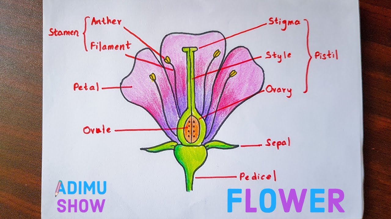
Draw and label the parts of hibiscus flower. Label the parts of hibiscus. Parts of a flower. Images Of How To Draw Hibiscus Flower And Label Its Parts Popular Drawing Images Of How To Draw Hibiscus Flower And Label Its Parts.
Individuals now are accustomed to using the internet in gadgets to view image and video information for inspiration and according to the name of the post I will talk about. At the bottom of every hibiscus bud is a green structure at the top of the stem. Youll recognize the pistil in a plant diagram because it looks like a small knob that protrudes from the flower.
Diagram of hibiscus flower with label. This is called the calyx. Draw and label the parts of hibiscus flower.
When the hibiscus begins to bloom the petals begin to grow which. The pinkish white hibiscus flower seen below is from one of my hibiscus plants. How To Draw A Hibiscus Flower Easy Step By Step With Color In 2020 Hibiscus Drawing Drawings Hibiscus Flowers.
It is a tough part of the flower because it houses the. Every flower has multiple petals which differ in color depending on the species. And it will teach you how to draw a flower very easily.
The female part of the flower the pistil is located at the center of the bloom. Inside the child parts of a flower hibiscus label the following diagram brainly in grass features and structure digital flowers hibiscus vectors photos and. Hand drawn illustration Roselle flower.
Download this image for free in High Definition resolution using a download button option below. Vector background with ornamental hibiscus flowers. Flower Diagram was drawn but not labelled properly.
A beautiful drawing of a flower. Draw And Label The Parts Of Hibiscus Flower is important information accompanied by photos and HD images sourced from all websites in the world. Examine a flower diagram and youll see that flowers consist of several parts.
Hibiscus flower drawing coloring page 671x623 hibiscus coloring page flower pages diagram of hibiscus flower parts stigma female style ovary. This slight curve also helps give the flower some weight. Vector illustration of hibiscus flower.
If you do not find the exact resolution you are looking for go for Original or higher resolution. Hand drawn illustration dry roselle flower. Draw hibiscus flower and label the parts free draw hibiscus flower and label the parts we manage to pay for you this proper as capably as simple pretension to acquire those all.
Labeled hibiscus flower parts drawing Indeed lately has been sought by consumers around us maybe one of you personally. Hello friendsIn this video Im going to draw labelled diagram of hibiscus flower China rose and its partsThis video is helpful for you in your exams. Hibiscus Flower Drawing Step By Step At Getdrawings Free Download The male parts of the flower are responsible for producing pollen.
How To Draw And Label A Flower Step By Step Tutorial Youtube Parts Of A Flower Flower Step By Step Drawings. Identify And Label Diagram Of Flower Reading Industrial. It is a tough part of the flower because it houses the young bud.
Watch the video and please be kind enough to thumbs up to my videos. At the bottom of every hibiscus bud is a green structure at the top of the. Labeled Hibiscus Flower Parts Drawing Parts Of A Flower And Their Functions With Diagram Trees Com.
The bud grows from this structure. Drawing still life of food and vegetable. Draw the hibiscus.
Hello welcome to my channel kids day a channel dedicated to the entertainment of children and their parents where you will find videos of play doh drawings colors. Biology parts of a flower hibiscus. The bud grows from this structure.
As the hibiscus begins to bloom the petals begin to grow. How to draw a hibiscus flower with parts step by stepdraw Flower Structure and its Partsi draw this flower and different parts for science subject project. This is called the calyx.
ᐈ Draw A Hibiscus Flower And Label Its Parts Stock Vectors How To Draw Flower Diagram Cross Section With Labelling See also Beautiful Flowers Wallpapers For Laptop. The corolla is the colorful section that attracts animals and insects. The bud grows from this structure.
Youll recognize the pistil in a plant diagram because it looks like a small knob that protrudes from the flower. The corolla has two different parts the petals and the corolla lobes. This is called the calyx.
At the bottom of every hibiscus bud is a green structure at the top of the stem. Set of two black labels with Roselle aka Hibiscus sabdariffa and Cocoa tree aka Theobroma cacao branch sketch. A beautiful drawing of a flower and it will teach you to draw an flower very easily.
For this section on the webquest you will given. You can also save this page easily so you can view. Petals come in a wide variety of colors.
Parts of flower diagrams labeled and unlabeled tulip test. The Parts of hibiscus flower are consists of the pointed ends of the calyx are the sepals. Hand drawn illustration Roselle flower.
Saved by Nova Gabbe.
It protects vital organs provides framework and supports soft parts of the body. Label 20 of the Bones of the Horse 5 points each.

Equine Skeletal Anatomy Chart Horse Anatomy Horse Bones Animal Skeletons
Some like the rib cage provide protection for softer body parts while other bones enable mobility by supporting the muscles.

Horse skeletal system label game. Its components are shown by the pictorial to be easily identifiable. Aaron Zagonel - Agriculture Education Instructor FFA Advisor. Is the least efficient diagram among the electrical wiring diagram.
The skeletal system of the horse has three major functions in the body. Learning Resources Map Quiz. Horse Of A Skeletal System Diagram To Label It is far more helpful as a reference guide if anyone wants to know about the homes electrical system.
_____ is the number of bones in a horses body. Three levels to try as you race the clock in this challenging game. A quick glance at this skeleton horse will show you where the vertebrae of the neck are placed.
Label the bones of the skeletal system. Your Skills Rank. This big skeletal system foldable will get your students excited about learning about the different bones of the skeletal system.
Head Core Arms Legs All. Identify and label figures in Turtle Diarys fun online game Human Skeleton Labeling. Horse Skeleton - Learn the names of the bones that make up the horses skeleton.
The main functions include. Aiding in locomotion 3. Providing a framework 2.
It provides structure to the body and each bone has a distinct purpose. Diagram Horse Skeletal System Label Full Version Quality Labeling Worksheet Pdf Posted in Worksheet October 22 2020 by Jennie Skeletal system worksheet packet 6 hands activities bones den labeling. Of the Dead.
_____ Name 2 reasons why the skeletal system is important to horses. Can you pick the Horse Skeletal Anatomy. The collection of bones in the human body is called the skeletal system.
Drag the given words to the correct blanks to complete the labeling. Test your knowledge on this science quiz and compare your score to others. Horses with longer necks have a slight speed advantage over horses with shorter necks.
Then try assembling a human skeleton. Protecting vital organs 4. Find the main bones of the horsess skeleton.
You must choose a section of the skeleton. Erie High Charter School. This science quiz game will help you learn 15 of the most important bones.
Storing minerals Calcium Phosphorus 5. Try upgrading your browser to the latest version. Students will be able to label all the major bones of the body in one large graphic organizer made up of either 2 or 3 ta.
You must choose a display type. Shinbone Ridge was named after the. This is an online quiz called horse skeletal system There is a printable worksheet available for download here so you can take the quiz with pen and paper.
The pelvic limb typically contains 19 bones while the thoracic limb contains 20 bones. Lecture Equine Skeletal System Return to Table of Contents Functions of the Skeletal System The equine skeletal system is a complex structure consisting of approximately 205 bones. In viewing the skull you can clearly see the bars of the horse.
Bonesetter Chondrocyte Endochondral ossification Intramembranous ossification Osteoblast Osteometric points Skeletal system of the horse Oxford where the line of traction is directly linked with the skeletal system of the horse allowing for nearly full exertion. Skeletal Anatomy game level 1. Horses typically have 205 bones.
Realbody work - Game 1. What is a browser. When giving vaccines you will avoid this area of the neck.
Skeletal system interactive notebook foldable. It was in universal use by the time in the US. States of Alabama and Georgia.
Test your knowledge by identifying the bones in the skeleton. Then play the Skeleton Viewer Game and Bone Viewer Game as you race the clock and improve your score. Your browser does not appear to support HTML5.
From the quiz author. Part of the horse representing a politician is not indicated Image by WikipedianProlific in Wikipedia. Explore skeleton parts and the insides of bones.
TurtleDiary Games and Flash.
Well labelled diagram of bacteria bacteria cell wall structure. Post Comments Atom.

Cell Structure Of Bacteria With Diagram
Apne doubts clear karein ab Whatsapp par bhi.

Well labelled diagram of bacteria cell. Draw well labelled diagram of bacterial cell- Get the answers you need now. Draw a labelled diagram of a bacterial cell. Cell Envelope of Prokaryote.
Some bacteria posses capsule which is the outer covering of cell wall. A Name and role No. Well labelled Diagram of Prokaryotic cell For board and NEET exams Bacterial cell.
Draw a labelled diagram of a bacterial cell. Structure of Cell Membrane. Basic Cell Structures Review Article Khan Academy.
On this page we will learn about what is a plant cell definition structure model labeled plant cell diagram its cell organelles and the difference between plant cell and animal cell. Labels are a means of identifying a product or container through a piece of fabric paper metal or plastic film onto which information about them is printed. Mahra Obaid Al swuaidi _ 2006M002.
They also contain thin short filaments called as fimbriae or pili. Bacteria Prokaryotic Cell Diagram Labeled Printable Diagram Prokaryotic Bacteria Diagram Unlabeled Structure Of Typical Bacterial Cell General Microscience Components Of Bacterial Cell 7 Components With Diagram 1000 Bacteria Cell Diagram Stock Images Photos Vectors Please Give Me The Well Labeled Diagram Of Bacterial Call Prokaryotic Cells Bioninja Bacteria Diagram. Newer Post Older Post Home.
LABELLED DIAGRAM OF BACTERIAL CELL STRUCTURE. Bacteria are unicellular prokaryotic microorganisms. It is mainly made.
Make your work easier by using a label. The nucleus consists of double-stranded circular DNA. Ultra-Structure of Bacterial Cell.
Loading DoubtNut Solution for you. Wide collections of all kinds of labels pictures online. Can boy apply foundation to his skin.
0 Response to Labeled Simple Bacterial Cell Diagram Post a Comment. The sizes of bacteria cells that can infect humans beings range from 01 to 10 micrometers. Structures Outside the Cell Membrane.
The types of bacteria and their labeled diagrams are shown below. The cell wall plasmid cytoplasm and flagella are clearly marked in the diagram. Bacteria Diagram representing the Structure of Bacteria.
Draw well labelled diagram of a Bacteria cell b Euglena. They possess long filamentous flagella protruding through cell wall which is used for locomotion. Find the members of the team ACPTU ACPTD APUDE APUTS Q.
Draw well labelled diagram of a Bacteria cell b Euglena. Some larger types of bacteria such as the rickettsias mycoplasmas and chlamydias have similar sizes as the largest types of. Well labelled Diagram of Prokaryotic cell For board and NEET exams Bacterial cell Diagram - YouTube.
Maths Physics Chemistry Biology. Shape and Arrangement of Bacterial Cell. The Plant Cell is the basic structural and functional unit found in the members of the kingdom Plantae.
Answers 1 P Priyanka Kumari. LABELLED DIAGRAM OF BACTERIAL CELL STRUCTURE. Please Give Me The Well Labeled Diagram Of Bacterial Call.
2 2 Prokaryotic Cells Bioninja. Hello Friends in this video I tell you about how to draw labelled diagram of bacterial cell step by step for beginners in easy wayso friends if you have pro. The bacterium despite its simplicity contains a well-developed cell structure which is responsible for some of its unique biological structures and pathogenicity.
What are viruses parasitising bacteria called. Many structural features are unique to bacteria and are not found among archaea or eukaryotesBecause of the simplicity of bacteria relative to larger organisms and the ease with which they can be manipulated experimentally the. Watch 1000 concepts tricky questions explained.
The bacteria diagram given below represents the structure of bacteria with its different parts. 19The people of India is friendly and welcoming. Maths Physics Chemistry Biology.
Draw a well labelled diagram of the same. Sudheerkher Fri Nov 14 2008 243 pm. They do not have nuclear membrane.
Sunilsharmaguruji sunilsharmaguruji 11022021 Science Secondary School answered Draw well labelled diagram of bacterial cell- 1 See answer sunilsharmaguruji is waiting for. 152 K views 700 people like this Like Share.
Iris optic nerve pupil cornea lens retina. Blank Diagram Of The Eye Unlabeled Diagram Of The Eye Best Of Unlabeled Muscle Diagram.

16 Human Eye Diagram Ideas Eye Anatomy Human Eye Human Eye Diagram
The cornea is shaped like a dome and bends light to help the eye focus.

Blank diagram of the human eye. Free Blank Eye Diagram Download Free Clip Art Free Clip Art On. As shown in figure 4 on the basis of removing jitter. Displaying top 8 worksheets found for - Eye Blank Diagram.
See 12 Best Images of Anatomy Human Ear Diagram Worksheet. Feel free to browse our website for more information on this particular topic. Stomach Gastroesophageal reflux disease Gastritis Therapy Blank Eye Diagram angle white png.
Diagram of the eye human diagram 28 images human eye diagram unlabelled human eye diagram stock vector 169 human eye diagram for anatomy organ blank human eye diagram unlabeled blank free engine eye anatomy worksheet human anatomy diagram. Anatomy Of The Eye How The Eye. Are Your Eyes Playing Tricks On You.
The iris reduces the size of the transparent zone or. Human Eye Diagram Images Stock Photos Vectors Shutterstock. BLANK 5 The diagram shows the control of.
Try these curated collections. Muscles eye eyeball diagram the human eye diagram diagrams of human eye. Thousands of new high quality pictures added every day.
The _____is also known as the white of the eye. AQA-BYA7-W-QP-JUN05pdf - Read File Online - Report Abuse. Free Blank Eye Diagram Download Free Clip Art Free Clip Blank Brain Diagram Get Rid Of Wiring Diagram Problem Grain Anatomy Worksheet Vector Illustration Blank Seed Free Printable Human Body Diagram For Kids Labeled And Quiz Label The Parts Of The Eye Proprofs Quiz Diagram Of The Eye Lions Eye Institute Label Parts Of The Human Ear Eye Anatomy Diagram.
The cornea is the clear outer part of the eyes focusing system located at the front of the eye. Vitreous fill in the blanks with the correct answers. 6513 human eye diagram stock photos vectors and illustrations are available royalty-free.
Here are descriptions of some of the main parts of the eye. The _____ is the transparent front part of the eye. Free Download Octopus Cephalopod Eye Human Eye Blind Spot Blank.
Mar 10 2019 - Blank Diagram Of The Eye Eye Diagram Blank Diagram Electricity Free Download Boss Od 1. The _____ is a gel-like substance that helps to keep the eyeball in its proper shape. Best viewed on 1280 x 768 px resolution in any modern browser.
Eye Anatomy Blank Worksheet Printable Test. For us to see there has to be light. Find eye diagram stock images in HD and millions of other royalty-free stock photos illustrations and vectors in the Shutterstock collection.
It lies in a bony cavity within the facial skeleton or is usually known as the bony orbit. Check Your Progress B. See human eye diagram stock video clips.
Question Leave Foundation Tier Number Blank. Diagram of the eye diagram of eyeball cross section of human eye section of human eye cross section eye cross section of eye anatomy of the eye eye parts human eye diagram of eye. The iris is the colored part of the eye that regulates the amount of light entering the eye.
Cornea the clear dome shaped tissue covering the front of the eye. Please click on the images to view larger version. The Human Eye Diagram Unique Blank Eye Diagram Blank Anatomy Dicot Flower Diagram Blank Printable Shopnextco Eye Diagram Simple Animal Cell Diagram Blank Daniel Prophecy Eyes Clipart Diagram For Free Download And Use In Presentations Blank Eye Diagram Png Download 600427 Free Transparent Eye Diagram Coloring Sheet Anatomy Worksheet Ask A Biologist Page Fill In Anatomy Worksheets Blank.
Diagram of the eye reviewed by umasa on 1432 rating. 2 The graph shows the distribution of rod cells and cone cells across the retina of a human eye. Visit an eye care unit or clinic with your friends and teacher to observe various charts displaying the anatomy of eye.
Fill in the blanks 1. The Human Eye Grade 8 Free Printable Tests And Worksheets. Blank Eye Diagram.
File Human Eye Diagram Sagittal View Nei Jpg Wikimedia Commons. Eye blank diagram. The diagram shows the outline of a human.
The retina in the human eye is. 1000 Human Eye Diagram Stock Images Photos Vectors Shutterstock. The lens is a clear part of the eye.
A Use the diagram to. Blank Diagram Of The Eye Blank Diagram Of The Eye Continue Reading. Human Eye Diagram Unlabeled Anatomy Physiology Human.
Oko Organ Za Gledaњe. Ks2 The Human Eye Labelling Activity Teacher Made. Some of the worksheets for this concept are 3 side view 7 The human eye Eye anatomy handout Light work 24 the eye pupil retina optic nerve See well for a lifetime parts of the eye Lesson 3 S2 topic 9 eye structure Bony fish anatomy work.
Label eye diagram printout. Eye Diagram Labeled Game Top Electrical Wiring Diagram. View Blank Diagram Of The Human Eye Pics.
1000 Human Eye Diagram Stock Images Photos Vectors Shutterstock Eye Doctors Lasic Surgery Chapel Hill Durham Nc Blog Blank Eye Diagram Free Download Clip Art Webcomicms Net Free Blank Eye Diagram Download Free Clip Art Free Clip Art On Blank Diagram Of The Ear Awesome Anatomy Eye See Worksheet Education Com How The Eyes Work National Eye Institute 1000 Human Eye Diagram. This article is about Blank Eye Diagram. Inspiring Anatomy Human Ear Diagram Worksheet worksheet images.
Ask them to color and label the pictures of a frogs life cycle. When light shines on an object a reflection is sent which passes through the eye lens and later. Blank Eye Diagram This short article displays Blank Eye Diagram.
Blank Ear Diagram Human Eye Diagram Unlabeled General and Special Senses Worksheet Male and Female Reproductive System. This is an exercise for students to label a simple blank eye diagram with the following parts. Blank Eye Diagram And 1000 Ideas About Human Eye Diagram On Images Of Human Eye Diagram Unlabelled Golfclub Blank Eye Diagram Png Download 468586 Free Transparent Png Blank Eye Diagram For Blank Human Eye Diagram Becuo Exercise Your Wonder October 1 2004 The Human Eye Diagram Unique Blank Eye Diagram Blank Anatomy.
File Eye Diagram Without Text Gif Wikimedia Commons. Parts of Human Eye A. FilePSM V45 D215 Diagrammatic section of the human eye.
Parts of a Computer - BlankLabel On this worksheet students label the major parts of a computer including the modemrouter monitor mouse keyboard CPU and printer. Some of the worksheets displayed are Name Computer parts labeling work Name word bank Use the words below to label the parts of a Label the parts of this desktop computer In this lesson you will learn about the main parts of a Whats in the box Inside a computer hardware and.

Computer Parts Labeling Activity 6 Worksheets By Techcheck Lessons
Report this resource to let us know if it violates our terms and conditions.

Label parts of a computer worksheet. They can think back to the last time they used a computer and its individual partsThere are three differentiated exercises. Use this set of differentiated worksheets with your students to emphasise the key elements of the topic Parts of a Computer. Image result part computer system unit worksheets coordinate graphing mystery picture.
Monitor computer speakers mouse keyboard Computers like ones in the picture are sometimes called workstations if they are attached to a network. Computer Parts Labeling Worksheet See how many of the parts of the computer you can label using the following key words. Parts worksheet google search modem computer hardware video card.
Some of the worksheets for this concept are Computer parts labeling work Use the words below to label the parts of a Name Computer basics for kids Student edition complete Work of std 3rd In this lesson you will learn about the main parts of a Introduction to the internet label. Worksheet Computer parts labeling and key displaying top worksheets found for this concept. A handy worksheet I have put together to get learners to label parts of a computer - can also go into whether it is input or output devices depending on ability.
Little ones be trained in several approaches and interesting them with coloring drawing routines and puzzles surely allows them grow their language skills. Label Parts Of A Computer. Label Parts Of A Computer - Displaying top 8 worksheets found for this concept.
Found worksheet you are looking for to click on. Some of the worksheets for this concept are Computer parts labeling work Monitor case Computer parts crossword puzzel work Module 1 handouts computer basics computers Lesson plan In this lesson you will learn about the main parts of a Whats in the box Computer. Computers like ones in the picture are sometimes called workstations if they are attached to a network.
Some of the worksheets for this concept are Computer parts labeling work Use the words below to label the parts of a Name Computer basics for kids Student edition complete Work of std 3rd In this lesson you will learn about the main parts of a Introduction to the internet label window parts internet. Use the words below to label the parts of a computer. Worksheet - Parts of a computer.
Fun revision activity students required fill missing teaching computers computer teacher classroom. Some of the worksheets for this concept are Computer parts labeling answers Computer parts labeling work answers Use the words below to label the parts of a Name word bank Whats in the box Computer hardware software work Computer computer Work. Label The Parts Of A Computer Displaying top 8 worksheets found for - Label The Parts Of A Computer.
A laptop has the screen keyboard and computer built together. Some of the worksheets for this concept are use the words below to label the parts of a computer keyboard practice work whats in the box adverb key answer computer labeling work with answers. From the USB port to the central processing unit children cut and stick the correct words and functions of computer parts to the relevant picture.
Label The Parts Of Computer. Found worksheet you are looking for to click on icon or print icon to worksheet to print or download. A laptop has the screen keyboard and computer built together.
Worksheet labeling waves answer key elegant light worksheets middle school electromagnetic spectrum computer basics template. Label parts computer worksheet labels database blank keyboard. Illustration of computer parts worksheet stock vector image art.
Grade2- activity 1-parts of a computer worksheet. 4 computer parts labeling worksheets activity activities school computers. Our customer service team will review your report and will be in touch.
Some of the worksheets for this concept are Computer parts labeling work Use the words below to label the parts of a Label the parts of this desktop computer In this lesson you will learn about the main parts of a Name Whats in the box Computer parts labeling work answers Work of std 3rd. Some of the worksheets for this concept are computer parts labeling work use the words below to label the parts of a name computer basics for kids student edition complete work of std in this lesson you will learn about the main parts of a introduction to the internet label window parts internet. Found worksheet you are looking for.
Having a short worksheet time in the course of your lesson enables students to have quiet time when doing some exciting person activities. Pin on ewwja kl animal body parts worksheets for grade skeletal system labeling worksheet. Computer Parts Labeling Displaying top 8 worksheets found for - Computer Parts Labeling.
List of Computer Parts Labeling Worksheet. Tes classic free licence. To downloadprint click on pop-out icon or print icon to worksheet to print or download.
Worksheet - Parts of a computer - 1ideas for teaching Use the words below to label the parts of a computer. Draw and label a laptop computer correctly. Parts computer coloring page lessons class pages.
Showing top 8 worksheets in the category - Label The Parts Of Computer. Labelling Parts Of A Computer Displaying top 8 worksheets found for - Labelling Parts Of A Computer. 2nd through 4th Grades.
Great for visual learning this can also work on childrens memory skills too. Some of the worksheets for this concept are Computer parts labeling work Computer parts labeling work answers Computer parts labeling work answers Computer parts labeling work answers Computer parts labeling answers Computer work and answer key In this lesson you will learn about the main parts of a Use the words below to label the parts of a. Label Parts Of A Computer Displaying top 8 worksheets found for - Label Parts Of A Computer.
Some of the worksheets for this concept are Computer parts labeling work Name Name word bank Use the words below to label the parts of a Computer parts diagram Label the parts of this desktop computer Whats in the box In this lesson you will learn about the main parts of a. Computer Parts Labeling Worksheet Worksheets are a very important part of studying English. Monitor screen speakers CPU CD ROM mouse keyboard Space bar power button disk drive URL Internet 5.
Displaying top 8 worksheets found for - Parts Of Computer. Draw and label a laptop computer correctly.
Draw a labelled diagram of the alimentary canal of a co. Draw a labelled diagram of Alimentary canal system of cockroach.

Draw A Labelled Diagram Of The Alimentary Canal Of A Cockroach
Draw a labelled diagram of alimentary canal of a cockroach.
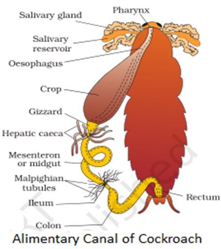
Draw a well labelled diagram of alimentary canal of a cockroach. Alimentary Canal Of Cockroach. Watch complete video answer for Draw a labelled diagram of alimentary canal of a co of Biology Class 11th. Well labelled diagram rhizome well labelled diagram of a cockroach diagram with well labelled virus well labelled diagram of the cervix in class 11 and class 12 this diagram is going to help you a lot it is well labelled diagram of cockroach of biology class 11 how to draw cockroach in easy way and color it simplest way of alimentary canal of.
Where is it found in human body. Q4- Draw a labelled diagram of alimentary canal of a cockroach. B What is peristaltic movement.
A cockroach answers com drawing and label cockroach charltonglaziers co uk drawing well labelled diagrams maple assumptions draw a well labelled diagram of cockroach science diagram of alimentary and digestive glands in cockroach class xi paper biology set b dissection of cockroach pdf download lekonachhu wixsite com alimentary canal of. Draw a well labelled diagram of alimentary canal of a cockroach. Click here to get an answer to your question Draw a well labelled diagram of alimentary canal in humans harjeetsingh8032 harjeetsingh8032 08102019 Biology Secondary School answered Draw a well labelled diagram of alimentary canal in humans 2.
I Liver ii Pancreas iii Small intestine iv Large intestine. It is the first body segment or buccal segement. A Draw a well labelled diagram of human alimentary canal and label the following parts.
Draw a labelled diagram of alimentary canal of a cockroach. How does it differ from adipose tissue. The digestion of food would take place in the cavities specialized or combined together.
Q1 Answer in one word or one line. It is the lobe which overhangs the mouth and is used as a wedge to force open cracks in the soil. Announcing Numerades 26M Series A led by IDG Capital.
Paurometabolous growth is a gradual metamorphosis. Cockroaches belong to the category of insects of the Blattodea order. Draw a labelled diagram of alimentary canal of a cockroach.
Draw a labelled diagram of the alimentary canal of a cockroach. What is meant by paurometabolous development in cockroach. Home NCERT Solution Class Biology Structural Organisation in Animals draw a labelled diagram of alimentary canal of a c.
Click hereto get an answer to your question Draw a labelled diagram of the alimentary canal of a cockroach. Draw a labelled diagram of the alimentary canal of a cockroach. Step by step video text image solution for Draw a labelled diagram of alimentary canal of a cockroach.
I Give the common name of Periplaneta americana. By Biology experts to help you in doubts. Draw a well labelled diagram showing the alimentary canal of cockroach and label any four parts.
Please scroll down to see the correct answer and solution guide. 325 OR Draw a well labelled diagram of areolar connective tissue and label any four parts. Get FREE solutions to all questions from chapter.
Around 4600 cockroaches are living aside human habitats. Draw a well -labelled diagram of the human alimantry canal and explain the process of digestion with the help of a flow chart - 27246661 vimalkumarsony1 vimalkumarsony1 31102020.
And the ovaries which. In biology class we got to discuss all these parts and how to identify them.
:max_bytes(150000):strip_icc()/male_reproductive_sys-5882446c5f9b58bdb398e5f3.jpg)
Male And Female Reproductive Systems
The human female reproductive system is a series of organs primarily located inside the body and around the pelvic region of a female that contribute towards the reproductive process.
:max_bytes(150000):strip_icc()/female_reproductive_sys-588244195f9b58bdb397fd62.jpg)
All parts of male and female reproductive system. They connect ovaries with the uterus. The uterus which hosts the developing fetus produces vaginal and uterine secretions and passes the anatomically male sperm through to the fallopian tubes. A pair of ovaries.
They each contain half the genetic information necessary for reproduction. Primary organs are the testes and the ovaries because they are responsible for the production of sperms ovum and reproductive hormones. The Human Reproductive Systems Introduction.
These external organs include the penis scrotum and testicles. The vulva which leads to the vagina the vaginal opening to the uterus. They are the site of fertilization.
It consists of the following parts. The sperm and the egg are gametes. Unlike the female reproductive system most of the male reproductive system is located outside of the body.
All parts of the reproductive system in either men or women have distinct roles to play and they can be grouped into primary and secondary organs. Do you think you understood the topics wholly. When sperm fertilizes meets an egg this fertilized egg is called a zygote ZYE-goat.
Take up the test below and find out for sure. 22 rows The male external genitalia include the penis the male urethra and the scrotum. The male reproductive system is mostly located outside of the body.
Internal organs include the vas deferens prostate and urethra. The parts of the human male reproductive system. And the ovaries which produce the anatomically female egg cells.
The human female reproductive system contains three main parts. When a sperm cell penetrates and fertilizes an egg that genetic information combines. The female reproductive system is active before during and after fertilization as well.
The uterus which holds the developing fetus. The female reproductive system contains two main parts. The human male reproductive system contains these parts.
The male reproductive system is responsible for sexual function as well as urination. Gametes the male and female sex cells are produced through meiosis in the ovaries and testes. The male gamete or sperm and the female gamete the egg or ovum meet in the females reproductive system.
In the human reproductive process two kinds of sex cells or gametes GAH-meetz are involved. The male and female reproduction system has all the components that are needed for reproduction to take place. Ovaries produce and store ovum in them.
They also produce a female hormone called estrogen. Here is my report on the human reproductive system it covers the structure and functions of the male and female reproductive systems the role hormones play during key developmental stages which include gametogenesis puberty pregnancy and the menopause. These external structures include the.
Be sure to be on the lookout for more tests that test you on the topic. The bladder empties into the urethra but is.
Parts of a Microscope 87. Can you name the different parts of an animal cell.
Animal Cell Part Labeling Quiz Questions Proprofs Quiz
To play this quiz please finish editing it.

Label cell parts game. Learn vocabulary terms and more with flashcards games and other study tools. This quiz is incomplete. Animal Speedsters by Continent 50.
Cell Parts ID Game. Drag the given words to the correct blanks to complete the labeling. Especially made for those needing to remember them for exams etc.
The names of animal cell parts can be hard to remember but you can use this quiz game to make it easy. Learning about animal cells can be tricky at first so why dont we start you off relatively easy with this animal cell part labelling quiz. Periodic Table of Elements 65.
5x5 Math Grid Puzzle 36. Identify and label figures in Turtle Diarys fun online game Animal Cell Labeling. This game involves labelling parts of animal cells.
You must choose a cell type. Tectonic Plate Boundaries 76. In this series of games your students will learn about the different organelles in the cell and the functions they perform.
Join group and play Just play. Identify and label figures in Turtle Diarys fun online game Human Body Parts Labeling. Name 10 in 30.
Animal Plant Fungus Bacterium. You need to be a group member to play the tournament. This is an online quiz called Label the plant cell game There is a printable worksheet available for download here so you can take the quiz with pen and paper.
Lets find out how many you get right. In both the animal and plant cells what is the letter B pointing to. Choose cell type s.
This game is part of a tournament. There is a printable worksheet available for download here so you can take the quiz with pen and paper. Test your knowledge on this science quiz and compare your score to others.
Centrioles the cytoplasm the rough and smooth endoplasmic reticulums the golgi complex lysosomes microfilaments mitochondria the nucleolus the nucleus the nuclear membrane pinocytotic vesicles the plasma membrane ribosomes and vacuoles. The Parts of the Cell learning objective based on NGSS and state standards delivers improved student engagement and academic performance in your classroom as demonstrated by research. The quiz above includes the following features of a typical eukaryotic cell.
The mitochondria are the cells powerplants combining chemicals from our food with oxygen to create energy for the cell. Standard Model Particles 55. Identify and label figures in Turtle Diarys fun online game Plant Cell Labeling.
Test your knowledge by identifying the parts of the cell. In this one well be giving you a question referring to a given diagram and asking you to label it. Take your knowledge of.
Play this game to review Cell Structure. This is an online quiz called This animal cell needs labelling. Drag the given words to the correct blanks to complete the labeling.
3 Letter Body Parts 42. Drag the given words to the correct blanks to complete the labeling. Your Skills Rank.
Start studying Label a cell Labeling parts of a cell Cells Structures and Functions. Peek inside the microscopic world of animal cells to learn about the endoplasmic reticulum the golgi apparatus and more. From the quiz author.
There are 11-12 base pairs per pitch. Helium is the region or the junction where ovule fuses with funicle.
Draw A Details Diagram Of Dna And Label It Properly Sarthaks Econnect Largest Online Education Community
Draw a neat and labelled diagram of a screw gauge.

Draw a neat labelled diagram of dna. Draw a neat labelled diagram of an antibody. To advance the screw by turning it till the object is gently held between the stud and the spindle of. Paternity disputes can be solved by DNA fingerprinting.
Now the helix geometry of A-DNA at a glance is. DNA fingerprinting is the technique developed by Alec Jeffreys it is used for comparing the DNA sequence of two individuals by identifying differences in the repetitive DNA sequences of the individuals. Draw a neat labelled diagram of the hair pin model of t-rna.
I In 1953 James Watson and Francis Crick proposed DNA structure based on X-ray crystallographic studies. 1 See answer aishucool8463 is waiting for your help. It is also known as B form of DNA.
The integuments are the protective envelopes for ovule that has a small opening known as. Below is the diagram of a screw gauge with all its parts labelled. Draw a neat labelled sketch of a replicating fork of DNA.
Iii Each strand consist of number of nucleiotides. Q-With a neat diagram explain the 7-celled 8-nucleate nature of the female gametophyte. Q-With a neat labelled diagram describe the parts of a typical angiosperm ovule.
This structure has two ends and three arms. It is made up of nitrogenous base deoxyribose sugar and phosphate. The main parts and their functions of a screw gauge are as follows Part Function.
Draw a neat labelled diagram of structure of an antibody. Asked Oct 22 2018 in Biology by Afreen 307k points molecular basis of inheritance. The angiosperm ovule is a small structure attached to the placenta by means of a stalk known as funicle.
The nitrogenous base deoxyribose sugar and phosphate link together forms the nucleotide chain. It is a non-impact printer. Ii DNA molecule consist of two long stands coiled around a common imaginary central axis to form a double helix.
Draw neat labelled constructional diagram of INKJET printer. The length of one pitch is 2815 A 0. Draw a neat and labelled diagram of antibody.
Name its main parts and state their functions. Add your answer and earn points. Applications of DNA Fingerprinting.
Give a brief account of oogenesis. The axial rise per base pair is 256 A 0. It was given by Watson and Crick model.
Draw neat and labelled diagram of Nucleosome. Lovepeople lovepeople The hairpin model of tRNA is also known as the cloverleaf model of tRNA because of its four arms. 200 000 Related Videos.
It look like a twisted ladder. Where and how does it take place. Describe the function of each block.
Q-Differentiate between a zoospore and a zygote. 3 Q24 Explain construction and working of Electric hot plates. Through this model one can easily understand the molecular structure of tRNA.
The central portion of the mitochondrion encloses the matrix. It provides letter quality printout than dot matrix printer. Draw a well labelled diagram of an antibody molecule.
Draw a neat labelled diagram of an antibody melocule. Draw a neat labelled diagram of a typical agarose gel electrophoresis. There is a tilting of base pairs from the axis of the helix of 202.
Draw a neat labelled diagram of a mature anatropous ovule before fertilization. 3 Q25 State the different types of lighting schemes and explain any one 3 Q26 Explain Sodium vapour lamp with neat labelled diagram. Maharashtra State Board HSC Science General 12th Board Exam.
DNA has a double-stranded helical structure. Name the nuclei involved in triple fusion. 2 Q21 Draw a neat labelled diagram of Kit Kat Fuse 3 Q22 Explain Direct method laying method of UG cables 3 Q23 Write a note on Neon sign tube.
000 300 135. Q-What is triple fusion. Its output is sharper than Dot matrix though its output quality is not to the same level of laser printer.
Evolutionary relation between the species. Draw a diagrammatic sketch of a portion of DNA segment to support your answer. Ii Z DNA or Left-Handed DNA.
Mitochondria called powerhouse of the cell are the organelles of cellular respiration. Is compatible in quality to that of a laser printer. The mitochondria are bounded by a double unit membrane space.
The outer membrane is smooth and the inner folded repeatedly into projections called cristae.
Im sure I made it for you guys in the first place but I cant for the life of me find it anywhere on the blog. Now onto the fun part where you can start creating the catcher itself.

Free Paper Fortune Teller Printable Templates Fortune Teller Paper Origami Fortune Teller Fortune Teller Game
Its time to fill in your cootie catcher.

How to label a cootie catcher. Flip the Cootie Catcher over so that the numbers are face up. They have different comprehension questions that can be. Typically there are three main sections of the cootie catcher that need to be labeled.
Download and print the template on a piece of paper. And since it resurfaced for me - I thought Id resurface it for you. Get a pen and write four words they dont have to be colors.
All you have to do is create your origami and add the desired fortune tellers to it. Alternatively they could be drawings of animals such as fish cat horse elephant. How to Make a Cootie Catcher - YouTube.
When you flatten out the cootie catcher inside tabs should be labeled on the outside with any number. To create a cootie catcher you will need a square paper of any size that you are comfortable with. How to Make a Cootie Catcher.
Usually the outside of the has a color. Normally there are 3 main sections that need to be labeled. You can use some of the examples above and have a great time with your partner friends your child or even yourself.
Place the square faceup and fold and unfold the square in diagonals from corner to corner so you end up with x shaped creases. With the numbers facing up insert fingers from both hands into the bottom of the cootie catcher and pinch it so the colored papers show. First pick a color.
Learn how to make a Cootie Catcher from scratch or with our printable template. Place the square facedown and fold each of the four corners in so the points meet in the center. 1-- Find a friend and ask him or her to pick a colour on top flaps 2-- Moving the flaps in and out and side to side in time with the lettersspellout the name of the colour 3--Stop on the last letter with the cootie catcher open to reveal the numbers ask your friend to pick a numberCount out the number and open the the flaps in and out and side to side like step 2When your done counting your first number get.
Follow the step by step instructions on the template to fold the cootie catcher together. Turn the paper over so the flaps are now facedown. Add Your Fortunes to the Cootie Catcher.
Fold it first then challenge your child to come up with labels for each of the sections. Its a reading comprehension cootie catcher that I TOTALLY forgot I made. Cut out your cootie catcher and color it.
Flip the paper over and write the numbers 1 through 8 on each of the sections. Open-ended creativity like this will challenge them to make choices and think sequentially as. Jul 7 2015 - Fill in and decorate this printable cootie catcher for fun interactive games with friends and family for different occasions or holidays.
On top of the flaps write a number. When you are holding the catcher each of the four corner tabs should be labeled with a color. Open out the flaps you drew the numbers on.
What you write depends on the type of Cootie catcher you have made. We filled our cootie catcher with markers and unfolded it to show you how it looked like. Pick one person to control the cootie catcher and another to make choices.
Finally on the outside flaps write the name of a color. Parts of the cootie catcher are labeled with colors or numbers that serve as options for a player to choose from. Bring the bottom left corner up to the top right corner and fold your paper the.
Playing cootie catcher is fairly easy. Open the close the cootie catcher to spell out the name of the color. Then open each flap and write a fortune or prediction.
Simple easy just takes a few trys so try it on your friends its fun. Have your cootie catcher flattened and with the outer flaps facing up. Using the perfect paper and stickers you can really transform this past-time into a festive activity.
Flip the Cootie Catcher over and write the name of a colour animal person or place on the flap. Use cootie catchers and fortune tellers at your next holiday party or gathering. You can change the texts and pictures accordingly.
Open each flap and write something on each triangle. When you flatten out the catcher the inside tabs should be labeled on the outside with a number. Theres actually 3 different versions.
Instead of writing colors on your cootie catcher use holiday stickers. Youve created your square piece of paper that will act as your catchers foundation. Unfold and reveal your cootie catchers square.
Master Cootie Catchers can use any piece of square paper to make their own. Free to download and print. When you are holding the cootie catcher each of the 4 corner tabs should be labeled with a color.
Lastly each of the inner tabs should be labeled with a fortunenote under a corresponding number to be read when opened up. This is the editable cootie catcher template for reading and speaking games.
Majority of the neurons occur in the brain. Human neural system has about 100 billion neurons.
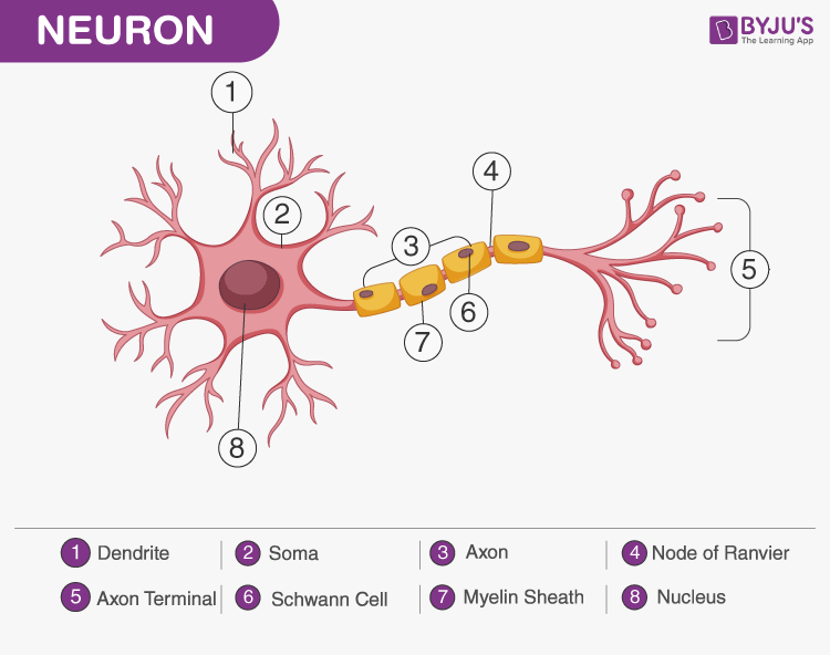
A Labelled Diagram Of Neuron With Detailed Explanations
Axons the cable of a neuron from the hippocampus are finely separated shown as the blue tracks and labelled with colourful toxins shown in green and red.

Labelled image of neuron. Jan 29 2018 - Explore Joni Seidensteins board Neurons followed by 144 people on Pinterest. Try these curated collections. Neurons also known as neurones and nerve cells are electrically excitable cells in the nervous system that process and transmit information.
Image by Dr Iris Wang in the Anggono lab who is exploring neural plasticity and how receptors for glutamate are controlled in communication between neurons. Thousands of new high-quality pictures added every day. A group of neurons forms a nerve.
Neurons are the structural and functional units of the nervous system. Drag and drop file orBrowse. Releasing of neurotransmitters through specific transport vesicles called synaptic vesicles from one neuron to the adjacent connected neurons called exocytosis.
2464 nerve cell diagram stock photos vectors and illustrations are available royalty-free. Certain neurons may almost equal the length of body itself. Illustration of anatomical peripheral organ - 61746125 Stock Photos.
VectorLabeled diagram of the neuron nerve cell that is the main part of the nervous system. Sensory or afferent carrying toward the brain neurons are specialized to be sensitive to a particular physical stimulation such as light vision sound audition chemical olfaction or pressure touch. See nerve cell diagram stock video clips.
In humans there are two types of neurons. Search 123RF with an image instead of text. A neuron is also known as the nerve cell.
See more ideas about neurons neuroscience brain art. Neuron Diagram Diagram Of A Neuron How To Draw A Neuron Step By Step For Beginners - YouTube. Illustration about Labeled diagram of the neuron nerve cell that is the main part of the nervous system.
The structure of a neuron varies with their shape and size and it mainly depends upon their functions and their location. Find Motor neuron detailed and accurate labeled Stock Vectors and millions of other royalty-free stock photos illustrations and vectors in the Shutterstock. Thus neurons with longer processes projections are the longest cells in the body.
It is formed between the axon terminal of one neuron and the dendrites of the next neurons. Axon and dendrites neuron myelin cell education neural cells nervous system diagram neuron flat vector neuron infographic the neuron nerv cell structure of neuron. Neurons are distinguished and categorized according to general function.
There are receptor or sensory neurons motor neurons and interneurons. Find Motor Neuron Detailed Accurate Labeled stock images in HD and millions of other royalty-free stock photos illustrations and vectors in the Shutterstock collection. To appreciate the enhanced image quality produced by the new technique the team first examined a conventional bright-field image of a metal-labelled neuron within a grasshoppers abdominal ganglion a type of mini-brain which even at that size presented out-of-focus structures.
Draw a labelled diagram of nerve cell neuron. Labelled Diagram of a Nerve Cell. 2D labelled diagram - Nerve cell.
Neurons are the structural and functional units of the nervous system. Try dragging an image to the search box. Synapse or synaptic cleft is the small space or gap between two adjacent neurons.
Neuron Diagram Diagram Of A Neuron How To Draw A Neuron Step By Step For Beginners. The Structure and Function of Sensory Relay and Motor Neurons The nervous system is composed of specialised cells called neurons. In vertebrate animals neurons are the core components of the brain spinal cord and peripheral nerves.
Complete neuron cell diagram. A neuron is a structural and functional unit of the neural tissue and hence the neural system.
While mealworm beetles are significantly easier to raise the Darkling Beetle is larger lives longer and looks quite nice. Once the eggs start to hatch after staying in the media for approx 3-4 weeks you can remove the beetles and put them in a new container with fresh media and start again.

Mealworm Beetle Stock Illustrations 16 Mealworm Beetle Stock Illustrations Vectors Clipart Dreamstime
Dry Food Mealworm Substrate Both beetles and mealworms need a mealworm substrate.

How to draw a mealworm beetle. A darkling beetle experiences complete metamorphosis which means that it has four distinct stages of life. Ideally separate mealworms pupae and beetles from the substrate and throw it away in an outside dumpster dont throw it in the kitchen trash can. Replace the substrate and return the mealworms pupae and beetles.
The adult beetles fly and are attracted to light. These eggs have a sticky exterior which helps to hide them as they become covered in dust and debris. Mealworms are the brown worm-like larvae of darkling beetles.
Although the grub looks a bit like a worm the mealworm has six small jointed legs. Mealworm to Darkling Beetle in under two minutes. Beetles will eat the substrate and use it as a substrate to lay their eggs in.
The four stages are egg larva pupa and adult. It is the larval stage grub of the yellow mealworm beetle also called the darkling beetle Tenebrio molitor. Throw out the old mealworm mixture and do not use any of it to start the new colony even if it means throwing out potentially viable eggs.
Every few weeks move both the pupa and. A mealworm substrate can be made from ground up cereals or meals grain and seeds. Vacuum the floor and surfaces around the mealworm farm.
Mealworms however will eat the substrate and use it as a bedding material. First the male beetle chases the female beetle till she gives in. Rotating Mealworms with These Easy Steps.
Their DIY instructions include an 8-inch x 11-inch tub that is around 6 inches deep. Stage I The Egg. Move any mealwormspupabeetles to a new clean container and start again.
Pick out the pupas and place them into the egg cartons of Container 2. The amount of time a darkling beetle spends in each stage can vary greatly due to environmental factors like temperature humidity food and water. From there they explain how to create your mealworm nest with fine wire.
Egg The first stage of life is spent as an egg. The mealworms will begin eating the substrate and reproducing. Clean the old container with soap and hot water or natural disinfectant.
The mealworm is NOT a worm. They are the second of four stages of life and exist to eat and grow until they have enough energy stored to begin transformation into pupae and then beetles. Put the lid on top of the bin.
It is worth mentioning that there are many species of Darkling Beetle like 20000 but the one that is sold in pet stores is usually Zophobas morio which is a tropical species native to Central and South America. Both the larva and the beetle are nocturnal active at. Slice up an apple a carrot or a potato and place the slices on top of the substrate to provide moisture for the mealworms.
Place your starter stock in the bin. The mealworm goes through a huge metamorphosis during its life cycle transforming from a tiny egg to a full-grown adult beetle. Again turn the pack into a separate breeding tray.
The beetles will crawl out of the egg cartons into the mealworm substrate below and lay their eggs. Facts about mealworm beetles The female mealworm beetle lays up to 500 eggs often in batches over a period of weeks. May also draw your interests on true spiders very cool to keep.
This will be where your beetles will continue to breed and lay eggs amongst the media. Wipe down containers and surfaces with hot soapy water. Mealworm beetles Tenebrio molitor are widespread throughout Britain.
Mealworms can be found throughout most of. Development in mealworm beetles occurs in three stages. I always feed off the excess mealworms etc off to native spiders most can handle prey alot larger than themselves Tegenaria Steatoda Amaurobius genus are all more than happy to tackle prey larger than themselves over there you also have Parasteatoda sp etc.
Mealworm to Darkling Beetle in under two minutes - YouTube. Place your mealworms into the first container Containers 1 until the pupas appear. The first stage is the laying of the eggs.
Homestead describes mealworms as buttery oaty toasty almost breakfast cereal-y.