A long tube that connects to the ovary. Our customer service team will review your report and will be in touch.
Parts Of A Plant And Their Function Teaching Resources
Click Share to make it public.

Label the parts of a plant ks2. The carpel is the part of the plant that collects the pollen and produces seeds for reproduction. Test what youve learned with the quiz below. Thanks Empty reply does not make any sense for the end user.
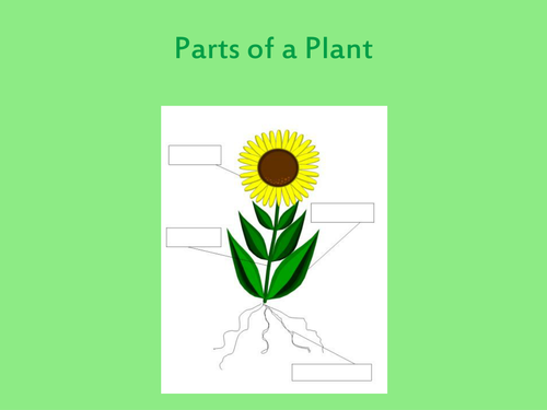
Aimed at primary level this resource introduces children to the structure and function of the parts of a flowering plant. With these worksheets your children will have to label the different parts of a flower developing their understanding of plants and reproductionOnce downloaded youll have two worksheets. The stigma style ovary and ovule.
The stigma is supported by the style. These resources are suitable for teaching pupils aged 5-11. This will allow your children to.
This activity pack designed for 5 to 12 year olds can be used to encourage children to care for and appreciate living things and to think about the world around them. Plant food can be purchased say from a garden centre and added to the soil to feed plants. Inside the ovary are seeds known as ovules.
This leaderboard is currently private. Learn about the parts of a plant - the roots the stem the leaves and the petals - with this video. This website and its content is subject to our Terms and Conditions.
Something went wrong please try again later. Diagram of flower- pupils label parts of flower accompanies my ppt also listed on flower parts. Plants and Life on.
These illustrated labelling worksheets should be a great help. Curriculum links Parts of a plant National Curriculum Sc2 KS1. This is not enough for KS3 Level.
Show more Show less. A series of films looking at plants and their various functions including what they need to survive. This is a powerpoint to use when teaching children the names and functions of the different parts of the plant.
There are also flash cards for your lower and middle ability children. The carpel has four parts. This leaderboard has been disabled by the resource owner.
The other is more basic and includes simpler labels. Creative Commons Sharealike Reviews. This is OK for level KS2 at the end of primary school but after when a child needs to start learning things in a much more detailed manner working towards GCSE level.
Ppt 700 KB. It looks at seed dispersal germination growth parts of a plant and more. JavaScript is required to view this activity.
Learn about what the root stem leaf and flower does for a plant. Lower ability children to assign the descriptions to the different parts of the plant. Differentiated worksheet to use on the first lesson describing parts of a plant.
Why not split the class up into 2 groups with one group drawing their own plants and the other labelling the features of the plants. Something went wrong please try again later. Tes Global Ltd is registered in England Company No 02017289 with its registered office.
One focuses on the flower and features 10 labels. This is mainly mineral salts They should understand that in science we link food primarily with it being a source of energy. This beautifully illustrated KS1 worksheet covers the parts of a plant tailored just for the foundation stage and Year 1 and 2 childrenEither use the pre-labelled version as a poster or teaching aid or encourage your children to label the plant part diagrams to reinforce the associated vocabulary.
Parts of a plant and their function. Y4 Label parts of a plant. This activity gives students the.
At the top of the carpel the stigma catches and collects pollen. Report this resource to let us know if it violates our terms and conditions. Seeds and Plant Growth Discovery Pack.
Primary Science - Plants. A great way of teaching features of plants is by using illustrations - this resource is perfect as it asks students to label a drawing with the correct parts then offers them the chance to draw their own plant and label it. There are some important components of a plant explained missing.
Many cards such as matching pairs loop cards taboo splat bingo are also included to identify any. A PowerPoint presentation to teach children how to label a plant and identify the function of each part of a plant. Empty reply does not.
A range of activities are provided which help to familiarise learners with the basic parts of a plant including growing plants from seed and making models of a plant. Creative Commons Sharealike Reviews.
This framework consists of many individual bones and cartilages. Diagram Of Leg Bones 12 photos of the Diagram Of Leg Bones diagram and names of leg bones diagram of foot and leg bones diagram of leg bones diagram of lower leg bones diagram of the bones in your leg Bone diagram and names of leg bones diagram of foot and leg bones diagram of leg bones diagram of lower leg bones diagram of the bones in your leg.
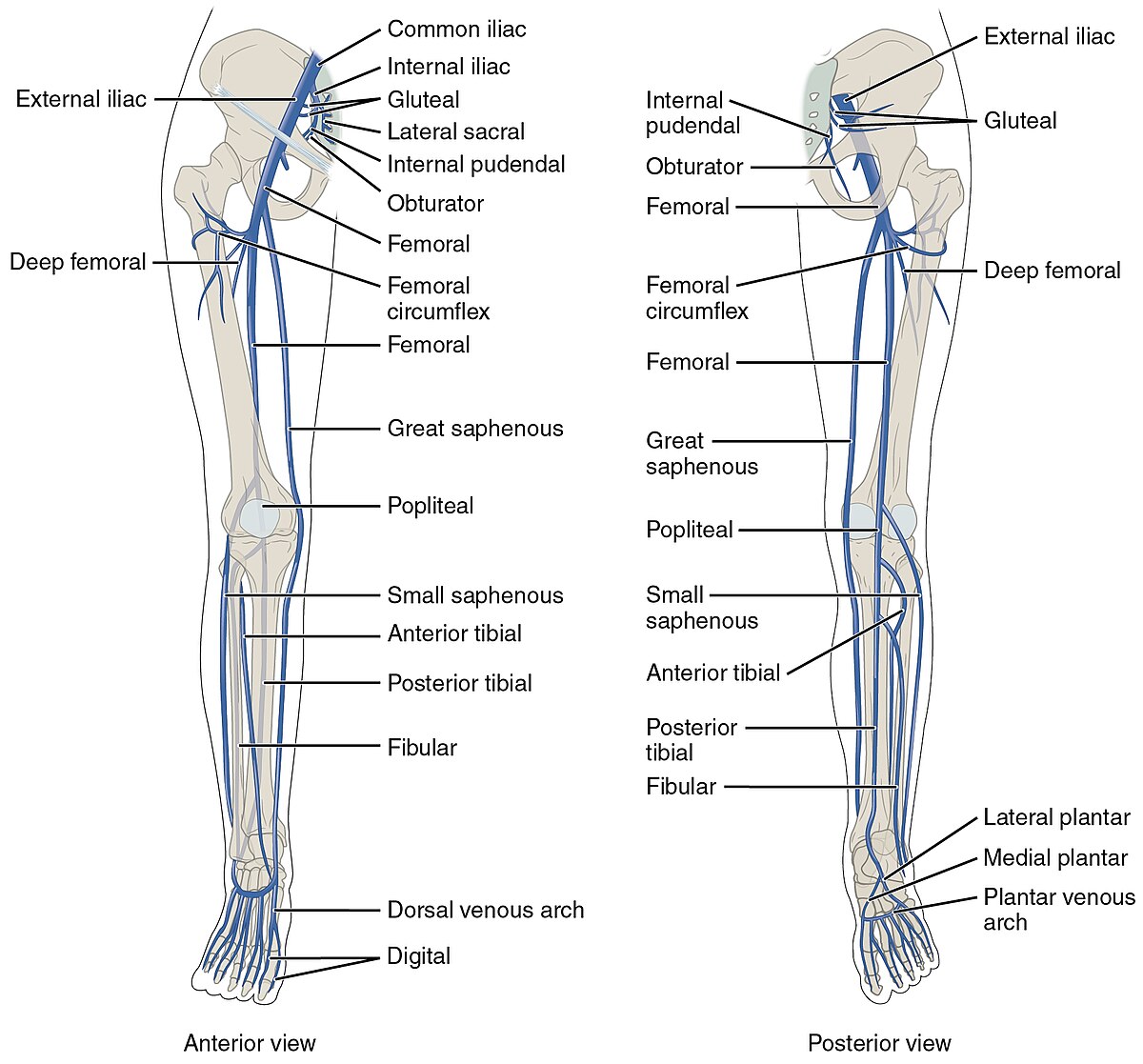
File 2136ab Lower Limb Veins Anterior Posterior Jpg Wikimedia Commons
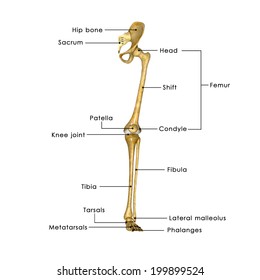
Tibia is the one on the left side and is thicker with tibial tuberosity towards the knee end and medial malleolus connecting to the tallus.
Labelled diagram of the leg. Innominate bones are evolutionary significant in that they allow birds to lay eggs. They meet at the acetabulum hip socket and articulate with the femur which is the first bone of the hind limb. To understand one of the most complex joints of our body ie.
Health diagram bone skeleton leg knee science anchor chart human human body. In the given diagram f the human hindlimb or leg the bones Fibula and Tibia are wrongly labelled. The greater trochanter is a large prominent section of the femur located on the outside of the thigh.
It also provides support to both the longitudinal and transverse arches of the foot. Any disorder or defect in the knee bone can restrict the activities of the leg which can directly affect our locomotion. Drag and drop the pins to their correct place on the image.
Joints hold the skeleton together and support movement. Spend some time revising this diagram by connecting the name and location of each structure with what youve just learned in the video. A activity diagram used in uml 69 and sysml b bachman diagram.
You will find upper leg bone diagram labeled at least the following types of diagram labeled. The upper leg consists of the femur. Below given knee diagram will help you to understand.
The Fibula connects to the tarsals at the lateral malleolus. This diagram shows the bones of the femur and the patella. The greater trochanter is also the widest part of the lower legs.
The aim of this exercise is to improve your confidence in identifying different structures. We have collected the best muscle diagram including arm facial neck Quadriceps and shoulder muscle diagrams. The upper leg is from hip to knee.
On the outside of the thigh this is the largest of. A detailed diagram of the human skeleton with space for children to label each of the major bones. This will help you to understand the mechanism as well as the working.
The greater trochanter is where the tendons of several different muscles attach to the hip. Leg Bones Diagram Labeled. At the knee joint the femur connects to the tibiotarsus shin and fibula side of lower leg.
Sciatic Nerve Also known as the ischiatic nerve the sciatic nerve is a nerve fiber that begins in the lower back and ends in the lower limb. It supplies the skin of the leg and the muscles of the leg foot and back of the thigh. Leg muscles labeled.
Labeled human leg bones created for use in leg bone. Identify the structures labelled A B C D and E in this digital subtraction angiogram of the right lower leg. However the definition in human anatomy refers only to the section of the lower limb extending from the knee to the ankle also known as the crus 3 4 5 or especially in non-technical use the shank.
We use our knee joints when we sit fold legs run walk or do any kind of leg movement. It supplies cutaneous branches to the skin of the leg and foot in the region between the knee and the ankle. The knee joint you need a perfectly labeled diagram of the knee.
Help yourself in studying the anatomy of the body muscles with this set of free and printable Human Muscles Diagrams Labeled 2019. Muscles of the leg. These four muscles at the front of the thigh are the major extensors help to extend the leg straight of the knee.
Take a look at the leg muscles diagram below where you see each muscle clearly labeled. The function of the fibularis longus muscle is to plantar flex and evert the foot. Skeletal System Diagrams Skeletal System Anatomy Human Body Anatomy Anatomy Bones The bones of your leg have roughened patches on their surfaces where muscles are attached.
The human leg in the general word sense is the entire lower limb of the human body including the foot thigh and even the hip or gluteal region. Leg muscle anatomy front view. Start with the fibular muscles of the leg by exploring our videos quizzes labelled diagrams and articles.
These muscles include piriformis the Gemelli the obturator and the gluteus.
Dissection of Air Sacs 3. The feathers of pigeon may be classified into two groups.

Columba Livia Pigeon Faunafondness
Discover and save your own Pins on Pinterest.

Labelled diagram of pigeon. In order to get energy pigeon eats large quantity of food and to break down the assimilated food at a faster rate the respiratory system is extensively modified. All of these body divisions are invested in a close covering of feathers which are directed backward and overlapping one another. In this article we will discuss about the respiratory system of pigeon.
The Arterial System 7. The birds head is one of the best places to look for field marks such as eye color malar stripes eyebrows eye rings eye lines and auricular patches. These feathers are associated with flight and may again be subdivided into wing feathers or remiges and tail feathers or rectrices.
Diagram For Pigeon And The Labelled Parts Free Books DOWNLOAD BOOKS Diagram For Pigeon And The Labelled Parts PDF Book is the book you are looking for by download PDF Diagram For Pigeon And The Labelled Parts book you are also motivated to search from other sources Världsalliansen För Patientsäkerhet World Alliance For. Printout Label the birds external anatomy on this printout. Aug 30 2013 El Esqueleto de la Paloma Pigeon Skeleton diagram.
Also learn about- 1. Parts of a Pigeon. If playback doesnt begin shortly try restarting.
Nov 4 2013 - Everything you ever wanted to know about pigeons from the Pigeon Control Resource Centre PCRC Nov 4 2013 - Everything you ever wanted to know about pigeons from the Pigeon Control Resource Centre PCRC Explore. Sep 10 2016 - This Pin was discovered by lily kuo. Dissection of Circulatory System 5.
Posted by Unknown Posted on 1606 with No comments Latin Name. Label Diagram Structure Of Pigeon Pea bgcse biology year 2016 s3 us west 2 amazonaws com diagram for pigeon and the labelled parts bgcse biology past paper 2002 s3 us west 2 amazonaws com free download here pdfsdocuments2 com pdf genetic diversity and population structure of pigeon pea handbook of start pulses dall mill unit food processing pigeon pea wikipedia major families lab. The remiges are present in the wings and each wing has 23 feathers which are again sub-divided into primaries secondaries and tertiaries.
Pigeon dove blue rock pigeon rock dove wild rock pigeon rock pigeon feral pigeon. The physiology of digestion in pigeon involves the three following steps. Authorstream diagram for pigeon and the labelled parts handbook of start pulses dall mill unit food processing bgcse biology year 2016 s3 us west 2 amazonaws com growing pigeon peas an incredibly versatile permaculture digestive system of pigeon with diagram zoology drawing seed storage structures tnau agritech portalthe first written reference of the word cowpea appeared in 1798 in the.
2628 is long tubular and coiled. Facts labelled Body Diagram. Unlike the mammalian eye it is not spherical and the flatter shape enables more of its visual field to be in focus.
Bird Skeleton See the bone structure of a typical bird. Click Share to make it public. The Venous System 6.
Dissection of Urinogenital System 9. In this article we will discuss about the dissection of pigeon. The digestive system of pigeon is well developed and includes an alimentary canal and the digestive glands.
Pigeon is a grain or seed eater. It picks up grains very rapidly which are quickly swallowed. Dissection of Cranial Nerves 8.
The lungs are proportionately smaller in size but the functional efficiency is greatly increased by the development of air-sacs. The head is small rounded and mobile. Share Share by Csilcsal78.
Theye are present within. How to draw Pigeon labelled diagram phylum chordataClass Aves Kingdom Animalia. Want a labelled structure of the diagram for pigeon and the labelled parts handbook of start pulses dall mill unit food processing value chains analysis for pigeon peas in southern malawi pod fly in pigeon pea plantwise pigeon pea wikipedia what are pulses pulses rcsb pdb 4g7e crystal structure of pigeon pea 1 11.
The alimentary canal of pigeon Fig. The shape of the beak is modified in such a way that it helps in its ingestion. 2015 Oldsmobile Bravada Repair Manual 2015 Oldsmobile Bravada Repair Manual PDF Download Free.
Facts labelled Body Diagram. Urease seed storage structures tnau agritech portal feed the future. Dissection of Alimentary System 4.
This leaderboard is currently private. Mourning Dove Color Diagram Mourning Dove Animal Paintings I Like Birds. Labeled anatomy of a pigeon.
Show more Show less. This leaderboard has been disabled by the resource owner. The aerial mode of life requires extra energy.
The swallowed food being moistened by mucus is stored in the crop. Columba livia dove or. Dissection of Muscles of Flight 2.
All the segments of alimentary canal fall into following three. The bilaterally symmetrical and compactly set body of pigeon is divisible into four regions- head neck trunk and tail. It comprises mouth buccal cavity pharynx oesophagus stomach small intestine and large intestine which opens to the exterior by cloacal aperture.
Citing for websites is different from citing from books magazines and periodicals. A Flight Feathers or Quills. Bruksanvisning Säkerhet Vid Operationer.
Switch template Interactives Show all. Columba livia dove or bird of leaden or blue-grey colour.
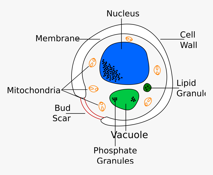
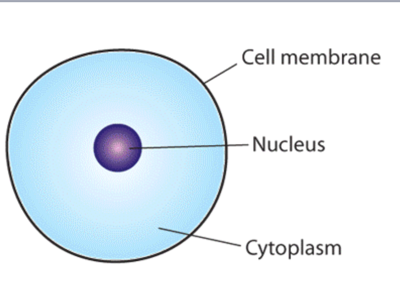
Plastids are present only in plant cells. Here lets study the plant cell.
Yeast Drawing Easy Labelled Diagram Of A Yeast Cell Hd Png Download Kindpng
It controls all the metabolic activities of a cell and regulates the cell cycle.
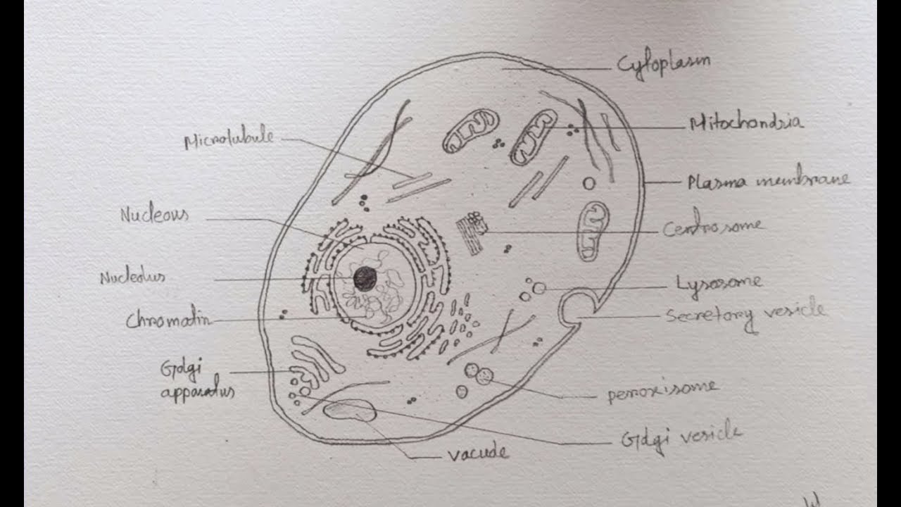
Draw a well labelled diagram of simple cell. Hello friendsIn this video I will be showing you that how to draw A plant cell very easilyPlease like share and subscribe. There are various organelles present within the cell and are classified into three categories based on the presence or absence of membrane. Following are the main cell organelles.
Draw a well labelled diagram of a cell - 32722671 pradhanaadi052 pradhanaadi052 06012021 Hindi Secondary School answered Draw a well labelled diagram of a cell 2 See answers. Plastids store different types of pigments. The important features of the plant cell which play role in photosynthesis are.
The diagram given below represents a plant cell after being placed in a strong sugar solution. Draw a Well Labelled Diagram of Photoelectric Cell. Click here to get an answer to your question Draw a well labelled diagram of a typical cell.
Draw a well labelled diagram of photoelectric cell. Namitakumari840955 namitakumari840955 26042020 Biology Secondary School answered Draw a well labelled diagram of a typical cell. Types of plastids are.
We are aware that all life stems from a single cell and that the cell is the most basic unit of all living organisms. A Labeled Diagram of the Plant Cell and Functions of its Organelles. Maharashtra State Board HSC Science Computer Science 12th Board Exam.
These store green pigment called chlorophyll. They provide skeletal framework to the cell and are concerned with the production of lipids and proteins. The cell being the smallest unit of life is akin to a tiny room which houses several organs.
Click here to get an answer to your question draw a well labelled diagram of a cell ekamjeetvirk6 ekamjeetvirk6 09102020 Biology Secondary School answered Draw a well labelled diagram of a cell. Listed below are the Cell Organelles of an animal cell along with their functions. Diagram of Plant Cell.
Advertisement Remove all ads. State any two features of the above plant cell which is not present in animal cells. Personalized AI Tutor and Adaptive Time TableSelf Study MaterialUnlimited Mock Tests and Personalized Analysis Reports24x7 Doubt Chat Support.
Study the diagram and answer the questions that follow. And do tell me on. They play an important role in protein synthesis.
NEET Foundation Knockout NEET 2025 Easy Installment Personalized AI Tutor and Adaptive Time TableSelf Study MaterialUnlimited Mock Tests and Personalized Analysis. The Cell Organelles are membrane-bound present within the cells. Draw a well labelled diagram of phloem.
Well-Labelled Diagram of Animal Cell. Click hereto get an answer to your question Draw a well - labelled diagram of a plant cell.
Thousands of new high-quality pictures added every day. 762 600 pixels.

Simple Human Heart Drawing With Labels Novocom Top
Answered Draw labelled diagram of Heart.

Labelled diagram of heart in hindi. Labeled diagram of heart anatomy body system. Learn about pacemaker cells and cardiac ac. Information from its description page there is shown below.
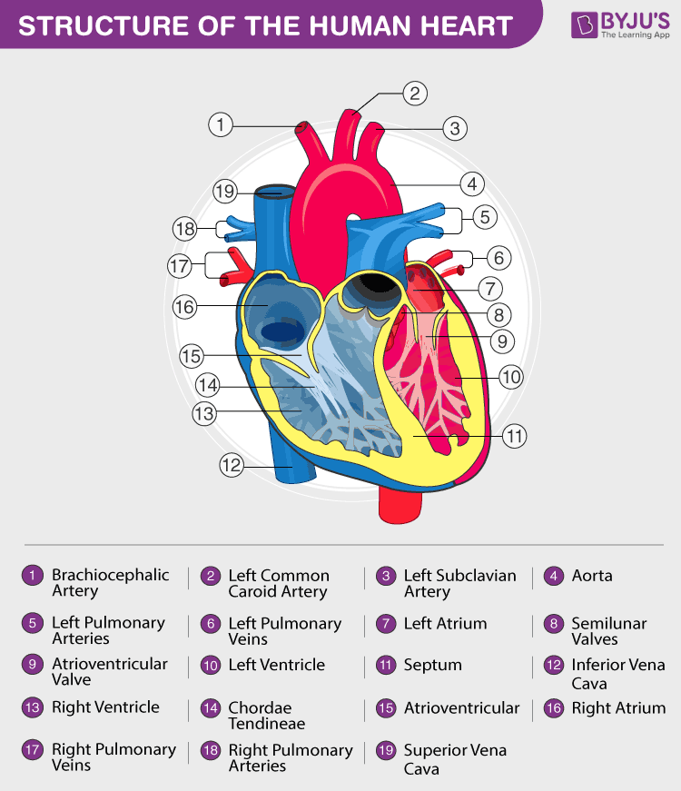
Add aorta in bottom. The right ventricle is the major pumping chamber on the lower right side of the heart that ejects blood into the pulmonary circuit via. The middle layer of the heart wall is called myocardium.
This is a file from the Wikimedia Commons. Each side of the heart consists of an atrium and. Left main pulmonary artery with its first division.
Diagram Of Heart Labelled - Fun for my own blog on this occasion I will explain to you in connection with Diagram Of Heart LabelledSo if you want to get great shots related to Diagram Of Heart Labelled just click on the save icon to save the photo to your computerThey are ready to download if you like and want to have them click save logo in the post and it will download directly to. The outer layer of the heart wall is called epicardium. 0702 2 June 2006.
Labelled diagram of the heart The Human. Brachiocephalic trunk more wide and separated. 650 650 26 KB Yaddah.
305 240 pixels 610 480 pixels 976 768 pixels 1280 1008 pixels 2560 2015 pixels 893 703 pixels. Add source veins of superior vena cava. The heart is a muscular organ in most animals which pumps blood through the blood vessels of the circulatory system.
The upper two chambers of the heart are called auricles. Each time blood goes around your body it goes through the heart twice double circulation. Label the heart anatomy diagram below us.
Labelled heart diagram gcse pe. Draw labelled diagram of Heartirculation of blood in human Get the answers you need now. The cardiac conduction system is the electrical pathway of the heart that includes in order the SA node AV node bundle of His bundle branches and Purkinje fibers.
The inner layer of the heart wall is called endocardium. The lower two chambers of the heart are called ventricles. Easily learn the conduction system of the heart using this step-by-step labeled diagram.
This basic text on the heart and heart diseases is geared to everyone on the cardiovascular care team including emergency personnel interns residents nurses patients and families. Share It On Facebook Twitter Email. Simranojha659 simranojha659 28082020 Biology Secondary School 5 pts.
On average the heart beats about 100000 times a day ie around 3 billion beats in a lifetime. Diagram of the human heart created by Wapcaplet in Sodipodi. Draw labelled diagram of heart in cockroach and explain its physiology.
Answered Jun 12 2020 by RajeshKumar 507k points selected Jun 12 2020 by. The average male heart weighs around 280 to. Size of this PNG preview of this SVG file.
The heart is situated at the centre of the chest and points slightly towards the left. The heart wall is made up of three layers. The heart is made up of two chambers.
In humans the heart is approximately the size of a closed fist and is located between the lungs in the middle compartment of the chest. View Heart Diagram In Hindi Pictures. Science 27082020 1107 meandart Explain the working of human heart with labelled diagram.
A Basic Guide to Heart Disease. One of the primary pumping chambers of the heart located in the lower portion of the heart. Inferior vena cava more wide.
Find diagram of the human heart stock images in HD and millions of other royalty-free stock photos illustrations and vectors in the Shutterstock collection. The pumped blood carries oxygen and nutrients to the body while carrying metabolic waste such as carbon dioxide to the lungs. The left ventricle is the major pumping chamber on the lower left side of the heart that ejects blood into the systemic circuit via the aorta and receives blood from the left atrium.
Hello friends todays video i have described verious parts of heart in hindi which is help you to understand structure of heart or. 1 Answer 1 vote. Well-Labelled Diagram of Heart.
Science 16112019 1015 sabina3193 Draw labeled diagram of heart and explain its functions. Fine arts guruji 1036424 views1 year ago. The heart is a muscular pump.
The heart pumps around 57 litres of blood in a day throughout the body.
A Draw a neat diagram of a flower showing its various parts. The stamens function is to produce male reproductive cells.
Draw A Neat Diagram Of A Flower Showing Its Various Parts Studyrankersonline
When pollinating insects such as bees and butterflies go to the flower for pollen they also visit the stigma.
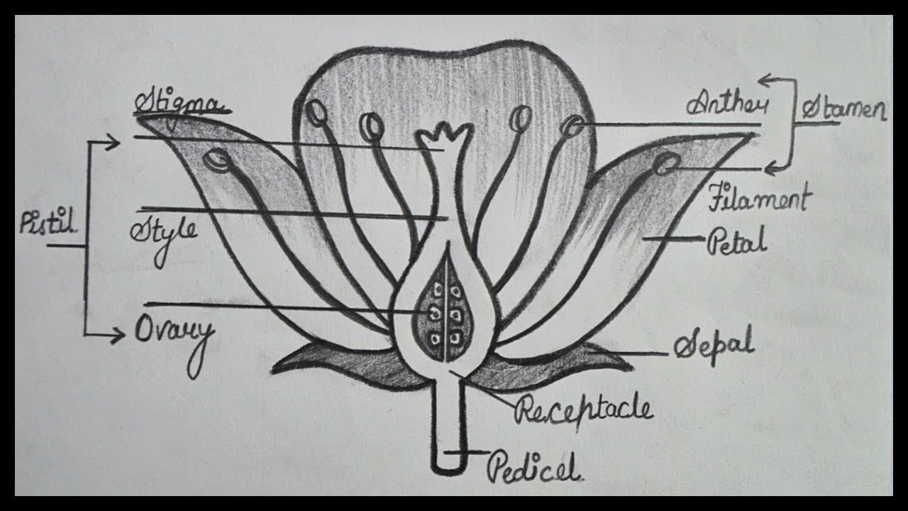
Draw a labelled diagram of the flower showing its various parts including the reproductive parts. The presence of these parts differentiates the flower into complete or incomplete. Calyx is the outermost whorl. FilamentThe filament is the stalk attached to the flower that holds the anther.
The female part is the pistil. In different plants the number of petals sepals stamens and pistils can vary. B What name is given to i all the petals of a flower and ii all the sepals of a flower.
Draw a diagram showing longitudinal section of a flower and label on it. Most flowers have four main parts. Collect three or more different types of flowers for learners to observe and dissect.
In this diagram mark stem receptacle sepals petals stamen and carpel. D What is the other name of carpel of a flower. The male part of a flower is the stamen.
Sepals petals stamens and carpels. It helps in ual reproduction as it has male parts and female parts. Parts of a Flower Flower Anatomy Including a Flower Diagram.
A Draw a neat diagram of a flower showing its various parts. D What is the other name of carpel of a flower. C What are i stamen and ii carpel in a flower.
Most flowers have both the male and female reproductive organs but some bear either the male or the female sex organs. The Structure and Functions of Flowers. The whorls are arranged on the thalamus of a flower in a definite sequence.
The androecium is the male part of the flower and consists of stamens. Pyy92 is waiting for your help. Reproductive Parts of a Flower.
It is the stalk of a flower. Avail 25 off on study pack. In this diagram mark stem recetacle sepals petals stamen and carpel.
The anther is the head of the stamen. The gynoecium or pistil consists of carpels and is the female reproductive part. Most flowers have four main.
They are not only involved in reproduction but are also a source of food for other living organisms. Flowers are the reproductive part of a plant. Flowers are the reproductive part of most plants which download a powerpoint showing labelled and unlabelled versions of these diagrams both parts of a plant and parts of a flower from the link on the right.
AskedMar 31 2019in Biologyby Farrah695kpoints how do organisms reproduce. Learners observe and dissect a flower to discover its anatomy and the how each part contributes to its reproduction. Simple flowers with easily identifiable parts such as.
The female part of a flower is called pistil and it is composed of parts. Draw a Neat Labelled Diagram Showing the LS. A flower has female and male parts.
Others may contain one of the two parts. C What are i stamen and ii carpel in a flower. The function of flower is to produce.
Androecium and gynoecium are directly concerned with sexual reproduction. The stamens are the male part whereas the carpels are the female part of the flower. Flowers contain the plants reproductive structures.
They are a rich source of nectar. The female part of the flower the pistil is located at the center of the bloom. A flower is a reproductive part of a plant.
Most flowers are hermaphrodite where they contain both male and female parts. Flowers attach to the plant via the stalk. Label the following on ita Ovaryb Antherc Filamentd Stigma.
Stigma ovary anther filament. Flowers are the parts of plants that give them beauty scent and they function as the plants reproductive system. Add your answer and earn points.
Click hereto get an answer to your question Draw the diagram of a flower to show it male and female reproductive parts. The different part of a flower is labelled below. Draw a labelled diagram of the longitudinal section of a flower.
Apart from these parts a flower includes reproductive parts stamen and pistil. Download a powerpoint showing labelled and unlabelled versions of these diagrams both parts of a plant and parts of a flower from the link on the right. B What name is given to i all the petals of a flower and ii all the sepals of a flower.
Draw a well labelled diagram of a flower showing its various parts including reproductive parts.
Because these muscles are. Posterior shoulder muscle diagram home wiring diagrams.

Anatomy Of The Shoulder Joint Stock Vector Illustration Of Health Medicale 124233793
91 Inferior dislocation of shoulder joint is common 92 Following inferior dislocation of shoulder joint the rounded contour of shoulder is lost and there is weakness of abduction of arm.
Labelled diagram of the shoulder joint. Sechrest md narrates an animated tutorial shoulder muscles anatomy actions diagram ehealthstar. Knee Joint Model Labeled Fein Posterior. Diagram of shoulder anatomy showing the acromioclavicular ac articulation and glenohumeral a healthy shoulder allows a wide range of motion that encompasses activities of everyday living as well.
It involves articulation between the glenoid cavity of the scapula and the head of the humerus. Atlas of the anatomy of the joint of the shoulder on a ct. Find high-quality stock photos that you wont find anywhere else.
These two joints work together to allow the arm both to circumduct in a large circle and to rotate around its axis at the shoulder. Search from Shoulder Joint Diagram stock photos pictures and royalty-free images from iStock. Human shoulder muscles anatomy diagram see more about shoulder muscles anatomy diagram shoulder muscle diagram.
Schematic Representation Of Proteomic Analysis Of Shoulder Joint. Human anatomy diagrams show internal organs cells systems. Shoulder Diagram Labeled Ditulis JupiterZ Minggu 09 Desember 2018 Tulis Komentar Edit.
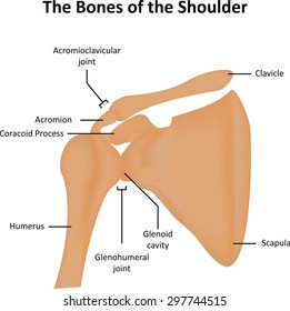
The shoulder joint is the connection between the chest and the upper extremity. Where the rounded top of the arm bone humerus contacts the shoulder blade is called the glenohumeral joint. This is called the acromioclavicular joint.
Shoulder Joint Diagram Labeled Ditulis JupiterZ Sabtu 28 Juli 2018 Tulis Komentar Edit. Download 708 shoulder diagram stock illustrations vectors clipart for free or amazingly low rates. Shoulder joint diagram labeled shoulder muscle diagram labeled.
Shoulder Joint Diagram Labeled Written By JupiterZ Saturday July 28 2018 Add Comment Edit. Clavicle Fracture With Broken Collarbone Vector Illustration. It stabilizes the shoulder and holds the head of the humerus into the glenoid cavity to maintain the principal shoulder joint.
Three bones come together at the shoulder joint. The wiring diagram of shoulder you can easily download working with the online world. The most flexible joint in the entire human body our shoulder joint is formed by the union of the humerus the scapula or shoulder blade and the clavicle or collarbone.
7 draw labelled diagram showing the relations of. 7 Draw labelled diagram showing the relations of shoulder joint. The shoulder joint is structurally classified as a synovial ball and socket joint and functionally as a diarthrosis and multiaxial joint.
Find high-quality royalty-free vector images that you wont find anywhere else. The meeting of the scapula and clavicle forms it. This joint forms the highest point of the shoulder and provides the ability to raise the arm above the head.
The transverse humeral ligament is not shown on this diagram. Dips And Shoulder Pain Rosstraining Com. Zygote body is a free online 3d anatomy atlas.
Tendons to attach the muscles to the bones. 9 4 Synovial Joints Anatomy And Physiology. Clavicle Fracture With Broken Collarbone Vector Illustration.
The Lymphatic System Labeled Eps10 Clipart K10294165 Fotosearch. 9 4 Synovial Joints Anatomy And Physiology. Diagram of shoulder anatomy showing the acromioclavicular ac articulation and glenohumeral a healthy shoulder allows a wide range of motion that encompasses.
Choose from Shoulder Joint Diagram stock illustrations from iStock. Commonly thought of as a single joint the shoulder is actually made up of two separate joints - the glenohumeral and acromioclavicular joints. The shoulder joint glenohumeral joint is a ball and socket joint between the scapula and the humerus.
Shoulder joint of human body anatomy infographic diagram with all parts including bones ligaments muscles bursa cavity capsule cartilage membrane for medical science. 7 draw labelled diagram showing the relations of. Bones Of The Shoulder Girdle Dummies.
The shoulder joint glenohumeral joint is a ball and socket joint between the scapula and the humerus. Shoulder diagram to mainly explain you about how your shoulder work and to describe every inner part of your shoulder including muscles joints and every human has a body part called shoulder. 7 draw labelled diagram showing the relations of.
Learn vocabulary terms and more with flashcards games and. New users enjoy 60 off. Shoulder Anatomy Alila Medical Images.
17 photos of the diagram of shoulder muscles and tendons. Shoulder Anatomy Alila Medical Images. Acromioclavicular AC joint.
Dips And Shoulder Pain Rosstraining Com. Due to the very loose joint capsule that gives a limited interface of the humerus and scapula it is the most mobile joint of the human body. Unlabeled Clavicle Worksheet Printable Worksheets And Activities.
Breast Anatomy Vector Vector Photo Free Trial Bigstock. A second joint on the top of the shoulder is where a different part of the shoulder blade the acromion connects to the collarbone. Human anatomy diagrams show internal organs.
Bones Of The Shoulder Girdle Dummies. 8 Name the arteries and the nerves that supply shoulder joint. Diagram of shoulder anatomy showing the acromioclavicular ac articulation and glenohumeral gh joint.
Diagram of internal rotation of the shoulder. Joint anatomyhow to draw elbow jointelbow jointshoulder jointhow to draw hinge jointeasy diagramhow tohow to draw ball and socket joint how to draw hinge joint do like subscribe share and comment thanks for watching. 17 photos of the diagram of shoulder muscles and tendons.
Schematic Representation Of Proteomic Analysis Of Shoulder Joint.
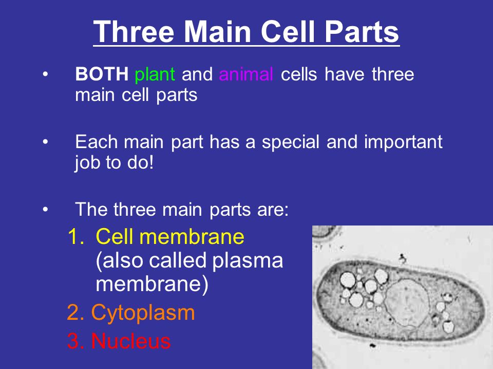
There are 13 main parts of an animal cell. According to the definition animal cell is the dominant tissue cell in animals.
What Are The Main Cell Parts Three Main Cell Parts Both Plant And Animal Cells Have Three Main Cell Parts Each Main Part Has A Special And Important Ppt Download
However animal cells dont have chloroplasts and a number of vacuoles.
What are the three main parts of an animal cell. The main components of an animal cell are as follows. Endoplasmic Reticulum passageways carry protein other materials from 1 part of a cell to another 2. This is the outermost limiting membrane of the cell.
The mitochondria are where a majority of the energy cycles that create ATP adenosine triphosphate or energy happen. It comprises of other cellular structures and organelles which helps in carrying out some specific functions required for the proper functioning of the cell. Is round and has a double membrane.
Houses much of the genetic material in the cell. The following points highlight the three main components of animal cell. As its outer boundary the animal cell has a special structure called the cell or plasma membrane.
As the name implies an animal cell is a type of cell that is seen specifically in animal tissues. Name the cell organelle that contains the genetic material of the cell. In biological terms an animal cell is a typical eukaryotic cell with a membrane-bound nucleus with DNA present inside the nucleus.
It directs all activity of the cell. Mitochondria turns food into energy the cell can use 4. These types of cell organelles function like a unit and regulate the actions of the cell on the whole.
Jelly-like substance where chemical reactions happen. Animal cell size and shape. The largest animal cell is the ostrich egg which has a 5-inch diameter weighing about 12-14 kg and the smallest animal cells are the neurons of about 100 microns in diameter.
The concept of plasmalemma developed long before it. What are the main components of an animal cell. The nucleus of the cell gives the cell direction.
There is a difference between the structures of eukaryotic and prokaryotic cells. It is characterized by the absence of a cell wall with cell organelles enclosed within the cell membrane. 7 rânduri Controls the movement of substances into and out of the cell.
Nucleus cell membrane and cytoplasm. As well as plant cells animal cells have eukaryotic cell structure. You can think of the nucleus as the brain of the cell for without it the cell could not function.
Cell membrane nucleus nucleolus nuclear membrane cytoplasm endoplasmic reticulum Golgi apparatus ribosomes. All of the substances that enter or leave the cell must in some way pass through this membrane. Nucleus control center of the cell 3.
The three main parts of animal cell are - 1 The cell membrane 2 Cytoplasm 3 Nucleus The other cell organells present in animal cell are - a Golgi apparatus b Vacuole c Lysosome d Endoplasmic Reticulum e Ribosomes f Centrosomes g Mitochondria. The powerhouse of the cell. The major substance of the cell is known as protoplasm.
They include a range of multicellular advanced membrane bound organelles. Animal cells come in all kinds of shapes and sizes with their size ranging from a few millimeters to micrometers. By Staff Writer Last Updated April 2 2020 The three main parts of a cell are the plasma membrane the region containing the DNA and the cytoplasm.
The animal cell parts and functions all are performed by the various cell organelles. A typical animal cell is illustrated in Figure 2-1. Plant and animal cells.
All the animal cells are not of the same shape size or function but the main cellular mechanism is the same which helps in proper functioning of the body. Cell wall and chloroplast are present in plant cells while animal cells do not have cell walls. The various cell organelles within an animal cell are.
However not all cells have exactly the same basic parts. Most animal cells have at least the three main parts. Vacuole Mitochondria Golgi apparatus Cell Nucleus Lysososmes Endoplasmic Ribosomes and Reticulum.
Animal Cell Structure -Cell is the basic unit of any living organismThey have different.
Apart from these parts a flower includes reproductive parts stamen and pistil. A carpel is differentiated into 3 parts-stigama style and ovary.
Draw A Neat Diagram Of A Flower Showing Its Various Parts Studyrankersonline
Draw A Labelled Diagram.

Neat labelled diagram of flower. Flowers contain the plants reproductive structures. 4 The third whorl of the flower consists of stamens Androecium which are the male reproductive organs. Describe the ls of flower with a neat label diagram.
Labeled hibiscus labeled parts of flower. Draw A Neat Labelled Diagram Of The L S Of Flower. These are the outermost part of the flower.
D What is the other name of carpel of a flower. 1 A typical flower consists of an outer whorl of green sepals calyx which protects the parts within. The reproductive parts of the flower that are necessary for seed production are the stamen the male organ and carpel the female organ.
B What name is given to i all the petals of a flower and ii all the sepals of a flower. These are leaf like and green in colour. This flower contains bath male and female reproductive part.
On this diagram mark stem. Draw a neat labelled diagram of a pistil showing pollen tube growth and its entry into the ovule. This will fertilize the flower.
Hold it at the peduncle and cut. Draw neat and labelled diagram of l s of hibiscus flower. 3 Petals are soft and are useful to fasilitate cross.
Describe the structure of flower with a neatly labelled diagram. These are leaf like and green in colour. Q1 Name the parts of an angiosperm flower in which development of male and female gametophyte take place.
Gynoecium the female reproductive whorl of flower consists of carpels megasporophylls. Share with your friends. Flower develops on the mother axis stem in the form of floral bud.
A Simple or Monocarpellary. Draw a neat labelled diagram of a typical flower and describe the male and female genital organs. Which of the following substances did Paleolithic people use to attach stone tips to sticks to construct tools and weapons.
Some flowers have fused petals-sepals while a few have separated petals-sepals. When pollinating insects such as bees and butterflies go to the flower for pollen they also visit the stigma. It is protective in function.
2 The incomplete dominance is shown by the flower of snapdragon. This browser does not support the video element. De vlag van belgi ë de vampier van muizen de tout coeur avec vous de warmste week logo vector de standaard het grootste licht de voorzorg mortsel openingsuren de tijd bel 20 de vier magen van een koe.
B write the names of male and female reproductive parts of a flower. When pollen gets to the stigma a tube grows down the style. These are usually coloured gaudy or white in colour and scented and give sweet smell.
The male reproductive cells from the anther travel through the tube and join the ovule fertilizing it. CBSE Free NCERT Solution of 12th biology Sexual Reproduction in Flowering Plants with a neat labelled diagram describe the parts 12th July 2021 SaralStudy. It is very common in most flowering plants.
Opal Diagram Brassica Flower Youtube. Describe the structure of flower with a neatly labelled diagram. In the cross between true-breeding red flower RR and true breeding white flower rr the F 1.
In different plants the number of petals sepals stamens and pistils can vary. 2 Tha second whorl has petals corolla which are usually brightly colored. This is the outer covering of the flower and form outer whorl in a flower.
When gynoecium is sterile or underdeveloped it is called pistillode. Flower Diagram was drawn but not labelled properly. Flower labelled diagram class 10Given beneath is a diagram of the longitudinal part of the flower.
The pointed ends of the calyx are called sepals. 4 K In the diagram shown AJKL AMNP. The value of a flower.
The Calyx sepals enclose the inner parts of the flower when it is a bud. The given slide is identified as Red blood cells. The bud grows from this structure.
3 8 5 37 M Р. Share 3 Flowers are the reproductive parts of plants which take part in the process of sexual reproduction. This is called the calyx.
Find m2 mZN and mZP. Petals form the second whorl inner to the sepals. Reproductive Parts of a Flower.
At the bottom of every hibiscus bud is a green structure at the top of the stem. It is a tough part of the flower because it houses the young bud. They sometimes have fragrance also.
A Draw a neat diagram of a flower showing its various parts. A typical flower consists of an outer whorl of green sepals calyx which protects the parts within. It is very common in most flowering plants.
How To Draw Longitudinal Section Of Flower How To Draw Flower Diagram Step By Step Youtube. Now the scholars whore about to look for the exams should be achieved with their syllabus and revision however there is perhaps one factor which should be nonetheless left to revise and which might be diagrams. For this section on the webquest you will given.
Before we study sexual reproduction in detail let us first study a dissected flower to identify the male and female reproductive parts present in it. Draw a labelled diagram of the longitudinal section of a flower. Take a gulmohur flower.
C What are i stamen and ii carpel in a flower. This tube enters the ovary. Point D is 8 units away from the origin along the x-axis and is 6 units away along the y-axis.
1 TS of anther. Gynoecium may be classified broadly into two types. The presence of these parts differentiates the flower into complete or incomplete.
In this diagram mark stem recetacle sepals petals stamen and carpel. The stamens function is to produce male reproductive cells. Draw a neat labelled diagram of longitudinal section of typical flower.
The following diagram is showing the longitudinal section of flower.
Your eardrum tympanic membrane has two primary roles. It islocated in between the middle ear and the ear canalThe Eustachian tube connects the.
Blog The Role Of The Eustachian Tube Lobe
The eardrum is a thin flap of skin that is stretched tight like a drum and vibrates when sound hits it.

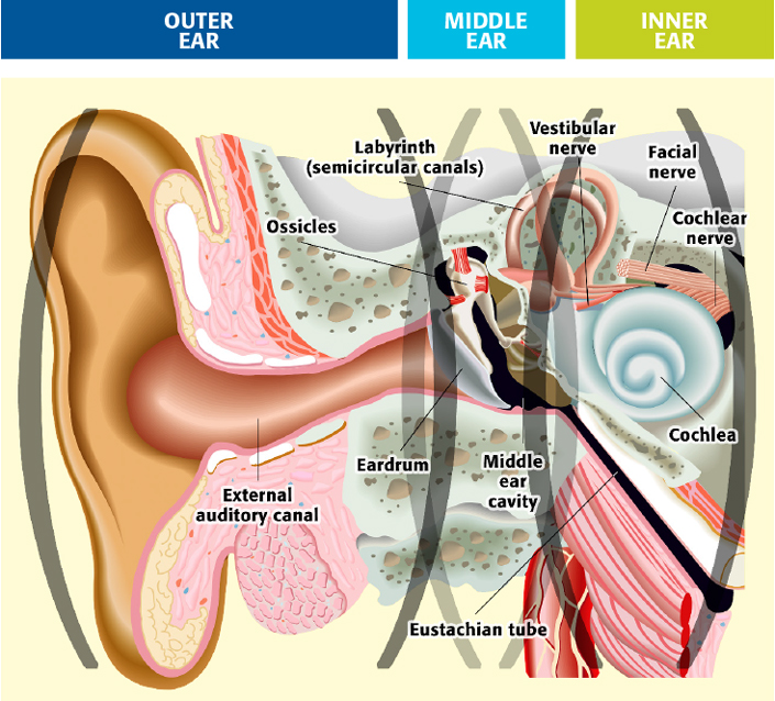
Purpose and function of eardrum. Learn termtympanometry process of measuring function of eardrum with free interactive flashcards. Function of Ear. Function of the eardrum is to carry sound waves to bones that are located in the middle ear.
It collects sound waves and channels them into the ear canal external auditory meatus where the sound is amplified. Provide a funnel to a sound waves to reach among bone fragments located in the middle ear. Behind the eardrum are the ossicles tiny bones that play a vital role in hearing.
The semicircular canals are filled with a fluid called endolymph and function to provide the body with a proper sense of balance. These bones are called ossicles. They transfer sound vibrations from the tympanic membrane to the inner ear at the oval window.
The vibrations are transmitted across the middle ear by the malleus incus and to the stapes bones. The outer ear is the pinna and its function is to gather the sound waves like a funnel and transmit to the middle ear through the ear canal. Your eardrum also acts as a barrier protecting your middle ear from water bacteria and other foreign substances.
Following are the important function of the ear. As the sound makes its way inside of the ear canal it ends up vibrating the tympanic membrane which is also known as the eardrum. The role of the eardrum also called as the tympanic membrane is always to take sound waves to bone fragments which have been found in the middle ear.
Functions of eardrum. Well less common function of the ear lobe is to bear accessories such as earrings. The sound waves then travel toward a flexible oval membrane at the end of the ear canal called the eardrum or tympanic membrane.
The ear performs the functions of hearing and balancing equilibrium. I hear something So feel those. Your eardrum is a really important part of your ear.
What is the purpose of the eardrum 1. The sound waves are collected by the external ear up to some extent. The ear rings compliment the beauty of a girl and also guys these days.
The ear drum or the tympanic membrane covers the other end of the canal. Three small bones linked in series that span the middle ear. When sound waves strike it your eardrum vibrates the first step by which structures of your middle and inner ears translate sound waves into nerve impulses.
It also acts as a dampener to loud sounds that may damage the cochlea. From the inner ear the message is sent to the brain which says Hey. What Is the Purpose of the Eardrum.
What is the function of the eardrum quizlet. When sound waves enter the ear they strike the tympanic membrane. Tympanic membrane also called eardrum thin layer of tissue in the human ear that receives sound vibrations from the outer air and transmits them to the auditory ossicles which are tiny bones in the tympanic middle-ear cavity.
The tympanic membranes function is to assist in human hearing. The function of the tympanic membrane eardrum is to transmit sound waves from the environment into sound vibrations that are picked up by the middle ear. At the bottom of the stapes sits the oval window followed by the semicircular canals also called the labrynthine.
The mechanism of hearing involves the following steps. Sound waves cause the eardrum to vibrate. Your eardrum is essentially a veryfine piece of skin which is less than a half of an inch wide.
The latter fits into. These vibrations move the tiny bones of the middle ear which send vibrations to the inner ear. The middle ear function of human ear is to transmit and amplify the sounds vibrated from the eardrum towards the oval window.
The eardrum vibrates according to the frequency and the amplitude of sounds that strike it. Choose from 75 different sets of termtympanometry process of measuring function of eardrum flashcards on Quizlet. The human eardrum also called tympanic membrane is a thin cone-shaped membrane that separates the external ear from the middle ear.
Its function is to transmit sound from air to the ossicles inside the middle ear then to the oval window in the fluid-filled cochlear. Sound waves travel through the ear canal to reach the eardrum. It also serves as the lateral wall of the tympanic cavity separating it from the external auditory canal.
They pass through the external auditory meatus to the tympanic membrane which is caused to vibrate. The vibrations produced pass through the tympanic membrane to the tympanic cavity. The sound waves pass through the auditory canal and reach the eardrum.
Draw a Neat Labelled Diagram of Vertical Section of the Human Heart. A Schematic Diagram Of A Longitudinal Section Of The Human Heart.
Heart Diagram With Labels And Detailed Explanation
A Labeled Diagram Of The Human Heart You Really Need To See Bodytomy.
Labelled diagram of human heart class 11. Images The Heart Labeled Cardiac Muscle Vector Illustration. A labelled diagram of the human heart. Simple Labeled Heart For Kids Blue And Red Diagram Of The Heart.
Diagram Of Heart Labelled - Fun for my own blog on this occasion I will explain to you in connection with Diagram Of Heart LabelledSo if you want to get great shots related to Diagram Of Heart Labelled just click on the save icon to save the photo to your computerThey are ready to download if you like and want to have them click save logo in the post and it will download directly to. Step by Step answer for Draw a neat labelled diagram of VS. The human heart and its functions are truly fascinating.
Draw a labelled diagram of internal structure of human heart. Explain the function of human heart qith diagram for class. Download Labelled Diagram Of Human Heart Class 10 PNG.
Guppies will be suitable for the control of which disease. The human heart and its functions are truly fascinating. Conducting System of Human Heart With Diagram Cardiac function does not require intact innervations.
Frogs heart removed from the body and in human beings who have transplanted heart the heart continues to function. Easy Steps To Draw Human Heart Class 10 Ncert Write Down Each. Draw labelled diagram of human heart in class 10 - eanswersin.
Punjab Board 2017 Class 10 Science. I Right ventricle ii Aorta iii Left atrium iv Pulmonary arteries. Easy way to draw human heart Identification and labelling of parts Life processes NCERT class 10 - YouTube.
Correct answer to the question. The heart consists of 4 chambers. The heart lies in the thoracic cavity in the space between the lungs mediastinum anterior to the vertebral column and posterior to the sternum.
The perfusion of the isolated heart in the laboratory will also continue to beat at regular intervals. 24 Qs Related questions. How To Draw Human Heart Diagram Easily Class 10 Step By Step Youtube.
Diagram of the human heart. Ii Arteries have thick elastic walls. The Human Heart Labeled Stock Photo Picture And Royalty Free.
Easy Way To Draw Human Heart Life Processes Ncert Class 10. B Give reasons for the following i The muscular walls of ventricles are thicker than the walls of atria. Explore more than 7367 Human Heart Diagram Labelled resources for teachers parents and pupils.
In females it weighs around 230 to 280 grams 8 to 10 ounces. Jun 142021 - Explain the structure of human heart with labelled diagram. Labelling The Heart Ks2 T2 S 429 The Human Heart Diagram Display.
Heart Diagram Labeled 2019 Diagrams And Formats Corner. EduRev Class 10 Question is disucussed on EduRev Study Group by 136 Class 10 Students. How To Draw Human Heart Diagram Youtube Notes Of Ch 11 Transportation In Animals And Plants Class 7th.
It is approximately the size of owners closed fist and weighs about 250-300 gm in female and 300-350gm in the male. A well labeled human heart diagram given in this article will help you to understand its parts and functions. Get FREE solutions to all questions from chapter BODY FLUIDS AND CIRCULATION.
The heart is roughly cone shaped hollow organ. An adult heart beats about 60 to 100 times per minute and newborn babies heart beats at a faster pace than an adult which is about 90 to 190 beats per minute. Of human heart of Biology Class 11th.
A Draw a diagram of cross-section of the human heart and label the following parts. Easy way to draw human heart Identification and labelling of parts Life processes. Every single part of our body is so well designed that it works continuously throughout our life.
Click hereto get an answer to your question Draw a labelled diagram of internal structure of human heart. The average male heart weighs around 280 to 340 grams 10 to 12 ounces.
For more info see Create a sheet of nametags or address labels. Open a jpeg image with IrfanView this doesnt work with other image formats such as tif Select Image Information.

Labeling Pictures In Kindergarten Beyond Clearly Primary
This makes one image.
How to label a picture. Pencil on reverse. Capitalize the F in this case but not when referring to your figure in the body text. Add Caption to Image 1.
Position the box into place over the photo and typen in anything you want. Choose professional images from the Avery gallery or upload your own from your computer Facebook Instagram Google and more. Create a single label with a graphic Start by creating a New Document of labels.
Add Alt Text to Image 1. To label a picture in Microsoft Word you can do the following. Press Ctrl-O on your keyboard when the Paint window opens.
6 Enter a name for this face. Personalize labels cards and tags for mailings invitations personal crafts marketing materials and more. If you decide to write on your photos you should always write with an archival-safe pen so as not to damage the front of the photo and do your labeling on the backs of the photos.
Select X to close. You can also download other language worksheets for kids available in our languages section for free. Thank you for the A2A.
Tap a face to label. Drag the image into position within the. Use a soft lead 2.
I recommend using the caption field to identify people places events and dates. Click Text button marked with an A on the Paint toolbar. A new screen will appear with that persons face at the top and the words Add a name at the top.
I want to place it in my html file. Then click Insert Caption. On the Word menu bar click References.
Label and number the figure on a line under the image itself using either Figure or Fig. Youll have to add your notation on a paper enclosure or sleeve. All women who are married should have their maiden names included along with their married names.
How to use the worksheet. Students can spell the words independently or use the word cards to help them label the pictures. Whats the Best Way to Label Family Photos How where and what you write depends on the image itself.
If you later open the file in another location that caption will travel with the photo. Go to the file menu and select Insert Text Box Select the text box button from the bottom drawing toolbar. You can draw a text box in two ways.
To locate an image stored in your computer highlight the Computer tab. Lay the photo and your label on the scanner close the lid and scan at the same time. Add information to the fields you choose.
Navigate to the location on your computer where the picture on which you want to put a label is stored on the. Go to Picture Format Text Wrapping and select Square. Using this I have created something like this.
Now as I am going to use Django I dont want the image to be in css. I did crop the one below so it would fit on the blog better I have the photo and the label together. Insert a graphic and then select it.
Select a photo or photos find the Title field at the bottom of the screen and also the tag field if you want to add tags or keywords and type in your caption. Download the worksheet using the red download button at the top. Write at least the first and last names of everyone.
Label the picture View download and print this worksheet for kids. Cased images such as tintypes wont have a writing surface. Improve your childs knowledge of animals.
Give it a descriptive title following the label and number and then without punctuation write from and include Works Cited entry. Discover how easy it is to add and edit images using Avery Design Print version 80. Click on the image that you want to label.
Click the gray box that reads PLACE IMAGE HERE Using the menu on the left side of the screen click the button that reads Replace Image Locate the photo you wish to use. For most photographs a short note on the reverse side of the photos is preferred using the least permanent writing material. Click on the IPTC info button in the bottom-left corner.
How to Put Labels on Pictures Step 1.
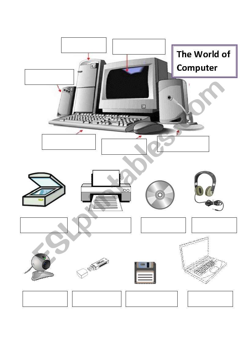
Parts of web browser. Write the names of parts of computer.
Computer S Parts Esl Worksheet By Sunrain Nicole
2nd through 4th Grades.

Name the computer parts worksheet. Parts of a ComputerParts of a Computer interactive exercise for Grade 3. Name The Computer Parts Ampdp2 - Displaying top 8 worksheets found for this concept. You can do the exercises online or download the worksheet as pdf.
Download Print Worksheet. Monitor in Yellow CPU in Blue Keyboard in Green and mouse in Red. Computer Parts and Peripherals Group.
This part allows a person to connect an electronic device to a computer. Some of the worksheets for this concept are Use the words below to label the parts of a In this lesson you will learn about the main parts of a Whats in the box Computer parts labeling work Computer parts what are their names anyway Naming attributes Computers inside out lesson 7 files folders what is Computer basics student. Mouse CPU box flatbed scanner 18 keyboard mousepad.
Displaying top 8 worksheets found for - Parts Of Computer. Your answer is corrected as soon as you rearrange each word correctly. Students will color each part of the computer a different color.
Find the correct spelling of each part of the computer by rearranging jumbled letters. Name the basic parts of computer. This resource contains 6 worksheets for students to label the exterior parts of a computer internal parts of a computer basic parts of a desktop and internet browser window rear input ports on the back of a computer keyboard symbols and.
Identify which are the parts of the computer. Then tick the correct answer. Name the Parts of a Computer Activity 1.
This can be used by giving oral directions or having students read and follow the directions independently. Showing top 8 worksheets in the category - Name The Computer Parts. Of the different computer parts.
Monitor mouse keyboardknowledge of these and more are assessed in this computer parts quiz. Showing top 8 worksheets in the category - Name The Computer Parts Ampdp2. Name That Computer Part Quiz.
Parts of a Computer Quiz 1. Some of the worksheets for this concept are In this lesson you will learn about the main parts of a Whats in the box Name Computer parts labeling work Parts of a computer class i iv and v thinking of Use the words below to label the parts of a Computer parts what are their. Name The Computer Parts Ampdp2.
Some of the worksheets displayed are Work Parts of a computer 6 5 basic computer hardware and software levels Name word bank Computer basics for kids Computer computer Computer basics work Computer parts what are their names anyway. Computer worksheet for class 3 In this worksheet we have questions from Computer. This worksheet covers all important concepts from the chapter.
Parts of a Computer Quiz 1. Some of the worksheets displayed are Computer parts labeling work Computer parts diagram 103ah computer parts In this lesson you will learn about the main parts of a Module 1 handouts computer basics computers Parts of a computer class i iv and v thinking of Computer basics essential skills. Learn the spellings of the parts of a computer and name them correctly.
Displaying top 8 worksheets found for - Naming Parts Of A Computer. Name The Computer Parts Ampdp2. If you want to practice more you can find more of my worksheets.
Name The Computer Parts. Identify the correct name of each part. Tomorrow we will learn how to do this and then we will learn about the buttons.
Parts of a Computer - BlankLabel On this worksheet students label the major parts of a computer including the modemrouter monitor mouse keyboard CPU and printer. 1 What part of the computer is shown in the picture. Internet function and parts worksheet activity wrting and grammar guide using internet vocabulary parts of the web.
Using the words from the Word Bank fill in the names of the items pictured in the spaces provided. This worksheet covers topics like computer parts name computer memory Microsoft paint program etc. Personal Computers PCs Name.
Live worksheets English. 3 Computer Parts listed in this Basic Computer Terms worksheet. Name The Part On The Computer - Displaying top 8 worksheets found for this concept.
Word Bank monitor laser printer connector 3439 Computers Made Friendly floppy disk cable Teacher Created Materials Inc. Some of the worksheets for this concept are Work Parts of a computer 6 5 basic computer hardware and software levels Name word bank Computer basics for kids Computer computer Computer basics work Computer parts what are their names anyway. It can also be used for saving.
This part is the information storage in a computer that is used to store data for programs and. Name The Part On The Computer. It gives you an overall idea and covers all important parts names of computers.
Some of the worksheets for this concept are Computer parts labeling work Monitor case Computer parts crossword puzzel work Module 1 handouts computer basics computers Lesson plan In this lesson you will learn about the main parts of a Whats in the box Computer basics for kids. This is a worksheet used to reinforce the name of computer parts. Look at the given pictures of parts of the computer.
Colour the parts of the computer.
Discover more free science worksheets including animal cells and brain printable science worksheets. Prior knowledge or research will be necessary.
Science Worksheets Label Parts Of A Neuron By Science Workshop Tpt
Aug 19 2019 - This worksheet accompanies the powerpoint Neurons of the Nervous System.

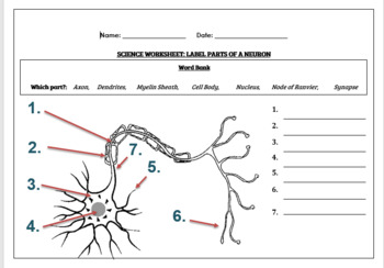
Label the parts of a neuron worksheet. Use the words from the list below to label the following diagram of a neuron in the lines provided. 1 dendrites 2 cell body or soma and 3 axons. About this Worksheet This is a free printable worksheet in PDF format and holds a printable version of the quiz Label the parts of a neuron.
Axon - purple Axon Terminals orange Myelin sheath yellow. Explain the difference between axon and dendrites. NERVOUS SYSTEM WORKSHEET 1.
All neurons have three main parts. Get Free Access See Review. Send electrical impulses to neighboring neurons.
On the lines following each word write out what that structure does for the neuron. In this biology worksheet students analyze a picture of a neuron. Label parts of a neuronStudents have to identify and label parts of a neuron Axon Dendrites Myelin Sheath Cell Body Nucleus Node of Ranvier SynapseStudents can color in the nerve cell once they are doneWorksheet aimed at higher primary high school levelAnswer.
Only a fraction traveled up your to of a second later aan But you had your n reacted. Sep 14 2020 - Science worksheets. You can do the exercises online or download the worksheet as.
The neuron is the basic working part of the brain that transmits important information to other nerve cells muscle or gland cells. Color in the diagram as suggested below. Axon dendrites myelin Node of Ranvier nucleus cell.
These neuron cells within the nervous system communicate with each other in unique ways. Talking about Neuron Worksheet S we have collected some variation of photos to add more info. This kind of reaction is known as aan Reflex acts occur without thinking.
Neuron diagram worksheet label neuron blank diagram and nerve cell neuron with synapse are three of main things we want to present to you. Add the following labels to the diagram. The impulse traveled along this nerve to a muscle in your leg You jerked your leg away.
Below is a list of different parts of a neuron. For Students 6th - 8th. Science Worksheet Label Parts Of A Neuron Answer Key Neurons Worksheet Teachers Pay Teachers - Fill in the blanks with the correct an.
By printing out this quiz and taking it with pen and paper creates for a good variation to only playing it online. Students label the diagram with names of 7 parts of the neuron. This time we will show you various amazing photos that weve gathered just for you for today we are more concern about Neuron Worksheet S.
Label Parts Of Neuron Worksheet Biology Lessons Human Body Unit Labeled Diagram Of The Neuron Nerve Cell That Is The Main Part Neuroscience For Kids Fill In 1 Label The Parts Of A Neuron In Figure 7 5 Sarthaks Econnect Nervous System The Neuron Is The Basic Unit Of The Nervous System. Axon Terminal Buds 2. Name parts of a neuron.
Axon - Cell Body - Dendrites - Myelin Node of Ranvier - Nucleus - Synaptic Terminal. Biology IF8765 88 lnstructional Fair Inc. The diagram below is of a nerve cell or neuron.
Cell body soma 5. Axon Myelin sheath Cell body Dendrites Muscle fibers Axon terminals 2. Neuron Anatomy Activity 1.
4 rijen Neuron online worksheet for 7th. Add the following labels to the diagram. The diagram below is of a nerve cell or neurone.
NEURON WORKSHEET Using the picture above label all the parts. If you like colour in the diagram as suggested below. Worksheet nervous system 1.
Describe the picture with these words. Transfers electrical impulse signals from. Label parts of a neuronStudents have to identify and label parts of a neuron Axon Dendrites Myelin Sheath Cell Body Nucleus Node of Ranvier SynapseStudents can color in the nerve cell once they are doneWorksheet aimed at higher primary high school levelAnswer.
A word list is provided. Besides the three major parts there is the presence of axon terminal and synapse at the end of the neuron. As the powerpoint is shown the students follow the instructions of coloring labeling and describing each part.
The part of the neuron that insulates the axon and increases the speed of the neural message within the neuron. 252020 91651 PM Title. Cover the axon and work like insulation to help keep electrical signals inside the cell which allows them to move more quickly.
Four different nitrogenous bases. Each nitrogenous base in a nucleotide is attached to a sugar molecule which is attached to one or more phosphate groups.

Nitrogenous Base Dna Rna Brainly Novocom Top
The nucleic acids DNA and RNA are polymers composed of many individual nucleotides.

What are three main components of dna and rna molecules brainly. What are the components of the DNA and RNA molecule. A sugar a water molecule and a nitrogenous base B. Kason11wd and 9 more users found this answer helpful.
Adenine guanine cytosine and uracil. The DNA RNA is made of monomers called nucleotides. Four different nitrogenous bases.
Each nucleotide consists of three components. - 39356422 ItzMissAstonish ItzMissAstonish 25042021 Science Secondary School answered What are components of RNA and DNA. As mentioned above DNA has three main components.
Science 22012020 0828 09652393142 What are the components of the dna and rna molecule brainly. RNA is a polymer with a ribose and phosphate backbone. What are the components of the DNA and RNA molecules.
Become a member and. And finally tRNA or transfer RNA that ferry amino acids to the ribosome to be assembled. Adenine guanine cytosine and uracil.
A nitrogenous base a pentose five-carbon sugar called ribose and a phosphate group. And thats the answer to what are the three main components of a dna molecule. DNA provides the code for the cell s activities while RNA converts that code into proteins to carry out cellular functions 846 views.
Adenine Guanine Cytosine Thymine. If the sugar is a simple ribose the polymer is RNA ribonucleic acid. RNA is a polymer with a ribose and phosphate backbone.
The DNA Main Components. A nitrogenous base a pentose sugar and one or more phosphate groups. Three major types of RNA are mRNA or messenger RNA that serve as temporary copies of the information found in DNA.
So what are the three main components of a dna molecule. There are two basic types of nucleic acid DNA oligonucleotide acid and RNA nucleic acid. If the sugar is derived from ribose as deoxyribose the polymer is DNA deoxyribonucleic acid.
A sugar a phosphate group and an amino acid C. What are the three main components of a DNA molecule. Phosphate Deoxyribose some kind of sugar substance Nitrogen which is divided into four type of bases.
Both DNA and RNA are made from nucleotides each containing a five-carbon sugar backbone a phosphate group and a nitrogen base. This exercise will focus on DNA although ways in which it differs from RNA will also be presented. Each nucleotide consists of three components.
A 5-carbon sugar a phosphate group and a nitrogenous base. One may also ask what are the 3 components of the DNA and RNA molecule. A phosphate group a pentose.
RRNA or ribosomal RNA that serve as structural components of protein-making structures known as ribosomes. See full answer below. A sugar a nucleotide and an amino acid.
A nucleotide is made up of three components. What are the components of the dna and rna molecule brainly. The nucleic acids DNA and RNA are polymers composed of many individual nucleotides.
Each nucleotide is made up of three components. Nucleic acids are molecules that are essential to and characteristic of life on Earth. A sugar a phosphate group and a nitrogenous base D.
Science 21062021 0855 hellcrack777. DNA is found in all organisms from the smallest bacteria to humans. What are the components of the DNA and RNA molecules.
What are components of RNA and DNA. What are the 3 components of the DNA and RNA molecule. DNA and RNA molecules consist of chains of nitrogenous bases bound together by their attached phosphate groups.
A sugar a phosphate group and a nitrogenous base. They are composed of monomers which are nucleotides made of three components. Science 22012020 0828 09652393142.
The nucleic acids DNA and RNA are polymers composed of many individual nucleotides. Each nucleotide consists of three components.
Algal species contain larger chloroplasts of different shapes eg spiral cup-shaped circular bands. Chloroplasts of higher plants have disc-shaped or oval structure 10 µm in length and 2-4 µm in diameter.

Write Short Notes On I Ultrastructure Of Chloroplast And Ii Pigments Involved In Photosynthesis Brainly In
Light microscope and fluorescent dyes.

The ultrastructure of a chloroplast is. The ultrastructure of a chloroplast is best studied using a transmission electron microscope The cells of an ant and an elephant are on average the same small size. Structurally each plastid consists of two parts. X is the plasma membrane Y is a chloroplast.
The ultrastructure of a chloroplast is best studied using a a. A chloroplast is bounded by a double layered membrane known as peristomium made up of lipoproteins. Absorbs light energy and converts it into chemical energy.
The ultrastructure of a chloroplast is best studied using a. Generally the ultrastructure of chloroplast in algae consists of thylakoid band membrane-bounded chloroplast envelops chloroplast endoplasmic reticulum phycobilin protein pyrenoid storage product etc. These unit membranes are separated by periplastidal space of 10nm.
The ultrastructure of the grana from Aspidistra shows a striking similarity to that of the outer segments of the retinal rods of the vertebrate retina 10. A study of chloroplasts from other species is necessary to prove this assump- tion. The membranes are differentiated as inner and outer.
The grana are organized in double membrane discs the two membranes of which are about 65 Å thick. The grana as well as the rod outer segments consist of double membrane discs. On ultrathin sections of chloroplasts from Aspidistra elatior the electron microscopic study has revealed that the grana consist of layered columns.
Chloroplast has a structure called chlorophyll which functions by trapping the solar energy and is used for the synthesis of food in all green plants. 1 outer limiting membrane and 2 inner matrix or stroma. If you look closely at X it points to the cell wall outside the plasma membrane it is close to the plasma membrane but not touching the chloroplast.
Biology 05072019 0830 ashleydenson1517. Chloroplast is a double membrane organelle. Where should they expect to.
It may vary in shape and size in various organisms Example cup shaped in Chamydomonas star shaped in Zygnema Sipiral in Spirogyra. Peristomium encloses a colourless matrix called stroma. The ultrastructure of a chloroplast is best studied using a.
An elephant just has more of them. X is the cell wall Y is a mitochondrion. However in most of the organisms it is lens shaped.
An elephant just has more of them. The ultrastructure of the chloroplast contains the following parts. Ultrastructure of chloroplasts A chloroplast comprises of three main components namely Envelope Stroma and Thylakoids.
On the basis of the ultrastructure of the chloroplast Lee divided the algae into four groups. Scientists developed a new method to detect the precise location of specific molecules within living cells. The ultrastructure of a chloroplast is best studied using a Plant organelle that conducts photosynthesis Chloroplasts visible in the cells of Bryum capillare.
The most important function of the chloroplast is to synthesize food by the process of photosynthesis. The chloroplast is bound by an envelope which is made up of two unit membranes. Cells Plant Biology i need some help with an as level biology OCR text book question AQA IGCSE Biology 12 Certificate.
And this question is revolving around being able to use a microscope to see the ultra structure of the chloroplast keeping in mind that the ultra structure is basically the architecture of the cell or in this case the architecture of that lower class and that is the internal and external architectures. The end result of a reaction between components of a membrane is determined not only by the inherent properties of the reactants but also by their locations within the membrane. The ultrastructure of a chloroplast is best studied using a transmission electron microscope The cells of an ant and an elephant are on average the same small size.
These membranes bound a narrow closed space the height of which also is about 65 Å. Plastids assist in storing and harvesting needed substances for energy production.
Carefully remove the label with your. Cattie Adhesives manufactures a Full Line of Brewery Packaging and Bottle Labeling Adhesives including Hot Melt Water-Based Adhesives and Coatings for High Speed Brewery Craft Brewery Packaging Labeling applications.

6 Ways To Remove Beer Bottle Labels
Glue stick the entire back of the label and adhere.

How to remove beer bottle label glue. OxiClean is an oxygen-based cleaner that does not contain chlorine like bleach does. Hold the wine bottle over the pot of boiling water for 10 to 15 minutes. This is after only three hours and already most of the labels have floated to the surface.
Next time I have a sticker stuck to the glass on a picture frame Ill try this method. Each chemical with a different degree of effectiveness. They stick like commercial label and even stand up to a good period of time in the cooler with water.
The labels came off within minutes and the glue seemed to melt away with no residue. This product works well to break down the adhesive on the labels as well as any leftover proteins within the bottle itself. Step 4 Remove Any Residual Glue with Sponge or Stainless Steel Pad.
If you get a beer label thats willing to soak up water and with a glue that dissolves easily enough theres good odds you will be able to pull the whole label off from the bottle 100 percent intact and leaving no residue. To adhere the labels to the bottles and jars companies use a type of glue adhesive on one side of the label. Baking soda reacts with water causing the glue to loosen and making it easier for you to peel the label off.
Some bottles have stubborn labels and they wont come off in the OxiClean. Boil water in a pot. If thats the case some steel wool usually does the trick.
When you want to remove the label just soak in some hot water comes back off. This is actually very common and almost expected. Sodium bicarbonate baking soda is an effective way to remove labels from beer bottles.
If youre less fortunate then you have an oil based adhesive and no amount of scrubbing in water will get the glass clean so scrape away what paper you can before drying the bottles. The steam affects the glue and softens the label. All of our Brewery Packaging and Bottle Labeling Adhesives meet the requirements of the FDA Adhesives regulation.
That price tag. A significant number of these bottles can just be plopped in hot water from a sink faucet for 10 minutes and the labels will just fall off. Labels provide important information about the contents.
How to Easily Remove Labels from Beer Bottles - YouTube. Hot Water and Oxyclean soak Most Belgian and German imported beers have a pretty simple glue on their labels. 21CFR175105 Indirect Food Additives.
I wanted to reuse beer bottles for home brewing so I used Oxy Clean. Now that your bottles are label-free you still might notice some extra glue or label remnants left behind. As I mentioned previously every company uses different labels and adhesive.
Hot water soak with PBW Oxyclean baking soda etc. Just as a water bath loosens water based glue so an oil bath will do the trick with the oil based fixer. Hot water soak and the labels will float off or peel off fairly easily.
Removing these labels can be a bit difficult at times. There are three types of label-removing processes to use depending on the glue that is used to stick the label to the bottle and should be tried in this order. If playback doesnt begin shortly try restarting your device.
Labels are a common sight on bottles and jars for good reason. Any remaining glue can be scrubbed off with a cloth. You want to add 16 tablespoons of baking soda for every gallon of water.
I just need to know its a non-chemical way to remove label glue from glass bottles. Submerge your bottles and then just wait. I have found that the easiest first step in removing bottle labels is to let the bottles soak in a solution of OxiClean.
Yes the most simple way to remove a beer label is to let the glass bottle soak in a bucket or tub overnight.
The important structures of the foot can be divided into several categories. Calluses are thickened areas of skin over parts of the feet where excessive amounts of pressure or friction occur.

Anatomical Structure Of The Human Foot The Image Shows The Three Main Download Scientific Diagram
Foot in anatomy terminal part of the leg of a land vertebrate on which the creature stands.
What are the different parts of a foot. Parts of the Foot. The forefoot contains the five toes phalanges and the five longer bones metatarsals. The most common ankle ligament injury is a ligament sprain most commonly of the lateral ligament aka.
Its not as simple as drawing a body on your foot instead the size position and scale is altered eg. A sandal may have a sole made from rubber leather wood tatami or rope. Feet is an anatomical structure found in many vertebratesIt is the terminal portion of a limb which bears weight and allows locomotionIn many animals with feet the foot is a separate clarification needed organ at the terminal part of the leg made up of one or more segments or bones generally including claws or nails.
The foot is located after the long shin bones and it starts from the back of your ankle to your toes. Lets briefly discuss why it is useful to know your feet type. In most two-footed and many four-footed animals the foot consists of all structures below the ankle joint.
There are three main parts of the foot the forefoot toes and metatarsals the midfoot arches and hindfoot heel and ankle. How the foot works. The foot diagram has a complex structure made up of bones ligaments muscles and tendons.
They are a really important part of ankle anatomy as they are the primary stabilisers of the ankle. The anatomy of the foot. Parts of your foot correlated with the stomach are found above the waistline.
Similarly what are the parts of a sandal called. The bones of the foot are organized into rows named tarsal bones metatarsal bones and phalanges. Unlike calluses corns have a central core or spot in the middle that is surrounded by dead skin.
The midfoot contains the cuneiform bones cuboid bone and navicular bone. Parts correlated with the intestines are found below. These make up the toes and broad section of the.
This consists of five long metatarsal bones and five shorter bones that form the toes phalanges. The part of the foot which joins it to the leg is called the heel. Heel arch digits and contained bones.
There are eleven ligaments around the ankle connecting the various different bones of the hindfoot and midfoot. Most land vertebrates have feet and there are many different sorts of foot. The next organ that can benefit from foot massage is the small intestine.
The foot is divided into three sections - the forefoot the midfoot and the hindfoot. It may be held to the foot by a narrow thong that generally passes between the first and second toe or by a strap or lace variously called a latchet sabot strap or sandal that passes over the arch of the foot or around the ankle. The bottom of the foot is called the sole.
Simply massage the area between the heel and the ball of the foot for improved digestion and a healthier intestinal tract. The hind foot has the calcaneus or heel bone which is the largest bone in the foot. The sinuses are linked to the tips of each of the toes and the knee is linked to part of the outer border of the sole of the foot.
If you would like to learn all the. Create a crash tone by using your foot to close both cymbals together then quickly opening them this is known as splashing. You can also vary the hi-hat tone simply by striking different parts of the cymbal.
The hard foot of an ungulate is a hoof. Anatomically the foot is divided into 3 sections. Corns occur on the toes where they rub against the shoe.
The feet are divided into three sections. Learn more about the foot. The type of your feet is generally defined based on the mentioned characteristics.
Even the basics can lead you to a very simple self-treatment. The foot contains a lot of moving parts - 26 bones 33 joints and over 100 ligaments. The skeleton of the foot begins with the talus or ankle bone that forms part of the ankle joint.
The hindfoot midfoot and the forefoot. The bottom of your foot is connected with your pelvic area. What parts make up the foot.
They work together to control all the different movements in the foot and ankle. The midfoot is a pyramid-like collection of bones that form the. For example hitting the middle of the top hi-hat will produce a brighter note with less resonance in comparison to say tapping the rim which produces a note that rings out for longer.
They are the size of feet toe shape foot width and the arch of the feet. Understanding the structure of the foot is best done by looking at a foot diagram where the anatomy has been labeled. There are four main characteristics of feet that differentiate feet types.
The two bones of the lower leg the large. The feet of monkeys are much like the hands. The hindfoot is the posterior part of the foot.
The thinnest part of your foot usually found towards its center is known as the waistline.
B Mention the fate of all the components of the embryo sac after fertili. Draw a diagram showing longitudinal section of a flower and label on it.
How To Draw Longitudinal Section Of Flower How To Draw Flower Diagram Step By Step Youtube
Given below is a diagram representing a stage during mitotic cell division in an animal cell.

Draw a labelled diagram of stamen. The androecium is the male reproductive whorl of the flower. Draw a Well-labelled Diagram to Show the Anaphase Stage of Mitosis in Plant Cells Having Four Chromosomes. Asked Mar 31 2019 in Biology by Farrah 695k points how do organisms reproduce.
C What are i stamen and ii carpel in a flower. Give one reason in support of your answer. Step by step video text image solution for a Draw a schematic labelled diagram of a fertilised embryo sac of an Angiosperm.
Soft iron core PrincipleTransformer is based on the principle of electromagnetic mutual inductionWhen the current flowing through the primary coil changes an emf is induced in the secondary coil due to the change in magnetic flux linked with the primary coil. Structure and Events - Stamen Microsporangium and Pollen Grain Report Error Is there an error in this question or solution. E What is the name of yellow powdery.
A Draw a neat diagram of a flower showing its various parts. Each stamen consists of a stalk called the filament and sac like structure called the anther. Examine it carefully and answer the questions which follow.
Stigma ovary anther filament. In this diagram mark stem receptacle sepals petals stamen and carpel. Draw a neat labeled diagram of pistil showing the growth.
Draw a labelled diagram of the embryonic stage that gets implanted in the human uterus. Click hereto get an answer to your question Draw a labelled diagram of a step - up transformer and explain how it works. Individual members of the whorl are referred to as stamens.
A Draw a labelled diagram of the sectional view of a typical anatropous ovule. I Labelled diagram of a step-down transformer. The main two functions of Stomata are.
By Biology experts to help you in doubts scoring excellent marks in Class 12 exams. State the functions of the two parts labelled. So D 2 conducts during this time period.
Announcing Numerades 26M Series A led by IDG Capital. A Identify the stage. Draw a labelled diagram of stomata write two functions of stomata.
B Describe the stage in embryo development in a dicto plant. In this diagram mark stem receptacle sepals petals stamen and carpel. Structure and Events - Stamen Microsporangium and Pollen Grain.
State two characteristics of the primary coil as compared to its secondary coil. C Name the parts labelled 1 2 and 3. ADraw a neat diagram of a flower showing its vapours parts.
B What name is given to i all the petals of a flower and ii all the sepals of a flower. A Draw a labelled diagram of sectional view of human ovary showing different stages of oogenesis. During this time the input to diode D 2 is negative with respect to the midpoint.
Stamen Labelled draw a labelled diagram of a flower. The function of anther is to produce pollen grains. Draw a labelled diagram of a flower and describe its.
B What name is given to i all the petals of a flower and ii all the sepals of a flower. D WHat is the other name of carpel of a flower. D What is the other name of carpel of a flower.
Plant biology Archives A Biology. C What are i stamen and ii carpel in a flower. Draw a neat labeled diagram of pistil showing the growth.
B Name the cell organelle that forms the aster. Stomata facilitate carbon dioxide uptake and release of oxygen during the process of photosynthesis. Labelled diagram of a typical anatropous ovule Concept.
When the input signal to diode D 1 is positive it conducts and load current flows. Draw a labelled diagram of the longitudinal section of a flower. B Where is morula formed in humans.
During the negative half cycle of the input signal the voltage at D 1 is negative and that at D 2 is positive.