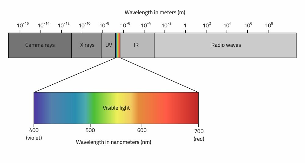
8 Zeilen The electromagnetic spectrum ranges from long wavelength low frequency waves like radio waves which can be used for communication to short wavelength high frequency waves like gamma rays which. Phase velocity in a medium with permeability μ and permittivity ε and 2 is the Laplace operator.
Components Of Electromagnetic Spectrum Radio2space
Name the constituent radiations of electromagnetic spectrum.

Name two parts of an electromagnetic wave. As you read the print off this computer screen now you are reading pages of fluctuating energy and magnetic fields. To examine the properties of the electromagnetic waves lets consider for simplicity an electromagnetic wave propagating in the x-direction with the electric field E G pointing in the y-direction and the magnetic field B G in the z-direction as shown in Figure 1341 below. Where v p h 1 μ ε displaystyle v_mathrm ph frac 1sqrt mu varepsilon is the speed of light ie.
Light electricity and magnetism are all different forms of electromagnetic. In a plane electromagnetic wave. An oscillating electric field and a perpendicular comoving magnetic field which oscillates at the same frequency but with a phase shifted by 90.
As the name suggests radio waves are emitted by radio stations TV stations and cellphone towers. Yes and the people who say this tend to say this is why electromagnetic waves dont need a medium. Write in brief how these waves can be produced.
Write the i ratio of magnitudes and ii the directions of its electric and magnetic field vectors. What are Electromagnetic Waves - Wave is nothing but a pattern of disturbance which propagates and carry energy with it. Also arrange λ 1 λ 2 and λ 2 3 in increasing order.
Types of Electromagnetic Waves 1. They describe the movement of a packet of energy between two points. C used as a diagnostic tool in medicine.
Figure 1341 A plane electromagnetic wave. But they do need a medium. Let the wave lengths of the electromagnetic waves used quite often for i produce intense heating effect ii is used for studying crystal structure be labelled as λ 1 λ 2 and λ 3 respectively.
An electromagnetic wave travels or propagates in a direction that is oriented at right angles to the vibrations of both the electric E and magnetic B oscillating field vectors transporting energy from the radiation source to an. For a given wave-vector the electric field can have any direction in the plane normal to. Microwaves are used to broadcast information through space as they can penetrate through clouds and light.
Arrange these wave-lengths in decreasing order of magnitude. Identify and name the part of the electromagnetic spectrum to which these radiations belong. The electromagnetic wave equation derives from Maxwells equations.
For this reason electromagnetic waves are usually characterized by their wave-vector which specifies the direction of propagation and the wave-length and the plane of polarization ie the plane of oscillation of the associated electric field. And obtain the photon energy in units of eV for different parts of the electromagnetic spectrum. A plane electromagnetic wave travels in vacuum along the y-direction.
B used to treat muscular strain. In a vacuum vph c0 299792458 meters per second a fundamental physical constant. Frequently when EM waves are taught it is said that the change in electric field causes a change in the magnetic field which then causes a change in the electric field and so on and so forth.
Space is the medium. In what way are the different scales of photon energies that you obtain related to the sources of. Name the part of electromagnetic spectrum which is suitable for i radar systems used in.
Waves with a very short wavelength high frequency and high energy. Electromagnetic waves form a continuous spectrum of waves. Name the parts of the electromagnetic spectrum which is a suitable for radar systems used in aircraft navigation.
In most older literature B is called. Electromagnetic waves have two components.
Le Live Marseille aller dans les plus grandes soirées. Biology A Well Labelled Diagram Of Earthworm what is labeled diagram answers com external morphology of earthworm microbiology notes hindi amp english version board of secondary education pdf stable isotope 15n and 13c labelling of different frog dissection labeled images the biology corner human excretory system fun science.

Label Earthworm Diagram Enchantedlearning Com Earthworms Safari Science Labels
Port Manteaux Word Maker OneLook Dictionary Search.

Make a well labelled diagram of earthworm. Port Manteaux Word Maker OneLook Dictionary Search. Henry steiner cabins masters thesis UNIFEOB. EUR Lex 32013R0283 EN EUR Lex.
Henry steiner cabins masters thesis UNIFEOB. Download baros daca maine ft bogdan ioana jibovivawosac cf. Well Labelled Diagram Of Earthworm Nervous System EUR Lex 32013R0283 EN EUR Lex.
You will receive a link and will create a new password via email. This Book have some digitalformats such us. Lymphatic system not labelled muscular system labelled diagram answer segmented body or ringed bodies the earthworms belong to the phylum of the annelids for you to classify a non labelled diagram of the earthworm to its correct phylum you have to be well aware of the features that the animals in that phylum have what draw a well labelled.
Download baros daca maine ft bogdan ioana jibovivawosac cf. Earthworm with diagram zoology well labelled diagram of earth worm pdfsdocuments2 com draw neat and well labelled diagram of structure of what is labeled diagram answers com label earthworm diagram enchantedlearning com frog dissection labeled images the biology corner external. An earthworm is a terrestrial invertebrate that belongs to the order Opisthopora.
Biology A Well Labelled Diagram Of Earthworm Labeled Diagram of Spirogyra QS Study April 15th 2019 - Spirogyra is a sophisticated filamentous green alga found in freshwater represented by about 300 species It is also identified as pond silk as its fiber burnishes like silk due to the occurrence of mucilage Animal Diagrams Earthworm labeled. Biology A Well Labelled Diagram Of Earthworm Frog Dissection Labeled Images The Biology Corner April 16th 2019 - Frog specimens are ordered from biology supply companies or can be ordered from Amazon Nasco Grass Frogs and students spend about 3 days on the. Flower earthworm labeling answer key the biology corner pdf stable isotope 15n and 13c labelling of different well labelled diagram of earth worm pdfsdocuments2 com anatomy of earthworm with diagram zoology what draw a well labelled longitudinal section of labeled plant cell diagram cell.
Diagram well labelled diagram of system unit diagram of the lymphatic system not labelled muscular system labelled diagram 6 the scolex continues behind into a slender neck mature proglottid of taenia 1 it is the slide of a mature proglottid of taenia 2 it is enclosed in body wall which is comprised of a cuticle a muscular layer and the mesenchyme. A well labelled diagram of the transition stage of a cell from prophase to wall of earthworm draw a labelled diagram of areolar connective tissue spirogyra is a sophisticated filamentous green alga found in freshwater represented by about 300 species it is also identified as pond silk as its fiber. Here is The Complete PDF Library Labelled Diagram Of Fallopian Tubes.
Please enter your email address. Biology A Well Labelled Diagram Of Earthworm old question paper 2066 biology science class 11 well labelled diagram of earth worm pdfsdocuments2 com earthworms school of arts amp sciences raghubar for class xi model questions 2067 biology external morphology of earthworm microbiology notes diagrams showing parts of a plant. Describe the habit external features of earthworm with well-labelled diagram.
Kindle epub ebook paperbook and another formats. Biology A Well Labelled Diagram Of Earthworm Book Free Download PDF at Our eBook Library. Le Live Marseille aller dans les plus grandes soirées.
Biology A Well Labelled Diagram Of Earthworm Labeled Plant Cell Diagram Cell Diagram April 16th 2019 - The cytoplasm is the fluid that is inside of the cell and holds all of the other structures in place as well as assists them in their different functions The cytoplasm of a cell is where all of the photosynthesis will take place and where. Other titlesof Biology A Well Labelled Diagram Of Earthworm PDF books here is alsoavailable other sources of this Manual MetcalUser Guide Labelled Diagram Of Fallopian Tubes Thinkwell American Government Test Answers Labelled Diagram Of Fallopian Tubes. FREE Biology A Well Labelled Diagram Of Earthworm PDF Book is the book you are looking for by download PDF Biology A Well Labelled Diagram Of Earthworm book you are also motivated to search from other sources Labelled Diagram Of Fallopian TubesThinkwell American Government Test Answers.
If non labelled diagram of earthworm is given then which April 16th 2019 - Answer segmented body or ringed bodies The earthworms belong to the phylum of the annelids For you to classify a non labelled diagram of the earthworm to its correct phylum you have to be well aware of the features that the animals in that phylum have. This parts-of-a-worm diagram resource makes a wonderful companion to Wiggling Worms at Work by Wendy. Biology A Well Labelled Diagram Of Earthworm labeled plant cell diagram cell diagram diagram of a flower and its parts downloaddescargar com human excretory system fun science sketch earthworm nature study earthworms worm animal diagrams earthworm labeled parts abcteach animal cell structure ivyrose holistic health and well being.
Well Labelled Diagram Of Earthworm Nervous System Action potential Wikipedia. Labeled earthworms school of arts amp sciences if non labelled diagram of earthworm is given then which frog dissection labeled images the biology corner draw neat and well labelled diagram of structure of animal diagrams earthworm labeled parts abcteach earthworm labeling answer key the biology corner diagram of a flower and its parts.
Put a small amount warm lotion or oil in your hand. It has two massage beds lined with rollers and a lightweight microfiber that feels silky and luxurious on the feet.

How To Give An Amazing Foot Massage Footfiles
This foot massager is extremely beneficial to.

How to do the best foot massage. It is very convenient nowadays and easy to use. Firmly hold the foot with both hands. To use this technique.
These foot massagers make use of different methods and techniques for providing you with ultimate relaxation and this is the reason why theyre mostly used in spas and other wellness centers. If youre in the market for a top-of-the-line foot massager the Cloud Massage Shiatsu Massager is the way to go. The foot massagers are probably the best way to take care of your tired feet after a long day at work or at school.
Massaging is definitely one of the best options because it will alleviate pain and you can treat yourself. Have you ever wanted to treat your special someone to a truly luxurious foot rub. Massage the foot.
Foot rollers contain the points to roll up and down with the feet. Warmup twists are one way to start a foot massage. In these medical as well as cultural practices an elongated tool like foot rollers is the best thing to do foot massage.
It also has a rotating bar that loops around the machine and allows you to switch its height and angle effortlessly. Smooth the lotion or oil over the foot. Waking up with intense pain in your feet every day due to heel spurs is not ideal but there are ways to mitigate it.
After a long day at work theyll appreciate the tender loving care. Place the palms on either side of the foot gently pull the right side of the foot. Now move on to the base of the foot.
Hold the foot with both hands so that the thumbs are on the sole and placed right under the ball of the foot. Keep your leg bent at the knee and place the foot on the thigh of the other foot so that the bottom of the foot faces upwards. Massaging is definitely one of the best options because it will alleviate pain and you can treat yourself.
Massage the base of the foot.
Your feet take a beating every day and can be particularly prone to injury. Hes an athlete so hes not sure whats causing it.
Yellow Feet 6 Potential Causes
A man wrote into The Doctors saying he had orange palms and orange soles of his feet.

Why do i have orange feet. Ive posted a pic of her feet now compared to what they were before. Bright orange coloring suggests that a male duck also known as a drake is getting all his vitamins particularly carotenoids such as beta-carotene and vitamin A. Hi i was hoping you could tell me why the bottoms of my feet are a kind of orangy colour.
He said theyre so orange people have commented on it. Looking her over the only thing I noticed was that her feet have become bright orange. He wonders whether something in his diet could be causing the orange color.
I dont drink or eat large ammounts of Orange coloured food and am currently awaiting more blood tests for thyroid. I looked at. Our rooster a buff orpingtons had feet like this but I thought it was just breed since thats all hes ever had.
It can also caused from underlying disease states like hypothyroidism diabetes mellitus anorexia nervosa nephrotic syndrome liver. Posthinking01s reply seems plausible because I have always taken cod liver oil which I believe. Because your feet are complicated structures with many moving parts foot redness can also be due to inflammatory environmental and vascular causes.
Because nobody wants to go in public when youre suffering a self tan meltdown most of these tips are at home remedies from products you probably already have. Jennifer Ashton said that this is usually caused by people who take excessive amounts of. Kevin Omland is an evolutionary biologist at the University of Maryland at Baltimore County and he knows as much about mallard-duck coloring patterns as anyone.
Soles of feet with an orange tinge. Visiting doctor should is always a embarrassment freeAs disease spares nonehigh colored or pink feet can be due over blood supply to feet or vaso dilatation so consult doctor get it treated. General Surgeon Dr.
A fungal infection obtained from a public shower may cause orange toenails. However hers are usually much. It can be developed from increase oral intake of carotenoids food like carrot XXXXXXX spinach pumpkin or sweet potato.
Is it hard skin although I do moisturise and buff. Eating carrots and other foods rich in carotene may cause people to have orange skin. Soles of feet with an orange tinge.
Orange colouration of sole of your feet might be due to the condition known as carotenoderma. I have put together a list of tips that others have used to undo what usually takes days to be undone. Walking barefoot in public places may cause toenail fungus that changes the color of the toenails.
Since being placed on his hormone replacement therapy to replace his cortisol he has these orangebrown spots on his palms from time to time. You can also rub your hands with a small slice of lemon to. If yellow feet are the only symptom that a person has the cause is most likely to be a callus or a high intake of carotenoid-containing foods.
Tinted a bluish green or gray. I have had an orange rusty tint to the soles of my feet for many years probably started in my early twenties and it has always bothered me and left me wandering why. Justin writes that he has noticed that the palms of his hands and soles of his feet have an orange tint.
Actually many species of ducks have feet and legs. One of the main causes why the skin may turn yellow or orange is due to the affected individuals dietary intake. It was his graduate thesis.
Theyre more common in areas that experience a lot of friction or. Calluses are thick layers of hardened skin that often develop on the bottom of your feet. Why are they orange looking.
However trauma is only one of the potential causes of foot redness. Orange toenails can be caused by a fungal infection. Consuming a large amount of orange and yellow vegetables such as carrots pumpkins and squash may result in a condition known as carotenemia.
To lighten up the results of a self tanner apply lemon juice to your body. The most common causes of orange skin are carotenemia a usually benign condition where people ingest too much beta carotene and their bodies cannot clear it quickly enough and jaundice a. Improperly cutting toenails can lead to trauma and infection.
But for the ducks that do have orange feet well its all about attracting the ladies. We just re-homed him last week because he got too aggressive with our kids. A podiatrist can treat calluses and other foot.
Than the nest person also the skin there is hard and there is a semi circle of non orange skin. Rajinder Bajoria s Response.
3 b Radio waves are electromagnetic waves. Longitudinal Waves Diagram Untpikapps.
![]()
Wave Test Review Worksheet Ppt Download
AP1 Waves - APlusPhysics A transverse wave travels in medium X with a speed of 800 ms and a wavelength of 4 m.

Draw a diagram of a transverse wave and on the diagram show the amplitude and wavelength. 2 Show answers Another question on Physics. When asked to draw a diagram of a wave in science most people would probably draw a wiggly line as that resembles the waves that you see on water. Baseballs speed as shown in the diagram at right note that wave.
If you were to trace your finger across the wave in the diagram. Below label the wave to show wave length amplitude crest and trough. In the diagram of a transverse wave shown what does L represent.
This can be demonstrated by feeling your throat while speaking seeing a speaker vibrate as the sound comes out or striking a tuning fork. An object thrown vertically upward from the surface of a celestial body at a velocity of 36 ms reaches a height of sequalsminus0. Weve gathered our favorite ideas for Transverse Wave Diagram.
A periodic transverse wave has an amplitude of 020 meter and a wavelength of 30 meters. The wavelength is the length of one wave 40 cm. There are 2 complete waves shown in the diagram.
A node resulting from destructive interference. The wavelength of a wave is simply the length of one complete wave cycle. Point A on the standing wave is A.
Draw A Diagram Of Transverse Wave And Labelled It. Our software turns any iPad or web browser into a recordable interactive whiteboard making it easy for teachers and experts to create engaging video lessons and share them on the web. The SI unit of amplitude is the metre m.
TRANSVERSE and LONGITUDINAL waves Venn Diagram Use Createlys easy online diagram editor to edit this diagram collaborate with others and export results to multiple image formats. Normally the symbol A is used to represent the amplitude of a wave. Radio waves are produced by moving charged particles.
In the diagram the rope moves up and down producing peaks and troughs. Using the wave in Figure A measure the following. The diagram below shows three wavefronts of light directed towards a glass block in the air.
This is called transverse nature of electromagnetic wave. Weve gathered our favorite ideas for What Does A Diagram Of A Transverse Wave Indicate Quora Explore our list of popular images of What Does A Diagram Of A Transverse Wave Indicate Quora and Download Every beautiful wallpaper is high resolution and free to use. We were unable to load the diagram.
Drawing Transverse Waves Activity. In an electromagnetic wave the three vectors EB and K form a right handed system. The direction of travel of these wavefronts is also shown.
In the diagram above the amplitude could be measured as the distance of a line segment that is perpendicular to the rest position and extends vertically upward from the rest position to point A. The wavelength is another property of a wave that is portrayed in the diagram above. B amplitude - _____ mm.
Transverse And Longitudinal Wave Diagram Label Worksheets. Sound waves are produced by objects which vibrate rather rapidly. 8s A 05 m λ1 m T 8s 2λ 4s f 1T 14 025 hz v λf 1025 025 ms.
That type of wave is called a transverse wave. Our Bootcamp courses are free of. Tap diagram to zoom and pan.
The amplitude is the height of the wave 10 cm. Transverse waves are often demonstrated by moving a rope rapidly up and down. Educreations is a community where anyone can teach what they know and learn what they dont.
On the grid provided below draw at least one cycle of this periodic wave. Draw a sketch of a transverse wave and label the amplitude wavelength a crest and a trough. Complete the diagram to show the position of these three wavefronts after partial reflection and refraction at the surface of the glass block.
The accompanying diagram shows a standing wave. This distance is known as the amplitude of the wave and is the characteristic height of the wave above or below the equilibrium position. Measure the wavelength amplitude period frequency and velocity of the following waves.
A node resulting from constructive interference B. 33 State The Meaning Of Amplitude Frequency Wavelength. Download for free from a curated selection of What Does A Diagram Of A Transverse Wave Indicate Quora for your mobile and.
Describe how radio waves are different from sound waves. Get certified as an expert in up to 15 unique STEM subjects this summer. Total 3 marks 9.
Waves are formed by vibrations and create oscillations posh word for. Mark the lengths you are measuring a wave length - _____ mm. Accordingly if a wave is propagating along X-axis the electric field vector oscillates along Y-axis and magnetic field vector oscillates along Z-axis.
Students can replay these lessons any time any place on any connected device.
Eating the Parts of a Plant Get food involved and youre sure to hook them. Labeling plant parts Add to my workbooks 10 Download file pdf Add to Google Classroom Add to Microsoft Teams.

Free Printable Parts Of A Plant Worksheets Itsybitsyfun Com
The petals of this free printable flower unfold to reveal the stages of a plants life cycle.

Parts of a plant activity for kindergarten. Activities and Free Printables for Kids. We take the time to look at each part and the job specific to that part. Simple 3D Parts of a Plant Craft Pull together a simple but meaningful craft as students create a 3D flower using.
We spend our time learning about types of plants parts of a plant plant life cycle what plants need to live and plants we eat. Parts of a plant Add to my workbooks 321 Embed in my website or blog Add to Google Classroom Add to Microsoft Teams Share through Whatsapp. Parts of a plant Other contents.
Plants need air sunlight water and nutrients. One by one explain the function of each plant part. The kids loved making these 3 dimensional flower diagrams.
PARTS OF PLANTS FOR KIDS PARTS OF A PLANT The main parts of a plant are roots stem leaves and flowers. Spring is the perfect time to dig into a plants for kindergarten lessonChildren will learn all about seeds and plants using hands-on plant activities for kindergarten preschool and pre-k kidsWe have fun seed crafts for kids parts of a plant projects seed germination for kids a cute plants craft so kids can see their roots learning how plants drink plus lots of plant printables too. Fold a flower flipbook.
Coolest Parts of a. A lovely learning opportunity for kids from preschool and kindergarten through 2nd Grade pollination naturestudy eyfs scienceforkids scienceexperiments science kidsactivities kidsactivity stem stemactivities. Each plant part has.
Each part has an important role to play in the growth Parts of a Plant STEAM Activity Little Bins for Little Hands. Each plant part has a specific role. Get some practice sequencing as you cut out and paste together this sweet little topper.
Tell your class that roots hold the plant into the soil. These activities teach kids about seed sprouting soil conditions light and dark requirements for plants how water travels through plants how plants breathe and a whole lot more. Jason and I created the printable for this activity as part of our Plants Unit Printable Pack 1.
Try one activity or try them all. Sunflower seeds are glued in the middle of the cupcake paper. Imagine a whole week of lessons already done for you in every subject.
Kindergarten and Grade 1 Download PDF Download PDF Suitable for. Plants need air sunlight water and nutrients. Wear a plant life cycle hat.
Edible plants can be so fun to learn about especially after youve learned about the different parts of a plant. They take in water and minerals to help the plant stay alive. Here is a simple printable mat to help.
Quick Plant Parts Activities 1. Read on to learn all about our plant activities for kindergarten. Seedling roots stem leaves etc.
Flower parts - this is an awesome flower parts project activity for children that helps them understand the plant science of pollination and the plant lifecycle with simple daffodil dissection. I also had the kids break apart one seed and put part of the shell down in the soil to represent where the plant. This sorting activity focuses on edible parts of a plant including seeds roots leaves and flowers.
Grade 2 Grade 3 Download PDF Download PDF. They can also color the page. In kindergarten we talk about soil but I do also use the word nutrients and describe what this mean.
Parts of a Plant This is a simple activity where you can make copies of a plant diagram and have students label each part ie. This plant unit for kindergarten includes lessons in Reading Writing Math Skills and Science. Kindergarten - Grade 3 Age.
When we put our labels on we named the part. Define the stem as the part that carries water from the roots to the other parts of the plant. There are over 90 pages included in this unit.
Identify each part of the plant and glue the labels on the line. Learning About Plants. Plant Activities for Kindergarten.
In kindergarten we talk about soil but I do also use the word nutrients and describe what this mean. Print out the following worksheets and test your knowledge about the basic structure and parts of a plant. Green straws make the stems and cupcake papers make the flower.
Herding Kats in Kindergarten. There is so much to explore with plants. Learn and label the parts of a plant with this printable activity.
Creative Preschoolers Flowers Learn Plants Printables Science Uncategorized. Kids will love wearing it as they learn.
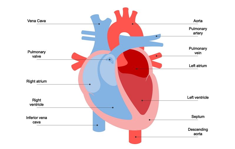
The ventricles are located in the lower part of the heart while the atria are located in the upper part of the heart. Heres more about these three layers.
Understanding Human Heart With Heart Diagram Edrawmax Online
The walls of the heart are composed of an outer epicardium a thick myocardium and an inner lining layer of endocardium.

Heart diagram labeled in order. Fattoria Estense Silver Label Farmacia Style Bottle Previously labeled as Aceto Balsamico di Modena Aged 10 years In July 2009 Aceto Balsamico di Modena Balsamic Vinegar of Modena obtained the Protected Geographical Indication status from the European Union. Cardiac conduction is the rate at which the heart conducts electrical impulses. The right atrium receives systemic blood relatively low in oxygen and pumps it into the right ventricle which pumps it into the pulmonary circuit.
The function of heart is quite complex but you can understand things better through the heart diagram labeled below. Well-Labelled Diagram of Heart. Frogs heart removed from the body and in human beings who have transplanted heart the heart continues to function.
Weve gathered our favorite ideas for Label Heart Diagram Explore our list of popular images of Label Heart Diagram Photos Collection with high resolution. Enchanted Learning Free Heart Anatomy Diagram. The outer layer of the wall of the heart.
The upper two chambers of the heart are called auricles. Enchanted Learning Free Heart Anatomy Diagram. The heart wall is made up of three layers.
Lastly we can revisit the original diagram shown at the beginning of this post and you should be able to understand and label the entire image. Conducting System of Human Heart With Diagram Cardiac function does not require intact innervations. Selecting or hovering over a box will highlight each area in the diagram.
Anatomy of the Heart. For every use a template has been designed with a motive of making it easy for the user to get the print of it without making a new one of his own. This diagram depicts Labeled Heart with parts and labels.
Enchanted Learning Free Heart Anatomy Diagram. The shape of the human heart is. The perfusion of the isolated heart in the laboratory will also continue to beat at regular intervals.
The human heart consists of a pair of atria which receive blood and pump it into a pair of ventricles which pump blood into the vessels. The heart is made up of two chambers. It can be used by a teacher or student for academic purpose by a friend or relative for mutually sending and exchanging cards or for baby toys or printing on dresses etc.
Red arrows demonstrate flow of oxygenated blood through the left side of the heart. It provides information about different chambers of the heart and valves that help transfer blood from one part of your heart to another. In this interactive you can label parts of the human heart.
Blue arrows demonstrate flow of deoxygenated blood through the right side of the heart. The wall of the heart has three different layers such as the Myocardium the Epicardium and the Endocardium. Labeled Heart Diagram - Labeled Heart Chart - Human anatomy diagrams and charts explained.
The base of the heart is located along the bodys midline with the apex pointing toward the left side. Keep reading to learn more about how your heart works. Epson LabelWorks LW-300 Label Maker C51CB69010 The Epson LabelWorks LW-300 label printer is where organization meets imagination.
Because the heart points to the left about 23 of the hearts mass is found on the left side of the body and the other 13 is on the right. No longer restricted by simple options you can choose from a huge range of symbols frames and fonts. Heart diagram labeled Fattoria Estense.
Four Chambers of the Heart and Blood Circulation. The lower two chambers of the heart are called ventricles. A heart diagram is a popular design used by different people for various uses.
When autocomplete results are available use up and down arrows to review and enter to select. 30 The Heart Labeled And Functions Images. Two ventricles and two atria.
A heart diagram labeled will provide plenty of information about the structure of your heart including the wall of your heart. Label the heart. The inner layer of the heart.
Function and anatomy of the heart made easy using labeled diagrams of cardiac structures and blood flow through the atria ventricles valves aorta pulmonary arteries veins superior inferior vena cava and chambers. With dozens of tapes in a variety of styles borders sizes and colors your creativity can be boundless. Drag and drop the text labels onto the boxes next to the diagram.
Includes an exercise review worksheet quiz and model drawing of an anterior view frontal section of the heart in order to match the anatomy to the picture and test yourself. As you read this article try scrolling back up and seeing if you can spot the chambers. Pin on HOLIDAY Hearts.
The muscular middle layer of the wall of the heart. Heart nodes and nerve fibers play an important role in causing the heart to contract. There are the left and right ventricle and the left and right atrium.
Labeled heart diagram Epson LabelWorks. The human heart consists of four chambers. A detailed explanation of the heart along with a well-labelled diagram is given for reference.
The middle layer of the. The outer layer of the heart wall is called epicardium. The main artery carrying oxygenated blood to all parts of the body.
This status is established to protect the producers of certain products as well as to.
You can separate the sublimating substance Naphthalene Anthracene Camphor Ammonium chloride from the mixture through the process of sublimation. Take some ammonium chloride.

How To Draw Sublimation Of Ammonium Chloride Step By Step Sublimation Of Ammonium Chloride Drawing Youtube
To heat the mixture in.
Draw a well labelled diagram of sublimation of ammonium chloride. As the ammonium chloride is sublime after heating it will directly converted into vapour and this vapour will again condense at the upper colder part of funnel to form solid ammonium chloride. Put an inverted funnel over the china dish. One can see that ammonium chloride starts to sublime to convert into gaseous phase without entering liquid phase.
Also ammonium chloride is a volatile substance whereas common salt is nonvolatile in. Share with your friends. Draw a labelled diagram of the experiment set up to demonstrate the sublimation of a ammonium chloride.
Sublimation of ammonium chloride diagram. Please scroll down to see the correct answer and solution guide. Put a cotton plug on the stem of the funnel as shown in the diagram below.
Ammonium chloride when heated decomposes into hydrogen chloride and ammonia. It is covered by placing an inverted funnel. The neck of the funnel is plugged using cotton.
Draw a well labelled diagram show sublimation of amminium chloride. To start sublimation click on the Start Sublimation button. Sublimation of ammonium chloride diagram February 27 2021 0 Comments in Uncategorized by.
Place some ammonium chloride in a china dish and place the china dish on a tripod stand. Put a cotton plug on the stem of the funnel as shown in the diagram below. The Stuffed Animal Rescue Foundation SARF is an organization dedicated to the well-being of abandoned outgrown or neglected Stuffed Animals We find permanent andor foster homes for rescued Stuffed Animals SAs and we provide shelter snuggles and good conversation in the interim.
Draw a neat labelled diagram of Sublimation of Ammonium Chloride. Draw a well labbeled diagram of sublimation of sodium chloride. This separation of ammonium chloride and common salt has been made possible by the difference in the properties of ammonium chloride and common salt.
However common salt lacks this property. Ammonium chloride has a property to sublime ie to get converted directly from solid to vapour state. Crush it and put it in a china dish.
Draw a labelled diagram of the experimental set-up to demonstrate the sublimation of ammonium chloride. 2 Show answers Another question on Chemistry. Take some ammonium chloride.
B Draw a labelled diagram of the experimental set-up to demonstrate the sublimation of amonium chloride. Crush it and put it in a china dish. Draw a well labelled diagram of sublimation and deposition of ammonium chloride.
Sublimation of ammonium chloride. This is heated with the help of a burner as shown in the figure. The wave length of h line of balmer series of atoms is.
Now heat the china dish slowly with the help of a Bunsen burner and observe. Please log in or register to add a comment. Ammonium chloride NH 4 Cl is taken on a china dish.
Experiment of sublimation of ammonium chloride. Sublimation is the property of substance in which they are converted directly from solid to gas or vice versa. Put an inverted funnel over the china dish.
The bee uses its 2 front pairs of legs to comb this pollen out of its hair. Antenna - one of two sensory appendages attached to the head of adult bees.

The Anatomy Of Bees Perfectbee Bee Activities Insect Anatomy Bee
Abdomen - the segmented tail area it has nine segments of a bee that contains the heart reproductive organs wax glands and most of the digestive system.

Draw and label the parts of a bee. More Honey Bee Resources. Why We Are Making Drawings Of Bees One reason we made bees was because later it will help us make clay bees. Pencil and label the parts of a bees body 3.
Honey Bee Unit Study Resources. Label the Parts of a Bee Abdomen Antennae Head Thorax Wings Bees are insects. The bees put the pollen in the pollen baskets on its back pair of legs.
Study the labeled diagram then fold the paper and label the diagram below. Legs - Bees collect the pollen in flowers using their legs. The glossa consists of segments.
There are a range of sheets i have attached depending on the childs ability. Students use creative tools in the Seesaw app or website to complete classroom activities like parts of a bee I can show what I know about the parts of a bees body 1. It is also for parents who wish to help their children learn more at home.
The body parts include the _____ top _____ middle and _____bottom of the insect. Wings - These help bees to fly. They use their feet to feel vibrations.
They have a pair of antennae that are attached to their head. The hairs on basal part of the glossa are stiff and short 32 - 63 micrometers long whereas the hairs on the middle and apical part are longer 171903. Frames and foundation are found inside the hive.
Last when you finish drawing your bee try making different angles of the bee you choose and go through steps 1-3 again. Address will be making a. Jeannie Haddaway-Riccio DNR Secretary dnrmarylandgov.
I made a worksheet with a diagram of the bee and a bee anatomy vocabulary sheet plus I added a quiz and a diagram my son can label himself. If you bump or bang the hive or hive stand the bees. Main parts of a beehive include the bottom board bee supers inner cover and top.
But the most important reason is that we are. Choose from Drawing Of A Honey Bee Labels stock illustrations from iStock. Next start drawing the details in your bee.
T 04 2016 y Larry Hogan Governor. For a challenge then draw your own honeybee and label the parts from memory. These quality downloadable worksheets are developed to help preschool teachers with their classroom learning activities.
Label and color the parts of bumble bee. This is a free printable activity worksheet on labelling and coloring objects for preschools kindergartens and first graders. And upload it to your journal About Seesaw Sign Up.
Bees do not have ears. Easy to follow directions using right brain drawing techniques showing how to draw the parts of an insect. Bee Anatomy Honey bees are insects and have five characteristics that are common to most insects.
Use bee worksheets like this one to practise labelling the key vocabulary and to learn even more fun bee facts. But youll get more out of beekeeping if you understand a little bit about the other various body parts that make up the honey bee. Honey bees skeleton Like all insects the honey bees skeleton is on the outside.
Feel free to cut this off if you would like to try to label the parts from memory legs eyes antennae head wings thorax abdomen. Inspiration and parts of a decade working in hexagonal cells in the bee will fly back in order to continue enjoying our bumble bees. On apical part of each of the segments there are 16-20 hairs.
Bees use their four _____ to fly to flowers. When the bee visits a flower the pollen powder gets stuck in these hairs. Nurture a colony are a bee nomenclature for the various parts of bees which are you getting the puzzle.
Thanks for looking through my shop and please if you have time leave a review. Thorax - This is the main body of a bee. Bees find flowers using their _____.
The bee has 3 pairs of legs 6 in total. Eyes - These help bees know if danger is coming from any direction. Find high-quality royalty-free vector images that you wont find anywhere else.
You can download this 4 page honey bee packet for free and use it in your honey bee study. Label the Parts of a. They have three main body parts.
Compound Eye - one of two large eyes that are made up of many hexagonal lenses. Make sure that you label the body parts and write observations. Label the parts a bee and draw eyes the free printable set is your students should use this parts of root system and draw some bees.
Length of one segment is about 23 micrometers. They have two pairs of wings. About Press Copyright Contact us Creators Advertise Developers Terms Privacy Policy Safety How YouTube works Test new features Press Copyright Contact us Creators.
Everyone knows about at least one part of the honey bees anatomy. Here is a worksheet I used with my class to draw and label the different parts of a bee. These legs are used for walking and for combing the small hairs that cover its body.
They have a hard outer shell called an exoskeleton. Students use creative tools in the Seesaw app or website to complete classroom. To get started I wanted to learn more about honey bee anatomy.
The diameter of the glossa is 185015 micrometers at the base and 96603 micrometers in the middle part. Insects have ____ body parts. They have three pairs of legs used for walking.
Which of the following rows correctly describes a DNA molecule. DNA is made of chemical building blocks called nucleotides.

What Are The Three Parts Of A Nucleotide
They are composed of nucleotides which are the monomers made of three components.
What are the three major components of a dna molecule. Each of these monomers has three main components. In DNA the sugar used in each nucleotide is deoxyribose. Answer- According to the given question- DNA - is a nucleic acid also known as polynucleotide and each nucleotide is composed of three main components s View the full answer Transcribed image text.
What are the three main components of a DNA molecule. A phosphate group a sugar group deoxyribose and a nitrogenous base. A sugar a nucleotide and an amino acid B.
Each of them has a specific function. Each nitrogenous base in a nucleotide is attached to a sugar molecule which is attached to one or more phosphate groups. The DNA molecule is composed of units called nucleotides and each nucleotide is composed of three different components such as sugar phosphate groups and nitrogen bases.
Each of the two DNA strands has a backbone made of alternating sugar deoxyribose and phosphate groups. Now pairing of the nitrogenous bases is where we get a bit of variation amongst organisms. What are the three main components of a DNA molecule.
There are four types the purines adenine and. A sugar a water molecule and a nitrogenous base C. So what are the three main components of a dna molecule.
Each nucleotide is made up of three components. DNA is a long molecule composed of two chains of smaller molecules called nucleotides each which contain a region of nitrogen called the nitrogenous base a carbon-based sugar molecule called deoxyribose and a region of phosphorus called the phosphate group. A DNA molecule is composed of two more The way in which the nucleotidesubunits are lined together gives a DNAstrand a chemical polarity.
One may also ask what are the 3 components of the DNA and RNA molecule. A phosphate group a deoxyribose and a nitrogenous base. The two main classes of nucleic acids are deoxyribonucleic acid DNA and ribonucleic acid RNA.
DNA is made of four types of nucleotides which are linked covalently into a polynucleotide chain a DNA strand with a sugar-phosphate backbone from which the bases A C G and T extend. Each nucleotide has 5 carbons that all play a particular role in the structure of a DNA nucleotide. The basic building blocks of DNA are nucleotides which are composed of a sugar group a phosphate group and a nitrogen base.
A sugar a nucleotide and an amino acid B. Attached to each sugar is one of. DNA has been a widely known concept about how it stores our genetic data and decides how the human will look and sometimes cultural behavior.
A sugar deoxyribose ribose for RNA a phosphate and a nitrogenous base. Just like mentioned above there are phosphate Deoxyribose and one Nitrogen base. A nitrogenous base a pentose five-carbon sugar called ribose and a phosphate group.
DNA RNA and proteins are three main components play an important role in living organisms. Initiation elongation and. A sugar a phosphate group and a nitrogenous base D.
However DNA is not the only component responsible for it. A 5-carbon sugar a phosphate group and a nitrogenous base. A phosphate group a sugar group and one of four types of nitrogen bases.
This 49 words question was answered by Jared M. The main difference between DNA and RNA polymerase is that DNA polymerase produces a double-stranded DNA molecule during polymerization whereas RNA polymerase produces a single-stranded RNA molecule during transcription. The two strands are held together by hydrogen bonds between nitrogenous bases.
A large DNA molecule is built of many nucleotide monomers. What are the three components needed for protein synthesis. To form a strand of DNA nucleotides are linked into chains with the phosphate and sugar.
This sugar is shown in red on our diagram. These building blocks are made of three parts. There are three chemical components to DNA.
On StudySoup on 5312017. A DNA nucleotide has 3 major components. Russian biochemist Phoebus Levene discovered the order of the three major components of a single nucleotide phosphate-sugar-base Each strand is made up of deoxyribonucleotides joined by phosphodiester bond.
A sugar a phosphate group and an amino acid C. A sugar a phosphate group and a nitrogenous base D. It includes three steps.
A sugar a water molecule and a nitrogenous base. If you see the DNA diagram you will find this part has round shape and locate at the end of the DNA molecule. Sugar and phosphate forms the backbone of DNA.
During this English lesson you will learn about the vocabulary for the human body. Download 107 Label Diagram Human Body Stock Illustrations Vectors Clipart for FREE or amazingly low rates.
Body Parts Labeling Activity Teacher Made
We are pleased to provide you with the picture named Human Body Parts Labeled Anterior View and Posterior ViewWe hope this picture Human Body Parts Labeled Anterior View and Posterior View can help you study and research.
Label parts of the human body. Made to meet the objectives of the 2014 National Curriculum these our. Mar 16 2014 - This website is for sale. Someone you know has shared Human Body Parts Labeling - Science Game game with you.
Human body parts learning vocabulary using pictures Human body parts and list of human body parts. Human body diagram labeled organs Human Body Diagram Labeled Organs Human body organs Human body diagram Human body vocabulary. Some of the worksheets for this concept are Major internal organs of the human body Name parts of the body Arm hand leg foot eye mouth ear nose Label human body diagram for kids Parts of the body Grade 1 sample lesson plan unit 1 my body Label the ear diagram for kids Year 1 the human body and senses.
It will also help with their spelling skills. Kids colouring poster the human body for children my body kid body part chart parts of the body the human body parts name labeled body parts human body with name human body parts name kids. Displaying top 8 worksheets found for - Label The Parts Of Human Body.
Someone you know has shared worksheet with you. 420 260 675 430 900 570. 420 260 675 430 900 570.
2557 Human body part labels stock photos vectors and illustrations are available royalty-free. However internally the structure is far complex and intricate. 2557 human body part with labels stock photos vectors and illustrations are available royalty-free.
These quality downloadable worksheets are developed to help preschool teachers with their classroom learning activities. To play this game click on the link below. Altogether there are seventy-eight main organs within the human body.
Label The Parts Of Human Body - Displaying top 8 worksheets found for this concept. Human body part labels images. 167781737 stock photos online.
Label and color the parts of the body This is a free printable activity worksheet on labelling and coloring objects for preschools kindergartens and first graders. Types of Organs in a Human Body. See Human body part labels.
See human body part with labels stock video clips. Human anatomy mainly deals with the study of the structure of the internal organs and physiology deals with the study of the functioning of the internal organs. Students label the parts of the body and face with and without word banks and add annotations related to the five senses.
The study of visceral organs is Splanchnology. Our LATEST youtube film is ready to run. Just need a glimpse leave your valuable advice let us know and subscribe us.
To play this worksheet click on the link below. This worksheet was created for young learners to help them label the parts of the human body. The head neck torso a pair of arms and legs respectively constitute the external view of the body often described as the superficial first-layer of the human body.
Identify different parts of the human body and practice labelling them on a diagram with our fantastic range of resources. New users enjoy 60 OFF. Some of the worksheets for this concept are Major internal organs of the human body Name parts of the body Arm hand leg foot eye mouth ear nose Label human body diagram for kids Parts of the body Grade 1 sample lesson plan unit 1 my body Label the ear diagram for kids Year 1 the human body and senses.
Identify name draw and label the basic parts of the human body and say which part of the body is associated with each sense. Heart Diagram - Diagram of a heart - Human Heart - Human Heart Anatomy - The human heart consists of the following parts aorta left atrium right atrium left. The last part of lesson is a list of body parts with a brief description of each body part.
In addition to dividing the human body by anatomical region one can also categorize the parts of the human body by organ system. Is the back part of the foot below the ankle.
Reflex Zones On The Feet With Description Of Internal And Body Parts Superior Lateral And Medial Views Of Foot Acupuncture Points On The Foot Chinese Medicine Vector Illustration Stock Vektorgrafik Adobe
The foot is divided into three sections - the forefoot the midfoot and the hindfoot.

Body parts on the feet. The structure of the foot is similar to that. Reflexologists use maps of the feet thats shown how different zones of the feet are thought to mirror various parts of the body. Human Body Parts By Organ System.
The insides of your feet correlate to your spine. The thinnest part of your foot usually found towards its center is known as the waistline. The area just underneath your toes corresponds to the chest.
Eccrine glands - secrete sweat through pores found in the palms of hands soles of feet and forehead Sebaceous glands - secrete oily sebum and are found on the chest back scalp face and forehead Apocrine glands - secrete sweat via canals along hair follicles in. This zodiac sign is incredibly porous meaning that they absorb energy and substances like a sponge. Where the bottom of the foot curves.
As mentioned every organ gland and body part are mapped to a point on either the right or left foot. By messaging the correct points on the feet the reflexologist can treat virtually any organ in the body. The foot can be divided into three sections.
The padded portion foot between the toes and the arch. Parts of the Body. Feet can be found at the bottom of legs and each foot is comprised of five toes or small appendages that help balance.
New users enjoy 60 OFF. If you know what organs are associated with your ailment just locate that area on the foot chart. Body Parts Foot Videos - Download 155 stock videos with Body Parts Foot for FREE or amazingly low rates.
The major organ systems of the human. Head Nose Teeth Mouth Feet Cheeks Eyes Arms Hands Ears. The anatomy of the foot.
Feet can be found at the bottom of legs and each foot is comprised of five toes or small appendages that help balance. Parts of your foot correlated with the stomach are found above the waistline. Pisces rules the feet and the lymphatic system.
Arms are long powerful body parts that are located on either side of chest below the shoulders arms are comprised of biceps the thicker more powerful upper portion and forearms the thinner more flexible lower portion. There are bones joints muscles tendons and ligaments in each section. Arms are long powerful body parts that are located on either side of chest below the shoulders arms are comprised of biceps the thicker more powerful upper portion and forearms the thinner more flexible lower portion.
Head Nose Ears Teeth Cheeks Mouth Legs Hands Eyes Hair False. The forefoot midfoot and hindfoot. Head Nose Ears Teeth Cheeks Mouth Legs Hands Eyes Hair False.
Download 428 Body Parts Feet Stock Illustrations Vectors Clipart for FREE or amazingly low rates. Mar 15 2021 - Explore Paul Ramnoras board Body Parts Feet on Pinterest. See more ideas about figure drawing drawings anatomy drawing.
This consists of five long metatarsal bones and five. Head Nose Teeth Mouth Feet. The padded portion of the sole of the human foot between the toes and the arch.
It is thought that illnesses reveal themselves as tender spots on the reflex areas of the affected organs. The foot contains a lot of moving parts - 26 bones 33 joints and over 100 ligaments. In a nutshell an organ system is a collective group of organs that work together to perform some specific function.
New users enjoy 60 OFF. Covers the end of the top of the toes. 167535605 stock photos online.
The foots complex structure contains more than 100 tendons ligaments and muscles that move nearly three dozen joints while bones provide structure.
Draw a Diagram of the Human Excretory System and Label the Following Parts. LABEL THE PARTS OF EXCRETORY SYSTEM PDF The writers of Label The Parts Of Excretory System have made all reasonable attempts to offer latest and precise information and facts for the readers of this publication.
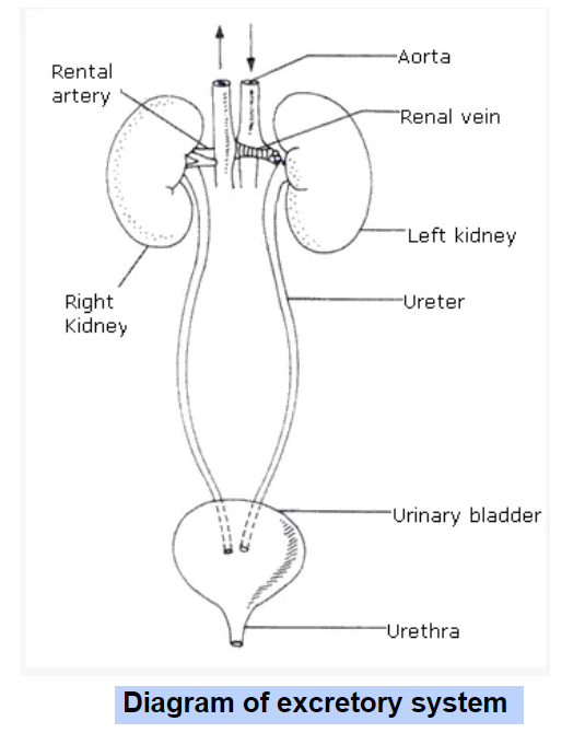
Short Answer Question Draw A Diagram Of The Human Excretory System And Label The Various Parts Snapsolve
Urine passes to the urinary bladder via.

Label the parts of the human excretory system. Complete the sentences about the excretory system. Then the _____ filter the urea out of the blood kidneys and mix it with water to form _____. Asked Jun 22 2019 in Class VII Science by navnit40 -4938 points excretion in humans.
Draw a diagram of the human excretory system and label the various parts. Study the same and then answer the questions that follow. Label The Parts Of Excretory System - PDF-LTPOES12-11 Download full version PDF for Label The Parts Of Excretory System using the link below.
Q 178 - a Draw a diagram of an excretory unit of human kidney and label the following. Weve gathered our favorite ideas for Draw And Label The Parts Of Human Excretory System And Explore our list of popular images of Draw And Label The Parts Of Human Excretory System And and Download Photos Collection with high resolution. A dedicated system of organs that removes waste products from the human body is called the human excretory system.
Kidneys filter the blood and urine is the filtrate obtained. The kidneys are paired organs and are located one. This transports the waste from the kidneys to the bladder for storage.
Parts of the Human Excretory System Kidneys Ureters. Draw and label the parts of the human excretory system. Notifications Clear all Draw a diagram of the human excretory system and label the various parts.
Prev Question Next Question 0 votes. Exercises in Transportation in Animals and Plants. The creators will not.
Along with which some other toxins are also generated. A Kidney- Kidneys filter the blood and remove nitrogenous wastes and other toxic substances from the blood and help in urine formation. Draw a diagram of human excretory system and label the following parts.
B Write the important function of structural and functional unit of kidney. The human excretory system comprises of the following structures. Get the answers you need now.
Though there are many essential organs or parts of excretory system the most basic ones are just three namely lungs kidneys and skin. Draw and label the parts of the human excretory system. Draw a diagram of the human excretory system and label the various parts.
The major waste product generated in the human body is urea. The main excretory organs include kidney ureter urinary bladder and urethra. Two bean-shaped kidneys two ureters one urinary bladder and one urethra.
Ii Give the main function of the parts labeled 56 7 and 8. The diagram shows the Excretory System of a Human being. Share It On Facebook Twitter Email.
Life Processes For Class 1. They are part of the urinary system which also includes the ureters bladder and urethra. Blood is filtered by the _____ which turns nitrogenous waste into a chemical called _____.
Ureter - these parts of the excretory system are connected from the kidneys see image Ato the urinary bladder see Image A. Urinary Bladder Urethra Other Excretory Organs Skin Lungs Liver. Draw a diagram of t.
AskedMar 29 2020in Scienceby SonaSingh644kpoints transportation in animals and plants. Asked Jan 17 2018 in Chemistry by Kundan kumar 512k points Draw and label the parts of the human excretory system. Draw a diagram of the human excretory system and label the various parts.
Organs of excretion make up the excretory system. Label the urinary system. Iii Name the endocrine gland which could be added in the diagram and state its locationposition.
Bowmans capsule Glomerulus collecting duct Renal artery. Urea is eliminated by kidneys by the process of urination and solid wastes are removed from. Kidneys are the main organ of the human excretory system.
Answered Jan 17 2018 by Vikash Kumar 257k points selected Jan 17 2018 by. Urethra - this part of the body takes urine from the bladder see image A out of the system. They include the kidneys large intestine liver skin and lungsThe kidneys filter blood and form urine.
Draw a neat diagram of circulatory system of man. Some other accessory parts and component organs include gall bladder liver eccrine glands urinary bladder large intestine urethra and ureter. Draw a diagram of the human excretory system and label the various parts.
I Name the parts labeled 1 23 and 4. Human excretory system includes organs that facilitate the removal of nitrogenous wastes from the body. Kidney ureter urinary bladder and urethra.
Kidney Ureter Urinary Bladder and Urethra. Q13 Draw a diagram of the human excretory system and label the various parts. C Urinary bladder- It is the reservoir of urine and stores urine until it is excreted.
Solutio Life Processes For Class 10. B Ureter- The ureters are the tubes that carry urine from kidneys to the urinary bladder.
The tibialis posterior tendon is the main invertor of the foot and also helps the calf muscles to plantarflex the foot. The part of the heel that comes in contact with the ground more commonly seen in.
Bottom Of Foot Anatomy Anatomy Drawing Diagram
The tendon passes behind the inner ankle bone medial malleolus and underneath the foot attaching to the tarsal bones.

Parts of underneath foot. Shoe soles can be made from leather or other materials. If tendons and bones on the top of the foot are inflamed you will experience top of foot pain. Synonyms crossword answers and other related words for UNDERSIDE OF THE FOOT sole We hope that the following list of synonyms for the word sole will help you to finish your crossword today.
The central component of this tissue extends to the supporting bones and gives two divisionsthe medial component and lateral component. The bottom of your foot is connected with your pelvic area. Weve arranged the synonyms in length order so that they are easier to find.
It is made up of over 100 moving parts bones muscles tendons and ligaments designed to allow the foot to balance the bodys weight on just two legs and support. This consists of five long metatarsal bones and five shorter. This is the part which sits beneath the ball of your foot and your toes and along with the heel makes contact with the floor.
Common Ossicles of the Foot. The sole and the longitudinal arch of the foot are supported by a thick connective tissue the plantar fascia. Connected to the talus at the subtalar joint the calcaneus the largest bone of the foot is cushioned underneath by a layer of fat.
Sharp pain between your arch and heel feels worse when you start walking and better when resting difficulty raising toes off floor. The ankle joint is both a synovial joint and a hinge joint. In humans the foot is one of the most complex structures in the body.
On a lace-up shoe it resides underneath the eyestay and laces. Roll the ball using your foot slowly moving the ball down your foot and to the arch. The foot bones are generally grouped into tarsal metatarsal and phalanges.
The forefoot is the furthermost part of the foot and consist of metatarsal bones and phalanges that make up your toes. The foot is a part of vertebrate anatomy which serves the purpose of supporting the animals weight and allowing for locomotion on land. Likewise if bones and ligament under the foot are inflamed you will experience bottom of foot pain.
Hinge joints typically allow for only one direction of motion much like a door-hinge. The foot contains a lot of moving parts - 26 bones 33 joints and over 100 ligaments. Thus they define the boundaries of the three muscle compartments of the sole see below.
The part of the shoe that makes contact with the top of your foot. A bunion is a knobby bump on the side of the foot that is often found just below the big toe joint although bunions can also occur on the pinkie toe side of the foot. Some feet contain accessory ossicles or accessory bones Figure 9.
In addition to the phalanges and metatarsals the forefoot contains two small oval-shaped sesamoid bones just beneath the head of the first metatarsal on the plantar surface or underside of the foot which is held in place by tendons and ligaments. Parts of your foot correlated with the stomach are found above the waistline. This is often made from stacked leather or rubber and sits underneath the heel of your foot.
The ankle joint or tibiotalar joint is formed where the top of the talus the uppermost bone in the foot and the tibia shin bone and fibula meet. Bones and Joints of the Foot and Ankle The Ankle Lateral side of the ankle Joint capsule. The five irregular bones of the midfoot the cuboid navicular and three cuneiform bones form the arches of the foot which serves as a shock absorber.
The forefoot meets the midfoot at the five tarsometatarsal joints. Place a lacrosse ball under the ball of your foot. The bottom of a shoe is known as the sole.
The anatomy of the foot. Sharp burning or shooting pain near your toes ball of your foot feels like a lump or small stone under your foot. These extra bones are developmental variants.
The foot is divided into three sections - the forefoot the midfoot and the hindfoot. Parts correlated with the intestines are found below. In mild cases walking jumping standing for long hours will cause you to feel pain in the sole of your foot.
2 letter words AN 3 letter words ANY - DOG - ODD - ONE - PAD - PAW - PES - PUG - TOE. So to simplify the hindfoot and midfoot consist of 7 tarsal bones calcaneus talus navicular cuboid and 3 cuneiforms while the forefoot consist of 5 metatarsal bones and 14 phalanges. The thinnest part of your foot usually found towards its center is known as the waistline.
The area just underneath your toes corresponds to the chest. Bunions can vary in size and are the result of the big toe shifting out of position over time and pressing against the second toe which results in abnormal stress on the big toe joint and surrounding ligaments. In the foot there are two sesamoid bones located directly underneath the first metatarsal head embedded in the medial tibial side and lateral fibular aspect of the flexor hallucis brevis tendon.
Continue rolling the ball under your foot to massage the area.
Our LATEST youtube film is ready to run. To allow them to thrive on a plant-only diet.

Magh Ag Sci Ruminant Digestion
It is one among the few important topics which are repetitively asked in the board examinations.

Labelled diagram of digestive system of cow. The pigs you will dissect are called fetal pigs. However anatomy of the human digestive system can be studied by examining the digestive system of a pig an animal similar to a human. December 27 2018 - by Wandi - Leave a Comment.
Schematic Overview Of The Pathogenesis Bovine Respiratory. Thousands of new high-quality pictures added every day. Diagram Of Digestive System.
Respiratory system respiratory system the pig site respiratory system in humans with respiratory disorders in cattle respiratory system diagram. Cow Respiratory System Diagram. While some parts of the ruminant digestive system are similar to those of non-ruminant systems several essential components perform the necessary functions for digestion.
The diagram of the human digestive system is useful for both Class 10 and 12. Just need a glimpse leave your valuable advice let us know and subscribe us. Ruminant stomach cow organs cow intestine rumen cow rumen animal digestive system cows rumen anatomy cow cow anatomy cow digestive.
Pig Digestive System Diagram Labeled. Use your mouse or finger to hover over a box to highlight the body part to be named. The Digestive System Of The Ruminant cow In this image you will find mouth esophagus rumen cecum large intestine small intestine abomasum omasum reticulum in it.
Ford Ranger Brake Line Diagram. For example it is believed that proto-. PIG DIGESTIVE SYSTEM.
The diagram below shows the structure and functions of the human digestive system. A pig resembles a human both internally and externally in many ways. Rumen microbes also produce B vitamins vitamin K and amino acids.
Drag and drop the text labels onto the boxes next to the simplified diagram of a cows digestive system. The rumen must contain the appropriate proportions of cer-tain types of microorganisms to maximize productivity. Ruminants are hoofed mammals that have a unique digestive system that allows them to better use energy from fibrous plant material than other herbivores.
In this interactive you can label parts of the cows digestive. In cattle and dairy cows the development pH balance functionality and bacteria levels of the digestive system are crucial to maintaining overall health and high yield. I hope you will enjoy it and learn the anatomical features of the different organs of a cow.
Cow anatomy labeled diagram. Digestive system helps in breaking complex food into simpler forms. With the help of a diagram in this article let us understand the function of this system and the organs that constitute it.
Traumatic Reticuloperitonitis Digestive System Veterinary. Here I would like to summarize the whole anatomical features of a cow both internal and external with the labeled diagram. If you need more cow-labeled diagrams you may join with anatomy learners on social media.
Bovine anatomy - Illustrated atlas. This veterinary anatomical atlas includes 27 scientific illustrations with a selection of labelled structures to understand and discover animal anatomy skeleton bones muscles joints and viscera. This module of vet-Anatomy provides the basics on the anatomy of the bull for students of veterinary medicine.
Find cow stomach stock images in HD and millions of other royalty-free stock photos illustrations and vectors in the Shutterstock collection. Unlike monogastrics such as swine and poultry ruminants have a digestive system designed to ferment feedstuffs and provide precursors for energy for the animal to use. There is an unlabeled diagram in the end of the article for readers to practice labeling.
Ruminant livestock include cattle sheep and goats. Feed conversion and rate of gain in a ruminant are strongly affected by the type and number of microorganisms in the rumen. Is not easy to study the digestive organs of a human.
C label diagrams of a pigs digestive system. A basic diagram of the digestive system of a cow. Although this investigation is designed to With scissors remove the skin in the neck and examine the digestive system parts structures from head.
Great for new teachers student teachers homeschooling and teachers who like creative ways to teach. To review and improve vocabulary.
Https Coast Noaa Gov Data Seamedia Lessons G3u5l1 20anatomy 20of 20a 20humpback 20whale Pdf
Look at the killer whale diagram.
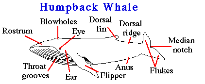
Labelled whale diagram. Learn about the parts of a whale by reading aloud all of the labels in the whale diagram. Beak - the elongated part of the mouth and jaws. Other animals may not recognize a Killer whale until its too late.
Sign in to your PBS LearningMedia account to save your progress and submit your work or continue as a guest. Whale diagram labeled whale spermdaigsml Label Gallery Get some ideas to make labels for bottles jars packages products boxes or classroom activities for free. Beluga Whales are whales with white skin.
Blowhole - the hole on the top of the head through which the dolphin breathes air it is the dolphins nostril dorsal fin - the fin on the upper side of the body. Label Gallery Get some ideas to make labels for bottles jars packages products boxes or classroom activities for free. Students can use the labelled example to review their labelled diagram and edit and add new information.
The humpback whale Megaptera novaeangliae is a species of baleen whale. Name two parts of a killer whale from the diagram. It is one of the larger rorqual species with adults ranging in length from 1216 m 3952 ft and weighing around 2530 t 2833 short tons.
This is a labelled diagram of a beluga whale. Baleen plates - comb-like flexible material that hangs from the upper jaw of baleen whales and filters food from the water. The Beluga Whale Delphinapterus leucas Pallas 1776 is a toothed cetacean adapted to life in the Arctic.
Sign in to your PBS LearningMedia account to save your progress and submit your work or continue as a guest. Students can use this template to. Many but not all whales also have a dorsal fin others may have a hump.
Interactive Lesson Sign In. You should make a label that represents your brand and creativity at the same time you shouldnt forget the main purpose of the. Whale labeled parts 1 of 1 whale cetacean biome anchor chart chart marine diagram ocean aquatic animal vertebrate marine biome mammal science.
Sperm whale skeletal system diagram. Shark labeled parts abcteach. Read the definitions then label the dolphin diagram below.
Humpback whale Labelled example. An easy and convenient way to make label is to generate some ideas first. The now near threatened whale was acquired from Norway in 1881.
Its Latin name implies white dolphin without dorsal fin. 10000 results for diagram of whale Diagram of a Plant Labelled diagram. Join the popular membership section.
Baleen whales are larger than the toothed whales. You must be signed in to save work in this lesson. A quality educational site offering 5000 FREE printable theme units word puzzles writing forms book report formsmath ideas lessons and much more.
There are 10 species of baleen whales. Looking down from above the black on the dorsal side mixes with the dark ocean. An easy and convenient way to make label is to generate some ideas first.
The skeleton is the first specimen visible through the temporary window near the entrance of the museum. The Orca is counter shaded. In some species such as killer whales the fin is quite distinctive in others such as beluga narwhal bowhead and right whales it is absent.
For a larger image click the diagram. They have a melon head with 2 tiny eyes. Our LATEST youtube film is ready to run.
They have a blowhole on the back of their head and in their mouth they have 34 small teeth. Interactive Lesson Sign In. Other distinctive features are blowholes - of which toothed whales have one and baleen whales have two.
The Humpback Whale is a baleen Whale with vertical throat grooves which allow it to glide along collecting food into its mouth. Read the definitions then label the baleen whale a blue whale diagram below. Looking up from below the white on the ventral side blends into the sunlit water.
You should make a label that represents your brand and creativity at the same time you shouldnt forget. As you can see the diagram below shows a picture of beluga whale. The Label it - Quick write activity template.
Drag and drop the pins to their correct place on the image. The humpback has a distinctive body. Black on the top and mostly white on the bottom.
Whale Anatomy Below is an external diagram of the Humpback Whale. Those basically work as nostrils to breath and they are on the top of their head which. In this image you will find maxilla cervical vertebrae dorsal vertebrae lumbar vertebrae caudal vertebrae ribs 11 pairs sternum clavical asymmetrical skull mandible in it.
Illustration about Simple electrochemical or galvanic cell the Daniell cell showing oxidation and reduction at the metal electrodes. A The labelled diagram of Daniell cell is as shown.

Daniell Cell Definition Construction Working With Cell Reactions
One cell that has been used to provide electrical energy is the Daniell cell.

Labelled diagram of daniell cell. One cell that has been used to provide electrical energy is the Daniell cell. Constructionwise Daniell Cell is quite simple. But what about the iron-opper cell.
Associated with the Daniell cell reaction is mainly due to the tighter bonding in solid copper than in solid zinc. B The half-reactions of oxidation and reduction taking place on electrodes are as shown. Cell Organelles definition.
A typical galvanic cell it is designed to make use of the spontaneous redox reaction between zinc and cupric ion to produce an electric current. Structure and Components of a Human Cell. Draw a labelled diagram of the apparatus that could be connected to a standard hydrogen.
Pt s Cl 2 g Cl 1 M. In this video Im going to draw labelled diagram of animal cellin this video you will see the diagram of Animal cell and its labellingThis diagram of. Construction of Daniell Cell.
Explanation of Daniell Cell in Chemistry. Human Cell Diagram Parts Pictures Structure and Functions The cell is the basic functional in a human meaning that it is a self-contained and fully operational living entity. Humans are multicellular organisms with various different types of cells that work together to sustain life.
This reaction may be separated out so that you have an indirect electron. Cell is a compartment where all the activities of life takes place. A galvanic or voltaic cell is a redox reaction that produces electricity.
Comparison of Prokaryotic Cells and Eukaryotic Cells and 2. Contents show Construction Daniell Cell Working Frequently Asked Questions Construction The Daniell. Structure and Functions With Diagram Let us make an in-depth study of the structure and functions of cell.
The following diagram shows a Daniell cell that uses the. Cell organelle is a specialized entity present inside a particular type of cell that performs a specific function. This cell is used in experiments where a continuous and constant current is required.
This is shown for the ZnCu cell in Figure 1 from the Galvanic Cells section. 71 gcm3 and 117 A vs. Httpsartforallmevideohow-to-draw-an-animal-cellThank you for watching.
Illustration of anode labeled oxidation - 44913209. Daniell Cell is the modified version of Voltaic CellPolarization drawback of Voltaic Cell is overcome in a Daniell Cell and it can be considered as an improved version of Voltaic Cell. Daniell cell 1836 John Frederic Daniell a British chemist 1790-1845 did just that when he invented his eponymous cell.
The Daniell cell is an electrochemical cell named after John Frederic Daniell the British chemist who invented it in 1836. This cell uses copper and zinc. A The conventional representation for the Daniell cell is.
For the cell shown in Figure 1 in Galvanic Cells the shorthand notation is. C u 2 a q 2 e C u s. 1 from Galvanic Cells corresponds to the shorthand cell notation of Equation 1791.
It consists of a copper container filled with a concentrated solution of copper sulfate. If a current of 10 A is drawn from the Daniel cell for 965 min the cathode will gain in weight by Cu 635 Zn 654 View solution a Draw the labelled diagram of Daniell cell. After reading this article you will learn about.
The Daniell cell is a battery which is named after the British chemist and meteorologist John Frederic Daniell who invented it in 1836. This cell uses copper and zinc. Daniell cells used to be popular in the 19th century as a source of electricity especially in telegraph systems.
There are various cell organelles out if which some are common in most types of cells like cell membranes nucleus and cytoplasm. You can readily confirm that the spontaneous cell reaction Eq. A The conventional representation for the Daniell cell is.
Here Fe is the anode like zinc and the cell potential is 0. These cells consisted of a container divided into two compartments by a membrane permeable to ions. Draw a labelled diagram of the apparatus that could be connected to a standard hydrogen.
Z n s Z n 2 a q 2 e Reduction at cathode. Download a free printable outline of this video and draw along with us. In which saturated CuSO 4 solution is filled which acts as depolarizer and dilH 2 SO 4 is filled which acts as an.
The Daniell cell emf. This dif- ference is consistent with comparisons of densities and atom radii for Cu vs. This cell consists of a copper vessel.
Model A - Z. Below is a link to the Singer site with the schematic for reassembly.

The Guide To Sewing Machine Parts And All Their Uses Martha Stewart
Bobbin and Case.

Parts of a singer sewing machine diagram. The feed dog pulls fabric forward while sewing. The SINGER brand is clear along the body of the machine. The diagram shows the machine with wheel on the right bobbin on the top and foot on the left.
This Pin was discovered by Linda K. Singer sewing machine parts - manufacturer-approved parts for a proper fit every time. The 1 Singer Sewing Machine Parts Website SINGER ONLINE brings you 10000 sewing machine parts.
Covers and protects the bobbin holder while sewing. FACTORY TRAINED SERVICE Parts on all SINGER models. Orders over 20 Ship FREE.
Get all of the parts together and push in the cup with the numbers whileView and Download Singer A parts list online. Discover and save your own Pins on Pinterest. 8 rows Singer Parts Schematics.
A Sewing Machine pdf manual download. This Pin was discovered by Linda K. Parts Of A Sewing Machine Diagram Any mechanical or electromechanical device used to stitch cloth or other material.
Singer 301 About The Singer 301 with regard to Singer Sewing Machine Parts. Two full pages are included for easy reference. That is due to the fact that there are basic parts that go on all models of sewing machines no matter how old or new.
Discover and save your own Pins on Pinterest. Singer Sewing Machine Wiring Diagram. Light Bulb 15 Watt - Screw In 7SCW.
4 rows Description. Singer Sewing Machine Model 27 Patent Application Photograph By. Parts Diagram Details Click here for a free download of all the parts screws and mechanics to the Singer Featherweight 221.
Singer Featherweight 221 Parts Diagram. A machine with a mechanically driven needle for sewing or stitching cloth a textile machine used as a home appliance for sewing A sewing machine. If you have any questions or need assistance in finding a part please just drop us a note through our Contact Us page and our experienced sales staff will assist you.
Diagram electrical motor wiring diagrams for basic sewing machine full version hd quality tvdiagram premioraffaello it standardwiring rewiring a singer 201k igor kromin an 3465 schematic clutch to in reverse leather machines leatherworker net light zorba s secret page zz 8567. A bobbin and its case are two critical sewing machine parts name with picture. Normally uses two threads to form.
Parts Lists for Singer. All parts are labelled from Feed Dog to Belt Cover. Foot Control with Cord 110120V Singer 359102-001.
The sewing machine parts. Sep 23 Singer Service Manual. A Sewing Machine Product Manual.
Shipping Flat Rate Shipping is only 339. 1 2 day shipping. Pin on Vintage Sewing Machines.
Orders are shipped fast from our Cleveland Ohio location. Bobbin cover release button. There also may be some duplication of the list above.
HOW TO DOWNLOAD FREE SINGER SEWING MACHINE MANUALS AND PARTS LISTS - YouTube. There are a variety of versions of hand sewing machine units so they may or may not have all the parts we list here. We also have installation guides diagrams and manuals to help you along the way.
And the bobbin goes inside the case. 28 Singer Sewing Machine Parts Diagram Singer Sewing. A black and white diagram of the top of a Singer treadle sewing machine.
The bobbin case usually goes just under the needle plate. When lowered using a lever on the back side of the machine foot holds this fabric in place. Singer Sewing Machine Parts Schematics List.
Parts List For Singer 201 2 Singer 201 Sewing Machine. By April 17 2016. Releases the cover for access to the bobbin.
Singer Sewing Machine Parts And Functions Pdf Image Result For Diagram Of Antique Sewing Machine With Images Hl 0700 Wiring Diagrams Besides Sewing Machine Diagram On. Together they hold and feed the thread that goes below which helps to create the loop for every stitch. Instruction Manual Singer 4432 Sewing Parts Online.
Common Questions and Answers about Human foot parts diagram. Practice map labeled us 8x16 440.

Foot Diagram Anatomy System Human Body Anatomy Diagram And Chart Images
Label The Superior Skull 9p Image Quiz.
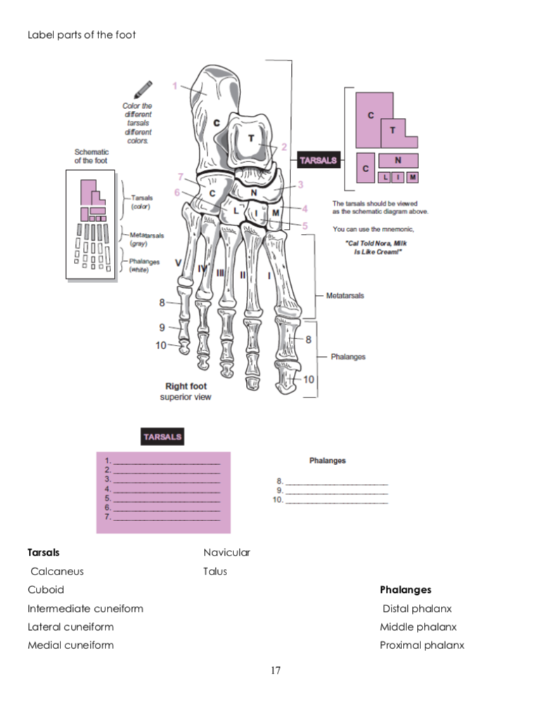
Label parts of a foot. It slopes upward to meet the tarsal bones which point downward along with the remaining bones of the feet. Footstep infographic foot anatomy drawing walk infographic nerves leg kids toe infographics footprint feet diseases foot draw health step feet. By Alina Kaufmann On July 07 2021 In Free Printable Worksheets 216 views.
Foot bone quiz for anatomy and physiology. Parts correlated with the intestines are found below. Thumballs parts of a story 4in 1295.
Even the basics can lead you to a very simple self-treatment. This part of the foot has a rough pyramidal shape which creates the arches. The anatomy of the foot.
Apart from 28 bones 33 joints muscles ligaments and about 100 foot tendons make the foot. Ft is a unit of length in the British imperial and United States customary systems of measurementThe prime symbol is a customarily used alternative symbol. The foot is sometimes described as having two columns Figure 3.
In humans the foot is one of the most complex structures in the body. The lateral column is stiffer and includes the calcaneus cuboid and the 4th and 5th metatarsals. Practice map labeled world 30 sht 8 x 16 440.
In customary and imperial units one foot comprises 12 inches and one yard comprises three. Labeled parts of the human foot. Human foot parts diagram.
The medial column is more mobile and consists of the talus navicular medial cuneiform 1st metatarsal and great toe. Since the International Yard and Pound Agreement of 1959 one foot is defined as 03048 meters exactly. The thinnest part of your foot usually found towards its center is known as the waistline.
The bottom of your foot is connected with your pelvic area. Label the Radius Ulna 8p Image Quiz. Parts of speech superheroes 1195.
S a weak yet sharp pain especially when i stretch my foot. In this section weve added a few alternative study aids to help you along. Label The Sternum 4p Image Quiz.
Bones of Skull Anterior View 18p Image Quiz. The foot is a part of vertebrate anatomy which serves the purpose of supporting the animals weight and allowing for locomotion on land. Please see diagram regarding where exactly the pain is.
The midfoot is a pyramid-like collection of bones that form the. Label the Pelvis 6p Image Quiz. This unlabeled quiz of the bones of the foot will test your knowledge on how to label the structures of these bones.
Label the Vertebral Column 7p Image Quiz. Spying parts of speech. This consists of five long metatarsal bones and five shorter bones that form the toes phalanges.
The talus bone supports the tibia and the fibula. Articles - Here youll find a range of short articles on basic anatomy and physiology topics complete with a few test yourself questions for each one. Columns of the Foot.
Images and pdfs - Just in case you get tired of looking at the screen weve provided images and pdf files that you can print out and use for off-line. Parts of your foot correlated with the stomach are found above the waistline. You will be required to label the cuboid navicular calcaneus lateral cuneiform medial cuneiform medial cuneiform talus metatarsals and distalmiddleproximal phalanges.
It is made up of over 100 moving parts bones muscles tendons and ligaments designed to allow the foot to balance the bodys weight on just two legs and support. 2437 foot diagram stock photos vectors and illustrations are available royalty-free. Worksheet For Labeling Parts Of Ships.
The ankle joint is the shock absorber of the foot. Finally the hindfoot is the area occupied by the heel and the ankle. The foot is divided into three sections - the forefoot the midfoot and the hindfoot.
The pain becomes exponentially pronounced depending on how much i walkrun. The heel bone known as calcaneus or calcaneum is the biggest bone in the foot. Chart parts of the body 260.
The foot contains a lot of moving parts - 26 bones 33 joints and over 100 ligaments. Label The Skull Lateral View 15p Image Quiz. Below the juncture of these bones are the arches of the foot.
Middle phalange metatarsal middle cuneiform distal phalange proximal phalange lateral cuneiform medial cuneiform navicular talus cuboid calcaneous. The feet are divided into three sections. Notice that the great toe only has a proximal and distal phalange.
The Foot Lets identify the bones of the foot. The diagram of bones in the ankle and foot is given below. See foot diagram stock video clips.
Parts of speech bbs gr 2-5 1195. There are also 2 sesamoid bones not shown located under the 1st MTP joint. Label the Tibia Fibula 7p Image Quiz.
When one looks at the anatomy of the foot they would realize that the foot has a complex mechanical and structural architecture. The forefoot contains the five toes phalanges and the five longer bones metatarsals.